Breast cancer usually occurs at an older age, when the animal has passed lactation. It has been scientifically proven that cats that have carried and fed kittens are less likely to suffer from malignant oncology than sterile pets. The reason for such changes is a hormonal imbalance, against which the glandular tissue or epithelium undergoes proliferation and then uncontrolled growth. Only by contacting a veterinarian in a timely manner can the animal be cured.
At the Zhivago clinic, the cat will undergo a comprehensive examination, based on the results of which treatment will be selected. It is important not to confuse oncology with inflammatory diseases, because the initial manifestations of pathologies are very similar.
Melanoma, what is it?
Melanocytes synthesize pigments responsible for coloring the skin, eye color, and hair. Pigmented formations filled with melanin are called moles and can appear throughout life. Certain causative factors of an exogenous (from the Greek “exo” - external) and endogenous (“endo” - internal) nature can cause malignancy of nevi. As a result, areas of the body where there are congenital or acquired nevi are at risk of developing melanoma: the skin, less often the mucous membranes and the retina. The altered cells are able to multiply and grow uncontrollably, forming a tumor and metastasizing. Most often, among benign “brothers”, a single malignant neoplasm is discovered.
The clinical picture is varied. The size, outline, surface, pigmentation, and density of the tumor vary widely. Any changes that occur with a mole should alert you.
Character traits
A melanoma tumor developing from a nevus is characterized by a prolonged increase in changes (up to several years) and subsequent aggressive transformation (1-2 months). Early self-diagnosis and timely examination by a specialist will help identify the symptoms of melanoma:
- Smooth mirror surface, with disappearance of skin grooves.
- Increase in size, growth over the surface.
- Unpleasant sensations in the area of the mole: itching, tingling, burning.
- Dryness, peeling.
- Ulceration, bleeding.
- Signs of an inflammatory process in the area of the mole and surrounding tissues.
- The emergence of subsidiaries.
The sudden appearance of subcutaneous lumps and nodules may also indicate a developing disease.
Squamous cell carcinoma in dogs and cats
Squamous cell carcinoma
(Squamous cell carcinoma - hereinafter referred to as SCC) is one of the types of malignant neoplasms that has local invasiveness (limited spread) and is characterized by slow tumor growth. This type of carcinoma arises from a type of cell called keratinocytes.
Almost 90% of epidermal cells are keratinocytes, which provide the protective function of the skin.
Despite the common cellular origin, squamous cell carcinoma (SCC) can manifest itself differently in cats and dogs, depending on the characteristics of the organism, genetic predisposition, immune status, and environmental factors.
Statistically, SCC is the fourth most common type of skin cancer and the most common oral cancer in cats. In dogs, squamous cell carcinoma accounts for 5% of all known skin cancers and is the second most common type of oral cancer.
Localization:
In dogs
Squamous cell carcinoma can most often be found on white or lightly pigmented areas of the skin, areas with short or completely hairless hair, and areas of skin with little movement. In the oral cavity, the tongue, gums, and tonsils are susceptible to SCC.
In cats
This carcinoma can be found on the temporal areas of the head, tips of the ears, eyelids, lips and nasal planum. The oral form of SCC in cats most often affects the gums.
The visceral or internal type of squamous cell sarcoma, affecting internal organs (liver, lungs), is quite rare in both animal species.
Despite the fact that the cutaneous form of squamous cell carcinoma is an aggressive type of cancer, it has a fairly low metastatic activity (the ability to spread throughout the body), and is also characterized by a low rate of locally invasive growth.
However, tumors that appear in the oral cavity are of a different nature. By affecting the soft tissue of the gums, they can change its shape and also go deeper into the underlying layers, thus penetrating the bone base and affecting the jaw.
More than 20% of oral SCC can metastasize to other organs through the lymphatic system adjacent to the affected areas.
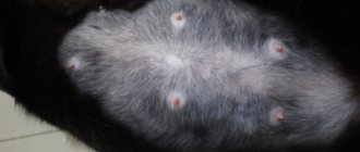
Often, squamous cell carcinoma can be found on the phalanges of one or more fingers. The growth rate of such neoplasms may vary.
The cutaneous form of SCC has a fairly low growth rate and practically does not metastasize to internal organs. The oral form of SCC, like the previous one, has a low level of metastasis, but a high growth rate, while destroying the jaw bones adjacent to the affected area.
Squamous cell carcinoma most often occurs in older dogs (8-10 years) and older cats.
No gender predisposition was found. That is, both males and females can be at risk in equal percentages.
Causes:
The causes of SCC can be varied, ranging from trauma to superficial tissues (skin) to genetic predisposition. A predisposing factor may be exposure to UV radiation, burns, or other physical factors affecting the skin. The infectious origin of this disease, which is associated with the papilloma virus, is also not excluded.
The appearance of oral squamous cell carcinoma in cats is most often associated with chronic inflammatory processes, such as periodontitis
.
The appearance of the internal (organ) form of SCC may be due to the presence of carcinogenic factors in the pet’s environment (tobacco smoke, etc.)
Forecast:
The prognosis for patients with this type of cancer can vary from favorable to severe, depending on the extent of the lesion, location, presence of metastases, and general condition of the patient. The time of detection of the tumor process also plays an important role. The sooner treatment begins, the higher the chances of a favorable outcome.
Symptoms and diagnosis:
Cutaneous forms of SCC are characterized by exophytic growth (growing outward). These are stable, raised formations in the form of plaques or nodules, often ulcerated. They can also look like warts or papillomas.
They most often occur on the abdominal wall, prepuce, inguinal area and scrotum in fair-skinned, short-haired breeds such as Dalmatians
,
bull terriers
,
beagles
. Clinical signs depend on the location of the tumor. Thus, in animals in which the tumor process affects the feet/toes, lameness may develop. The skin form can be detected by visual examination: erosions, plaques, papillomas.
Lesions in the oral cavity are characterized by decreased appetite in the pet, hypersalivation (excessive salivation), and bad breath (halitosis). Upon examination, you can detect erosive and/or ulcerated areas of the gums and tongue. In later stages of the disease, when the tumor process has affected the bone tissue, the animal may feel severe pain, which negatively affects its general condition. This is a complete refusal of food, lethargy, apathy, a desire to hide in a secluded place.
Other clinical signs include bleeding from the mouth, cough, swelling of the facial part of the muzzle, and loose teeth.
Diagnostics:
It includes a set of studies, starting with a blood test and ending with special diagnostic methods.
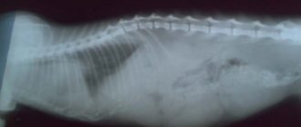
• general blood test • biochemical blood test • urine test • chest x-ray • biopsy of regional lymph nodes • smear impressions of affected areas • CT scan of the head
A complete diagnosis is carried out to determine the degree of damage to the body, the quantitative and qualitative involvement of internal organs.
The final diagnosis can be made by conducting a histological examination of material taken by biopsy from the damaged areas.
Breed predisposition:
Although all breeds of dogs can be affected, especially light-skinned and short-haired dogs, there are a number of breeds that are more prone to developing squamous cell carcinoma.
These are: Wolfspitz, Basset Hound, Standard Schnauzer, Collie.
The oral form of SCC is more common in large dogs: poodle, Labrador retriever, Samoyed.
No breed predisposition was identified in cats.
Treatment:
Like any other disease, squamous cell carcinoma requires a complete diagnosis before any treatment is prescribed. The diagnostic methods indicated above make it possible to determine the degree of development of the pathological process and prescribe adequate treatment, which may include both surgery and the use of chemotherapy or radiation therapy. Sometimes, a complex of these measures is required with excision of the affected tissue and the use of chemotherapy.
The choice of treatment depends on the location of the tumor and the degree of its infiltration into surrounding tissues. For example, if the soft tissues of the oral cavity are damaged and the tumor process invades the bone tissue, part of the bone base of the jaw can be removed. Unfortunately, this does not guarantee the animal’s complete recovery, but it can increase its lifespan with subsequent maintenance therapy. To summarize this material, we can say that timely and early diagnosis, as well as the vigilance of owners, can stop the pathological process at an early stage, which will greatly facilitate the treatment and subsequent recovery period of the pet.
Clinical classification. Types of melanoma
Melanoma manifests itself in various forms, there are 3 main types:
- Superficially widespread.
Tumor of melanocytic origin. The most common disease (70 to 75% of cases) among middle-aged Caucasians. Relatively small, complex in shape with uneven edges. The color is uneven, reddish-brown or brown, with small patches of bluish tint. The neoplasm tends to become a tissue defect, accompanied by discharge (usually bloody). Growth is possible both on the surface and in depth. The transition to the vertical growth phase can take months or even years.
Is it possible to get cancer?
In recent years, a lot of information has appeared that cancer is viral, but this is not true. Cancer cannot be infected, even if the affected cells are transplanted into a healthy body, as has been proven by numerous studies and experiments.
The experiments were carried out by scientists in Britain, the USA, Japan, France, Germany - and everywhere the result was the same - not a single sick subject.
Indeed, cat cancer by its nature is not much different from human cancer, but it is impossible to become infected with it.
What does melanoma look like in a photo?
- Nodal.
Nodular (diminutive from the Latin “nodus” - node) formation is less common (14-30%). The most aggressive form. Melanoma cancer is characterized by rapid growth (from 4 months to 2 years). Develops on objectively unchanged skin without visible damage or from a pigmented nevus. Growth is vertical. The color is uniform, dark blue or black. In rare cases, such a tumor, which resembles a nodule or papule, may not be pigmented.
- Malignant lentigo.
The disease affects older people (after 60 years) and is detected in 5-10% of cases. Open areas of the skin (face, neck, hands) are covered by dark blue, dark or light brown nodules with a diameter of up to 3 mm. Slow radial growth of the tumor in the upper parts of the skin (20 years or longer before vertical invasion into the deep layers of the dermis) can involve the hair follicles.
Causes of diseases
- Genetics. The main cause of cancer, as in humans, is genetics. Cancer is a mutation of certain genes, and they occur independently of us. In some cases, mutations are benign, and sometimes lead to the death of the organism.
- Carelessness. Cancer can develop from neglected or incompletely cured diseases, due to hormonal imbalance in the functioning of the body.
- Lack of care for the pet. In any case, if you do not follow the correct diet for your animal, or do not provide it with an active lifestyle, sooner or later it will get sick. Cancer is just one of the possible diseases.
The first signs of melanoma
Melanoma is the acquisition by cells of unfavorable signs of malignancy (malignancy properties), expressed by various symptoms.
To make it easier to remember the signs of melanoma, use the “FIGARO” rule:
Shape – swollen above the surface;
And changes - accelerated growth;
Borders are openwork, irregular, indented;
And symmetry is the absence of mirror similarity between the two halves of the formation;
P size – a formation with a diameter of more than 6 mm is considered a critical value;
About paint - uneven color, inclusion of random spots of black, blue, pink, red.
In widespread practice, the English version is also popular, summarizing the main, most typical features - the “ABCDE rule”:
A symmetry is an asymmetry in which, if you draw an imaginary line dividing the formation in half, one half will not be similar to the other.
B order irregularity – the edge is uneven, scalloped.
Color is a color that is different from other pigment formations. Interspersed areas of blue, white, and red colors are possible.
D iameter – diameter. Any lesion larger than 6 mm requires additional observation.
E volution – variability, development: density, structure, size.
Without special studies, it is difficult to determine the type of nevus, but timely changes in the nature of the spot will help detect malignancy.
Lifespan
With effective treatment, the survival rate of patients, depending on the type of tumor, is:
- Stage 4 liver cancer – about 5% of patients live more than 5 years.
- Stage 4 intestinal cancer – 5% of patients live more than 5 years.
- stage 4 lung cancer – 10% of patients survive for more than 5 years.
- Stage 4 stomach cancer – 6% of patients live more than 5 years.
- stage 4 pancreatic cancer – 10% of patients live more than 5 years.
- Stage 4 breast cancer – 15% of women survive more than 5 years.
- Stage 4 breast cancer – 15% of patients live more than 5 years.
- Stage 4 uterine cancer - depending on the spread of the process, the survival rate ranges from 3 to 9%.
- Stage 4 cervical cancer – 8% of women live more than 5 years.
- Stage 4 prostate cancer has a high five-year survival rate, about 30%.
The Oncology Department of the Yusupov Hospital in Moscow provides treatment for cancer at all stages of the disease. The diagnostic center and doctors of the oncology department make every effort to provide timely assistance to cancer patients. Seeing a doctor promptly can save your life. You can make an appointment for a consultation by phone.
Diagnostics
- Visual method. Examination of the skin using the “rule of malignancy.”
- Physical method. Palpation of accessible groups of lymph nodes.
- Dermatoscopy. Optical non-invasive surface examination of the epidermis using special devices providing 10-40x magnification.
- Siascopy. Hardware spectrophotometric analysis, which consists of intracutaneous (depth) scanning of the formation.
- X-ray.
- Ultrasound of internal organs and regional lymph nodes.
- Cytological examination
- Biopsy. It is possible to collect both the entire formation and its parts (excisional or incisional).
Prevention
A simple way of prevention would be to simply take care of your pet and be attentive. Take the animal 2-3 times a year for examination to a veterinary clinic to identify diseases. Provide your pet with a proper diet and active lifestyle. Monitor environmental conditions and do not allow your pet to consume excessive amounts of chemical additives. Little things like this can save your pet's life.
Treat your cat like you would a person. Pets cannot talk about their problem, and their health depends solely on the attentiveness of the owner.
Treatment
- Treatment of local local injuries consists of timely detection and surgical intervention. Removal is most often performed under infiltration anesthesia. For excision of large formations, general anesthesia may be used. In addition to malignant tumors, there are a number of pre-melanoma diseases in which the surgical method is indicated.
- Local-regional damage. Treatment includes wide-area excision and lymph node dissection of the affected lymph nodes. Types of unresectable, transiently metastatic tumors are subjected to isolated regional chemoperfusion. In certain cases, a combined approach has proven itself to be effective, with additional therapy that stimulates the immune system.
- Treatment of distant metastases is performed with monomodal chemotherapy. Certain types of mutations are targeted by targeted drugs.
Hemangiosarcoma
Hemangiosarcoma is a tumor that develops from the cells lining blood vessels (endothelial cells). This tumor usually affects middle-aged and older dogs of any breed. There is an increased risk of hemangiosarcoma in golden retrievers and German shepherds. Hemangiosarcoma develops slowly and does not cause pain to your dog for a long time. Often the disease is asymptomatic until a late stage, when internal bleeding develops due to tumor rupture. The organ most affected by the disease is the spleen, which can lead to extreme blood loss. The dog will show signs of severe shock - weakness, pale gums, shortness of breath. Hemangiosarcoma also often affects the heart, liver and skin.
Because symptoms do not appear until the disease has entered its final stages, complete recovery is unlikely even with extensive and thorough treatment. Treatment usually involves surgical removal of the spleen and bleeding, followed by chemotherapy after 2 weeks. Even with invasive treatments, most dogs once diagnosed with hemangiosarcoma die. With surgical treatment alone, the survival time is 2-3 months, with chemotherapy - 5-7 months.