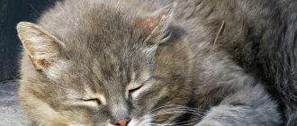
If a cat has an enlarged spleen, this may be a sign of diseases such as a malignant tumor, infectious anemia, liver disease, parasite infection, traumatic injury, eosinophilic gastritis. The condition is dangerous due to consequences in the form of splenic rupture, hemorrhagic shock, and cancer intoxication. At the first symptoms of splenomegaly, the owner should take the animal to the veterinarian, who will prescribe medications or perform surgery.
According to veterinarians, in 75% of cases, an enlarged spleen in cats is caused by a malignant tumor.
Reasons for the development of pathology
Oncological formations
Spleen cancer is rarely diagnosed in cats. Often, tumors in an organ form against the background of existing foci of cancer. The most common is hemangiosarcoma, in which cancer cells form in blood vessels. Factors in the appearance of spleen tumors include the presence of an animal in an area of increased radiation, prolonged contact with chemicals, hormonal imbalance, severe infectious diseases, and gene mutation. Symptoms:
- temperature increase;
- lethargy, refusal to play;
- the appearance of small hemorrhages on the gums and skin;
- vomit;
- poor appetite;
- intestinal disorder;
- frequent urination;
- bloating;
- deterioration of kidney function;
- sudden weight loss.
Infectious anemia
This infection causes yellowing of the mucous membranes of the animal.
The causative agent is Hemobartonella felis, an obligate parasite that develops in red blood cells. According to veterinarian T. Khleborad, in 50% of cases young animals under 3 years of age are affected, and about 75% of cats are carriers. The microorganism is activated due to decreased immunity, poor nutrition, against the background of a tumor of the spleen and other organs, and when infected with helminths. Hemobartonellosis manifests itself as follows:
- a decrease in the level of hemoglobin in the blood and its appearance in the urine;
- yellowness of mucous membranes and skin;
- increased heart rate and breathing;
- lethargy, fatigue;
- deterioration or lack of appetite.
Liver diseases
The causes of splenomegaly in cats lie in disruption of the gland due to hepatitis, which is provoked by viruses and leptospira bacteria. As well as chronic liver damage - cirrhosis. The pathology develops due to the use of certain medications by the owner of the cat, with heart failure, obstruction of the biliary tract, or due to hereditary diseases. Signs:
One of the manifestations of the disease may be severe thirst in the animal.
- unquenchable thirst;
- yellowness of the sclera, mucous membranes and skin;
- salivation;
- lethargy;
- intestinal disorder;
- vomit;
- increased urine production;
- ascites;
- splenomegaly.
Parasite infestation
The body of an adult cat and kitten is affected by roundworms, tapeworms, and toxocara. Worms migrate with the bloodstream and settle in various organs. Animals walking outside are more likely to get sick. Infection occurs during washing, when the cat licks helminth eggs from its paws, or when eating raw meat and fish. Symptoms:
- lethargy, apathy or aggression;
- abdominal pain;
- vomit;
- unstable stool;
- enlarged liver and spleen;
- loss of appetite or refusal to eat;
- change in taste preferences (the animal eats inedible objects).
Traumatic injuries
The condition is accompanied by internal bleeding, which poses a threat to the animal’s life, so the cat should be urgently taken to the veterinary clinic.
Similar injuries can be sustained by an animal when it falls from a height.
A bruise or rupture of the spleen provokes a fall from a height, a blow to the stomach with a heavy blunt object. In addition to this organ, the liver, kidneys, brain, and limbs are injured, so injuries manifest themselves as a complex of symptoms. The most common:
- severe pain in the body, the animal meows loudly;
- lameness or deformation of paws;
- breathing, heart rhythm, swallowing disorders;
- vomit;
- urinating or passing bloody stool;
- refusal to eat;
- loss of consciousness.
Eosinophilic gastritis
Characterized by the accumulation of eosinophils in the gastric mucosa. It mainly affects older cats. The occurrence factors are low-quality food, allergies to food additives, uncontrolled use of medications by the cat by the owner, infection with worms. Manifestation:
- vomit;
- weight loss;
- loss of appetite;
- black color of stool due to coagulated blood.
Spleen tumors in dogs
Damage to the spleen is often observed with tumors of the hematopoietic system.
But the spleen as an organ itself can become the site of localization of primary tumors and metastasis of other malignant tumors. An enlarged spleen in dogs (splenomegaly) is found quite often. In 43-75% of cases, the cause is tumors.
What types of tumors and mass formations of the spleen occur in dogs?
1) Primary:
- - hemangioma
- - hemangiosarcoma
- - sarcoma (various types)
2) Secondary or multicentric:
- - lymphoproliferative or myeloproliferative diseases (for example, lymphoma)
- - malignant histicytosis, histiocytic sarcoma
- - hemangiosarcoma
- - mastocytoma
- - other malignant tumors with distant metastases (for example, melanoma).
3) Non-tumor causes of splenomegaly:
- - nodular hyperplasia
- - hematoma
- - thrombosis or heart attack
- - stagnant changes
- - extramedullary hematopoiesis
- - torsion of the pedicle of the spleen.
The most common tumor of the spleen in dogs is hemangiosarcoma. This is a high-grade tumor with hematogenous metastasis in the early stages of the disease. Rupture of the primary tumor can lead to acute and fatal hemorrhage. It develops in dogs aged 9-10 years.
What are the reasons for the development of spleen tumors?
The etiology of malignant tumors of the spleen is unknown. Their high prevalence among dogs of certain breeds (German shepherds, retrievers, Labradors) indicates the presence of genetic factors. Mutations of the PTEN gene may be involved in the mechanism of initiation and development of hemangiosarcoma.
What are the manifestations of splenic tumors?
Benign tumors of the spleen do not cause any clinical manifestations, even if they reach a significant size. The reason to consult a doctor is an increase in the volume of the abdomen, which occurs due to the growth of the tumor. Or such a tumor is discovered during a routine examination.
Animals with splenic sarcomas may develop nonspecific symptoms (eg, malaise). They are detected during examination, x-ray or ultrasound examination, and during diagnostic laparotomy.
Hemangiosarcoma may have the following manifestations:
- - apathy
- - weakness
- - pallor
- - anorexia
- - fainting
- - hemorrhagic diathesis (spontaneous recurrent bleeding and hemorrhages of varying duration and intensity)
- - heart rhythm disturbances.
And other more severe manifestations:
- — acute collapse after rupture of a primary space-occupying lesion
- - abdominal bleeding (into the abdominal cavity)
- - acute vascular insufficiency.
However, hemangiosarcoma may not give any clinical manifestations and may be an accidental finding by a veterinarian.
How to diagnose splenomegaly or splenic mass?
- General clinical blood test. Hemangiosarcoma gives a number of hematological abnormalities: anemia (decreased hemoglobin), acanthocytes (damaged red blood cells) and schistocytes (red blood cell fragments), thrombocytopenia (increased bleeding due to a decrease in the number of platelets).
- X-ray examination. Allows you to identify a tumor or fluid (in case of bleeding) in the abdominal cavity.
- Ultrasound. Allows you to obtain information about the structure of the neoplasm and its location in relation to normal spleen tissue.
- Needle biopsy (there is a risk of bleeding) - tissue is taken using a syringe with a thin needle and examined under a microscope.
- Excisional biopsy (diagnostic surgery involving removal of the entire tumor being examined). It is used if there is a clearly visible tumor in the spleen.
- To detect metastases, chest x-ray and ultrasound of other abdominal organs are used.
How to treat spleen tumors?
Treatment involves:
- Surgical removal of the tumor. Unfortunately, in the case of a malignant tumor, surgery does not provide a cure.
- Postoperative chemotherapy to prevent or delay the progression of micrometastases. Monotherapy or combination chemotherapy is carried out. However, survival is relatively short. For combination chemotherapy protocols, it is on the order of 141-179 days, and only less than 10% of dogs survive more than 1 year.
What's the forecast?
The prognosis for dogs with splenic hemangiosarcoma is poor. Metastasis in the early stages of the disease is typical for this type of tumor. In most cases, micrometastases are already present at the time the primary tumor is diagnosed. They progress rapidly and cause low survival - 15-86 days after tumor removal.
For other types of spleen sarcomas, the prognosis is also unfavorable. Survival is about 4 months. The cause of death of the animal is metastases.
Histiocytic sarcoma has an extremely unfavorable prognosis. Most animals are presented for euthanasia or die at the time of diagnosis from extensive metastases.
What are the consequences?
If splenomegaly is not treated, the following complications occur:
The pathology can result in a state of shock for the animal.
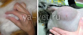
- Splenic rupture. When an organ is injured, a hematoma forms, which can rupture a few days after the injury. The condition threatens the animal's life, so the spleen is removed.
- Hemorrhagic shock. Appears with internal bleeding. The animal falls into a stupor, blood pressure drops, and death may occur.
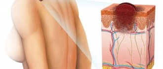
- Cancer intoxication. Occurs due to the breakdown of tumor cells. The released toxins enter the bloodstream and spread throughout the body.
Spleen tumors in cats
Spleen tumors are less common in cats than in dogs. As in dogs, damage to the spleen is possible as a result of leukemia and lymphoma.
Classification of tumors and space-occupying formations of the spleen of cats
1) Primary tumors:
- - mastocytoma
- - hemangiosarcoma
- - sarcomas (various).
2) Secondary or multicentric tumors:
- - lymphoproliferative and myeloproliferative diseases (for example, lymphoma)
- - hemangiosarcoma
- - other malignant tumors with extensive metastasis (for example, adenocarcinoma).
3) Non-tumor causes of splenomegaly (or splenic masses):
- - nodular hyperplasia
- - hematoma
- - stagnant changes
- - extramedullary hematopoiesis.
About 15% of tumor pathologies of the spleen in cats are lymphoreticular and visceral mastocytomas.
Symptoms of visceral mastocytoma
- Malaise.
- Anorexia.
- Chronic vomiting.
Presumably, these symptoms are associated with the formation of ulcers in the stomach and duodenum due to the influence of histamine on H2 receptors in the stomach. As the disease progresses, ulcers perforate, peritonitis and death of the animal occurs. Cases of splenic rupture have been recorded.
- Anemia due to blood loss from gastric or duodenal ulcers (or as a result of bone marrow infiltration).
Treatment and prognosis of mastocytoma
Treatment consists of surgical removal of the tumor. The prognosis is unfavorable.
How is the treatment carried out?
Drug therapy
If the disease is caused by worms, then the pet is prescribed the appropriate drug.
Any drug must be prescribed by a veterinarian; self-medication is prohibited. Antibiotics are used for infectious diseases. To reduce the possible development of allergies, it is appropriate to prescribe antihistamines. To strengthen the body, treatment is supplemented with vitamin and mineral complexes. Chemotherapy is used for cancer. Parasites are expelled by anthelmintic drugs.
How should you prepare an animal for an ultrasound of the spleen?
The fasting diet for dogs is 8-10 hours, for cats 6 hours with free access to water. At the examination site, the hair is mechanically removed (with a clipper), and a special gel is applied. In emergency cases, the examination is carried out without preparation. The patient is placed on the right side or on the back. Ultrasound of the spleen is performed with 5 and 7.5 MHz sensors. Normally, on ultrasound, the spleen is elongated and homogeneous. The contour is smooth, clear, sometimes the capsule is visible. Vessels are visualized. In dogs it reaches a width of 3 cm, in cats 1.5 cm.
In veterinary clinics in Vitebsk, Mogilev, Novopolotsk, Minsk (Belarus) and St. Petersburg, Smolensk, Sevastopol (Russia) of the Veterinary Center of Dr. Bazylevsky A.A. You can get detailed advice on performing an ultrasound scan of the spleen, undergo an examination and receive the necessary recommendations for the treatment and prevention of spleen diseases.
Hello student
PERIPHERAL LYMPHATIC TISSUE
In the mucous membranes of the digestive, respiratory and genital organs there are accumulations of round cells, mainly lymphocytes.
These clusters are considered to be local islands of the immune system. These infiltrates occur not only in the own layer of the mucous membrane, but also in other places, for example, in the form of so-called “milky spots” in the greater omentum and in the form of arachnoid cell accumulations in the pia mater. Spherical colonies of lymphocytes lead to the formation of primary follicles in young animals. By the time of the first contacts with the outside world, secondary follicles develop from them. In the center of the secondary follicle there are light differentiating reactive cells; on the outside they are covered with a layer of dark mature lymphocytes. Individual secondary follicles located in the mucous membrane become solitary follicles, folliculi lymphatici solitarii. They are visible to the naked eye, and their appearance has diagnostic value when examining the mucous membranes (nasal and oral cavities, foreskin, vestibule of the vagina, conjunctiva, etc.
Rice. 5. Schematic structure of lymph nodes
A primary follicle; Into a secondary follicle with a reactive center and a lymphocytic dome directed towards the epithelium. (Image of the reactive center is based on the results of studies by Gorgollon/Krsulovic, 1973)
Secondary follicles can unite into complex structures - Peyer's patches, folliculi lymphatici aggregati. They are located in the opposite-mesenteric side of the intestinal walls, their number and shape depend on the diet and composition of the intestinal flora, as well as on the age of the animal.
According to May (1903), in dogs, single lymph nodes are found along the entire length of the intestine, but in the small intestine they are almost invisible, and in the large intestine, on the contrary, they are clearly visible as whitish compactions the size of lentils. The number of Peyer's patches in the small intestine is 20 - 25 pieces, they have a round, oval or ribbon-like shape. Their length varies between 7 and 82 mm, width - between 3 and 11 mm. The last plaque is located in front of the opening of the ileum. There are no Peyer's patches in the large intestine.
In cats, individual lymph nodes are also found throughout the intestine. Peyer's patches are present in only 4 - 6 pieces. Their length is 4 - 30 mm. The last plaque, ending near the opening of the ileum, has a ribbon-like shape and is very long - 40-100 mm. At the apex of the cat's cecum there is a large accumulation of lymphatic follicles.
TONSILS
In the mucous membrane of the pharynx and in the submucosal layer around the entrance to the esophagus and the lower respiratory tract there is an accumulation of well-organized lymphatic tissue, generally referred to as the “lymphatic pharyngeal ring”. Its task is to recognize pathogens entering the body with food or air, and transmit this information to the organs of the immune system. The name of the individual tonsils is related to their position. The lingual tonsil (1), palatine tonsil (2), tonsil of the soft palate (3) are grouped around the oropharynx (a), and the pharyngeal tonsil (4) is located in the nasopharynx (b). In addition, the periglottic tonsil is found irregularly in the cat at the base of the epiglottis.
The lingual tonsil, tonsilla lingualis, is a collection of individual lymph nodes and diffusely located lymphatic tissue in the mucous membrane of the root of the tongue.
The palatine tonsil, tonsilla palatina, is located in the fold of the mucous membrane of the oropharynx. A fold of mucous membrane covers the depression, fossa tonsillaris. When you fold back the fold, you can see the tonsil itself, cylindrical in shape in a dog and hemispherical in a cat. Removing this tonsil, which is clearly visible with the throat wide open, is not particularly difficult.
The tonsil of the soft palate, tonsilla veli palatini, is formed by individual lymph nodes and diffusely located lymphoreticular tissue that penetrates the mucous membrane from the oropharyngeal side of the soft palate.
The pharyngeal tonsil, tonsilla pharyngea, is located on the posterior wall of the nasopharynx. In the direction of the openings of the auditory tube, the lymphatic tissue passes into separate nodules.
Peri-epiglottic tonsil, tonsilla para-epiglottica. It is a paired plate located at the base of the epiglottis and is found irregularly only in cats.
The pharyngeal and palatine tonsils are separated from the surrounding tissue by a thick connective tissue capsule. The epithelium covering them has transformed into spongy reticulum-like tissue and is permeable to lymphocytes. Under the epithelium in the lymphoreticular tissue there are numerous secondary follicles with an eccentrically located reactive center and a lymphocyte cap shifted towards the epithelium. The tonsils are well vascularized; they contain only efferent lymphatic vessels and no afferent ones.
SPLEEN
Spleen, lien's splen. is an organ of both the circulatory and immune systems. It monitors the state of blood cells and can serve to store blood. In addition, the spleen is involved in lymphopoiesis and can capture and accumulate certain metabolic products
Rice. 6. Palatine tonsil of a dog, transverse section, microscopic photograph (but Vollmerliaus, 1959. from Schunimer, 1987)
a velum palatinum; b radix liguac; with arcus palatoglossus 1 fossa tonsillaris; 2 subepithelial aggregated lymph nodes; 3 glandulae tonsillares
The external shape of the spleen in a dog and a cat is similar. It is flat and elongated, with a narrow dorsal end, extremitas dorsalis, and a wider ventral end, extremitas vmtralis. Its parietal surface, facies parlccalis, is smooth; on the visceral surface, facies visceralis, there is an elongated gate of the spleen, hilus lienis. In the area of the hilum, the spleen is relatively loosely connected to the greater omentum.
The spleen is always adjacent to the left abdominal wall. Its position largely depends on the fullness of the stomach. However, the own functional state of the organ, which can accumulate up to 16°o of all blood in a dog, influences the displacement of the ventral end to the right half of the abdomen. When the stomach is empty, the spleen in a dog is completely located in the hypochondrium; in a cat, in this case, its ventral end is always located outside the hypochondrium. With a moderately full stomach and in a dog, the ventral end of the spleen protrudes caudally beyond the border of the costal arch. When the stomach is full, the spleen lies in the iliac region and sometimes reaches the entrance to the pelvis.
Rice. 7. Dog spleen, visceral surface (according to Schummer, 1987)
a hilus lienis with the place of attachment of the greater omentum; b facies intestinal is; with facies gastrica 1st branch a. licnalis; 2 branches v lienalis
Rice. 8. Location of the spleen in a cat with a moderately full stomach, view from the left side (but Schummer, 1987)
A costal arch and min. intercostales; B diaphragma, open; With m. longissiinus; Dm. iliocostal is. E os ilium; F os ischii; G os pubis; G' azetabulum; Hm. bacrococcygeus dorsalis lateralis; Jmm. intcrtransversarii caudae, cut off; To m. sacrococ cygeus ventralis lateralis; Lm. iliopsoas; Mm. adductor sinister, cut off; N m. gracilis dexter; About m. rectus abdominis a aorta thoracica; a'n. phrenicus sinister; b. b' hepar: b lobus sinister lateralis, b' lobus sinister mrdialis; with omentum majus; d, d' ventriculus: d fundus ventriculi, d' corpus ventriculi; a»' oesophagus; e lien, facies parietalis; f colon transversum; g colon dcsccndcns; h a. et v. testicularis before entering the ostium vaginale; h' a. testicularis el plexus pampiniformis in canal is vaginalis; i ligamcntum vesicac medianum; i' ligamcnium vesicae laterale sinistrum; k vesica urinaria; l ren sinister; m ductus deferens; n canalis vaginalis; p scrotum; q corpus vagmale, open; r testis sinister, s cauda epididymidis: t praeputium; u penis; v m. ischiocavernosus; v' m. retractor penis; w m. sphincter ani externus; x rectum; in sinus paranalis; z Inn scrotales.
Rice. 9. The position of the dog’s spleen at different fullness of the stomach, schematically
a position with an empty stomach, b with a moderately full stomach, c with a very full stomach; the dotted line, dashed line and solid line show the corresponding contours of the diaphragm and abdominal wall
5, 7, 9, 11, 13 corresponding edges
Rice. 10. Diagram of the structure of the dog’s spleen (according to Vollmerhaus, 1984)
a peritoneum; b splenic capsule; c red pulp of the spleen; d lymphatic follicles of the spleen, white pulp of the spleen; e trabeculae
1 splenic artery; 2 trabecular artery; 3 pulp-paired artery; 4 central follicular artery; 5 brush arterioles; 6 sleeve capillaries; 7, 8 terminal capillaries with mouths in (7) splenic sinus and (8) parasinus reticular skeleton; 9 splenic sinus; 10 pulp veins; 11 trabecular veins; 12 splenic vein.
It is noteworthy that the dorsal end of the spleen is less mobile compared to the ventral one, since in this part the spleen is quite firmly attached by the gastrosplenic ligament to the greater curvature of the stomach. The possibility of palpating it in a living animal also depends on the size and position of the spleen. The spleen in dogs and cats is clearly visible on x-ray and ultrasound images.
Rice. 11. Segmentation of the spleen in a dog (after Heisse, 1989)
1 a. lienalis; 2 ramus dorsalis; 3 ramus ventralis; 4 a. gastroepiploica sinistra
Roman numerals indicate segments; at the top is the basic type with four segments, at the bottom are options with three or five segments.
The spleen is covered on all sides by a serous membrane - the visceral layer of the peritoneum; in the area of the hilum of the spleen and in an area located at right angles to it at the wide ventral end of the spleen, the fold of the visceral layer of the peritoneum passes into the greater omentum. Beneath the serosa is a thin capsule of the spleen. The pulp filling the space between the trabeculae is not differentiated with the naked eye. However, it is worth noting that a characteristic feature of red pulp in a dog is the formation of splenic sinuses, while in a cat these sinuses are very poorly developed or absent altogether (Schmidt et al., 1983 a, b).
The spleen is one of the organs with a very good blood supply. Blood flows from the celiac artery through the splenic artery, a. lienalis. Further branching of the arteries depends on the segmentation of the organ, which is the most important factor when performing a partial spleenectomy. Partial excision reduces the negative consequences of the operation (weakening of the immune system). The splenic artery passes in the splenic omentum medial to the dorsal quarter of the spleen. In dogs and cats, even outside the spleen, it is divided into dorsal and ventral branches, ramus dorsalis et ramus ventralis (dog: Thamm, 1941; Gupta et al., 1978; Heisse, 1989; cat: Pott, 1949). However, according to Godino (1964), this was observed in only 80% of the dogs examined.
The dorsal branch, together with two extrasplenic segmental branches (the cat has 3-5 of these branches, Pott, 1949) supplies blood to the dorsal third of the organ and often additionally gives off a small branch to the left leg of the pancreas. The ventral branch continues the caudo-ventral direction of the splenic artery and stretches to the border between the middle and ventral thirds of the organ. There it is also divided into two segmental branches, which, in turn, branch - also outside the spleen - into 4 - 5 subsegmental branches and supply blood to the middle and ventral thirds of the organ. In most cases, dogs have a four-segment type of spleen structure (Heisse, 1989), with both dorsal segments together making up only the dorsal third of the spleen, and the two ventral segments coinciding with the other two thirds. In most cases, the left gastroepiploic artery arises in an arcuate manner from the dorsal segmental branch of the ventral branch in the ventral direction. In other cases, the ventral branch, after separating the subsegmental branches for the ventral third of the spleen, continues as the left gastroepiploic artery along the greater curvature of the stomach.
The splenic veins run parallel to the arteries both in the area of the extrasplenic branches and within the segments, so a dog can have a partial ectomy of the spleen (for example, with ruptures or neoplasms), primarily of the ventral and middle third, since inside the spleen there are very rarely anastomoses between vessels and they are usually small (Godino, 1963; Gupta et al., 1981). Only in some cases do these segmental lines in the ventral half of the organ shift towards the ventral end, for example, with the five-segment structure of the spleen.
The rather large splenic nerves belong to the autonomic celiac plexus. Their functions primarily include control of the smooth muscles in the capsule and trabeculae of the spleen, the contraction and relaxation of which is directly related to the degree of filling of the spleen with blood.