One of the most common diseases that more than 80% of cats encounter throughout their lives is helminthiasis. They are characterized by a long and protracted course, and also have a wide range of clinical manifestations. Parasitic diseases occur not only in street cats, but also in domestic cats.
Read on to learn how to recognize a worm infection in your pet, what measures need to be taken, and whether parasites are dangerous for humans.
Read in this article
- Types and routes of infection
- Signs and symptoms
- Diagnostics
- How dangerous are worms?
- Is there a danger to humans? Probability of infection
- Anti-worm medications Tablets
- Suspensions
- Drops
- Injections
Types and routes of infection
Worms in cats can be round, tape or flat. The most common are roundworms and toxocaras, growing up to 3-5 cm in length. They are localized and multiply in the small intestine. Less commonly, hookworms (no more than 2 mm), heartworms and heartworms are detected in cats. They affect the intestines, lungs, muscles and cardiovascular system.
The most common routes of infection:
- drinking milk from an infected mother cat,
- eating small rodents (mice, rats, etc.),
- ingestion of worm eggs (for example, with dust and dirt from the soles of shoes),
- contact with other infected animals.
Infection with parasites is possible at any time of the year, but most often pets become infected in spring and summer. This is due to the fact that the most favorable conditions arise for the reproduction of helminths and the spread of their eggs. During this period, traveling with a cat to the country or hunting birds can cause infection.
Brief characteristics of parasites
Note that sometimes the term “pulmonary helminths” refers to the presence of migratory larvae of parasitic nematodes in a pet. Yes, the clinical picture is very similar, but still it is somewhat different. Lungworms are not short-term “guests” of a cat’s lungs. They live and reproduce there constantly, throughout their entire life cycle. And this cycle, by the way, can be very complex, since several intermediate hosts are involved in it.
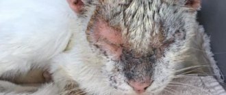
Signs and symptoms of helminthic infestation
It is difficult to recognize helminthiasis. If large individuals (5-10 cm) can be seen in feces, then small ones (up to 1-2 cm) go unnoticed. Note : in appearance, worms are long white “laces” curled into a tight knot.
You can identify helminthiasis in your cat by a number of signs:
- swollen belly (tight, full, barrel-shaped),
- digestive and stool disorders (recurrent vomiting, diarrhea, constipation),
- rare single but severe cough,
- appetite disorders,
- blood in the stool,
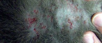
- dullness and matting of fur,
- increased licking of the area under the tail,
- rolling on the floor due to itching in the anal area,
- in especially severe cases, the pet becomes lethargic and depressed.
One of the signs of helminthiasis is anemia. Accumulating in the pet’s body, parasites adhere to the intestinal walls and provoke severe bleeding. Anemia in an animal can be determined by its gums: in a healthy cat they are pink, but in a pet with parasites they are white or grayish.
Have you noticed one or more signs of worm infestation in your pet? Do not hesitate - seek professional veterinary help at a veterinary clinic. Only an experienced specialist will be able to determine the type of parasites and prescribe an effective course of treatment.
Epidemiology
All cats can be infected with Aelurostrongylus abstrusus, regardless of their habitat, lifestyle, breed and gender. Infection with A. abstrusus has been detected in stray, wild and domestic animals [30, 47, 72, 73]. Of course, domestic cats kept primarily in houses and apartments that rarely go outside are much less at risk of becoming infected with A. abstrusus, while cats living outdoors with access to a large area in which they can roam and hunt, have more opportunities to eat shellfish and/or hunt. Studies conducted in Brazil [39] and Italy [73] showed that stray and free-roaming young cats were significantly more likely to be infected with A. abstrusus. In contrast, a study conducted in Australia found a high prevalence in older cats (in a semi-feral urban population), this was explained by the fact that during their lives the animals had more opportunities to hunt and ingest third-stage larvae than younger animals [47]. .
Also in Eastern Europe, the infection is more common in rural areas and in adult cats [17, 46, 55]. The nematode is distributed throughout the world, as confirmed by reports of cases of infection in almost all European countries, often in Australia and America, and sometimes in Asia and Africa [6, 27, 30, 39, 78]. The geographic range of A. abstrusus continues to expand, although the proposed reasons for this phenomenon are theoretical. The development and survival of gastropods depends on temperature, humidity and water availability, so one of the most important factors contributing to the spread of vector-borne pathogens is global warming. Although no specific information is available on A. abstrusus, it has been suggested that the spread of shellfish-borne parasitoses is driven by climate change [59, 78].
Therefore, given similar biological cycles, the same factors that promote the spread of other shellfish-borne parasites are likely to also influence A. abstrusus [78].
Table 1 provides data on the frequency of infection or isolated clinical cases of elurostrongylosis (and pulmonary capillariasis, see the corresponding section) in different countries.
Diagnostics
The first stage of treatment is the diagnosis of helminthiasis. It is aimed at determining the type of parasites, since further treatment depends on their characteristics. In a veterinary clinic, various methods are used to diagnose helminthiasis.
Among them:
- stool analysis,
- sputum analysis,
- X-ray examination of the thoracic region,
- ultrasonography,
- blood analysis.
After diagnosis, a course of treatment and specific medications are prescribed, aimed at destroying and removing parasites from the animal’s body, as well as relieving symptoms that arise.
How dangerous are worms?
Helminths seriously weaken the cat’s body, slowly depleting it. In advanced cases, in particular if left untreated, severe infection can lead to the death of the pet due to clogging of the intestines with lumps of parasites.
Worms provoke:
- loss of appetite,
- sudden weight loss in the pet (especially in the spine area),
- disruption of the digestive system,
- weakening of the immune system, etc.
To avoid these consequences, it is necessary to regularly carry out deworming, and at the first signs of infection, seek help from a veterinarian.
How to understand that a kitten has worms. Symptoms of helminth infection
Signs of the presence of worms in kittens are determined by their type and location. Common manifestations of infection include:
- partial or complete loss of appetite;
- digestive disorders with signs of diarrhea and vomiting;
- changes in behavior (apathy, lethargy, nervousness);
- deterioration in appearance.
Remember: similar symptoms can occur with other diseases. Giving your pet deworming medication for kittens without laboratory confirmation of helminthiasis may worsen its condition.
In some cases, other signs of a pet being infected with helminths may appear. When they are localized in the gastrointestinal tract - thirst, abdominal pain, pallor of the mucous membranes. When worms settle in the liver and pancreas of a kitten, it faces health problems such as yellowness of the sclera, skin, and pain in the right hypochondrium. When helminths are localized in the animal’s heart, it develops shortness of breath, rapid heartbeat, wheezing, and coughing leading to vomiting. When endoparasites form their colonies in the lungs of kittens, the latter experience symptoms such as hemoptysis, wheezing in the chest, and increased body temperature.
Is there a danger to humans? Probability of infection
About 32 varieties of worms can live in a cat’s body, which pose a danger to human health. The main cause of infection is non-compliance with hygiene rules. When feeding a cat by hand or playing with it, parasite eggs land on the skin of the hands and then penetrate into the oral cavity.
In addition, worms can enter the human body by touching surfaces on which eggs remain (for example, a tray).
The danger of infection with worms for humans (without proper treatment):
- enterobiasis,
- Iron-deficiency anemia,
- dyspeptic disorders (heartburn, diarrhea, etc.),
- damage to internal organs, etc.
To avoid dangerous consequences, it is necessary to regularly examine your pet, and if the first signs of helminthiasis occur, seek veterinary help. The specialist will conduct a thorough diagnosis of the cat’s condition and tell you whether they are dangerous to the human body.
Contraindications for deworming
You should stop taking antihelminthics in the following cases:
- if the product is not intended for use at this age, for example, in kittens;
- prohibited for use in sick and/or elderly pets;
- Contraindicated during pregnancy and nursing kittens.
For preventive purposes, it is recommended to carry out deworming once every 3 months.
Medicines for worms
External and internal drugs are used to treat helminthiasis. They not only eliminate the signs of the disease (diarrhea, bleeding, digestive disorders, etc.), but also help to completely get rid of parasites.
It is strongly recommended to abandon traditional methods in the treatment of helminthiasis. Remember that only after diagnosis and a complete examination can your pet be prescribed anthelmintic drugs.
Drug formats:
- pills,
- drops,
- suspensions,
- solutions for injections.
The veterinarian prescribes single-component drugs, as well as broad-spectrum drugs. The latter are aimed at destroying different types of parasites at the same time. Keep in mind that drugs and their dosage are prescribed by a specialist in each specific case individually.
Pills
Tablets are the most common format of drugs used in the treatment of helminthiasis. They are crushed and then added in crushed form to your pet's food or dissolved in water. Among the most effective drugs in tablet form:
- Polyverkan,
- Kanikvantel,
- Milbemax,
- Drontal,
- Dirafen,
- Alben.
Suspensions
To remove worms from a cat, various suspensions are also used. They need to be given to the pet through a special dispenser syringe (orally), and then after 6-8 hours you need to give activated charcoal. This is the most optimal solution for kittens 3-4 months old.
Among the most popular suspension preparations:
- Dirofen,
- Prazicide,
- Pirantel,
- Prazitel.
Drops
Another convenient and effective format of anti-parasite medications for cats is drops. Usually they have a complex effect, so they cope not only with worms, but also with other problems (fleas, lice, ticks, etc.). They must be applied to the withers, since there the cat will not be able to lick them off.
Among the most effective drops are:
- Dironet,
- Bars spot-on,
- Advocate,
- Stronggold,
- Profender.
Injections
In the treatment of helminthiasis, not only tablets, drops and suspensions are used, but also injections. They are more effective, but keep in mind that each time you will need to go to a veterinary clinic. It is strongly not recommended to give injections to your pet at home.
Among the most effective injection drugs:
- Novomek,
- Barmek,
- Ivomek.
Diagnosis
Feline elurostrongylosis is difficult to differentiate in modern veterinary practice because other overlapping conditions must be considered when making a differential diagnosis, such as the presence of other parasites, mycoses, viral and bacterial infections, nasopharyngeal polyps, allergic bronchitis, foreign bodies and neoplasms in the respiratory tract [ 19, 28, 78]. Veterinarians typically misdiagnose elurostrongylosis and treat the condition as an allergic respiratory disease or chronic bronchitis/asthma in cats [28]. Because symptomatic treatment is used, the infected cat may show improvement in clinical signs after administration of corticosteroids and bronchodilators, and therefore clinicians have no reason to suspect that they have made an error in diagnosis [18, 28]. X-ray data depend on the number of helminths in the body and the chronic nature of the disease. The earliest changes observed 2-3 weeks after infection include thickening of the bronchi and small, poorly defined nodules in the lung area, mainly in the caudal lobes. Changes in alveolar pattern can occur 5–21 weeks after infection, and after recovery, thickening of the bronchial wall and increased interstitial opacity usually persist for several months [52, 53]. Changes in bronchial and interstitial patterns (Figure 3), usually observed after partial improvement in alveolar disease, complicate the diagnosis of the disease as they overlap with changes that occur in other feline bronchial diseases [52, 53].
Figure 3. X-ray of a short-haired cat with elurostrongylosis, lateral view of the chest: in the caudal lobes of the lungs there is intense shading of the bronchi and interstitial tissue, which makes it difficult to visualize the vascular system of the lungs.
Computed tomography (CT) provides higher resolution images of the lungs than conventional radiography and may be useful in identifying poorly defined bronchial walls and nodules [60]. However, when evaluating CT images, it is difficult to differentiate elurostrongylosis from other lung diseases, and CT equipment is often unavailable.
The use of classical serological tests (eg, indirect fluorescent antibody test, IFAT) is limited by cross-reactivity with antigens of other endoparasites and an unsatisfactory ability to distinguish past from existing infection [36]. Antibodies to A. abstrusus determined by the IFAT method in the sera of both experimentally and naturally infected cats have shown promise in terms of sensitivity and specificity [14].
Suspicion of elurostrongylosis based on clinical signs is confirmed by the results of microscopic examination of feces, carried out to detect the first stage larvae (Figure 4) in the feces of an infected cat. Methods such as fecal smears and classical sedimentation and flotation methods are not very effective for diagnosis due to the inability to guarantee the presence of larvae in the sample, insufficient sample size and insufficient sensitivity of the methods [8, 78]. The reliability of the classical method of microscopic examination of feces depends on the solution used and the period of time, since concentrated solutions with a high specific gravity cause osmotic damage to the larvae and their dehydration. Dehydrated and/or drowned larvae (Figure 5) may lack morphological details and are therefore very difficult to detect and identify [67, 78]. The Baermann migration method is the gold standard for diagnosing feline elurostrongylosis, with a sensitivity of approximately 90%, although for detection of larvae it takes 24-36 hours, and recognition of first-stage larvae requires some skill [47, 78]
Knowledge of the characteristics of the first instar larvae of A abstrusus (Figure 4) is critical for correct diagnosis in clinical practice. According to data obtained from scientific literature sources, the larvae of the first stage of A abstrusus have a length from 360 to 400 microns [8, 10, 11, 13, 44, 47, 51, 60, 61, 67], a rounded head with a mouth opening at the end and a curved (S-shaped) tail with distinct nodular or small spine-like structures at the ends of the cuticle projections [13, 31, 43, 78] (Figure 4).
Larvae of shorter length, up to approximately 300 µm, have also been reported, and these cases of Elurostrongylosis have been confirmed by histological analysis [25] and genetic evaluation [43]. Therefore, the identification of first-stage larvae of A abstrusus should also be based on morphological features (i.e., head and tail shape), and not only on the results of measurements of their size [13, 57, 81]. First-stage larvae of A abstrusus must be distinguished from those of other lung nematodes (see relevant sections), from hookworm larvae, which may be present in samples left for incubation, and from free-living nematodes, which may be present in samples collected from the soil surface [ 78]
Figure 4. First stage larvae of Aelurostrongylus abstrusus.
Microscopic examination of feces does not allow identifying the pathogen during the period of parasitic incubation, as well as during the period when larvae are not released from the host’s body, even in the presence of serious clinical signs. In fact, first-stage larvae are periodically shed in cats with chronic infection, which alternate between cycles of shedding and non-shedding of larvae [34, 66]. Consequently, false-negative samples are common and repeat testing is necessary [67, 78].
First-stage larvae may be found in other specimens, such as tracheal swabs or washings, bronchoalveolar lavage fluid (BAL) (Figure 6), pleural effusion, expectorated material, and transtracheal lavage fluid. These approaches are invasive, have potential risks, and do not always detect first-stage larvae due to lack of significant lung tissue damage, limited numbers of first-stage larvae during parasitic incubation, or poor sample recovery [78].
BAL testing is a less sensitive method than the Baermann migration method, but the simultaneous use of both methods can be proposed to detect inconsistent excretion of first-stage larvae in feces [47].
Figure 5. Dehydrated and drowned first-stage larvae of Aelurostrongylus abstrusus.
To overcome the limitations of classical diagnostic methods, a nested PCR method based on genetic markers in ribosomal DNA (rDNA) was evaluated. This assay showed 100% specificity and a sensitivity of 96.6% when used on pharyngeal swab specimens, and was able to diagnose the disease in cats that were negative using classical diagnostic methods [74].
Prevention of helminthiasis
Try to do everything you can to prevent your pet from becoming infected. A number of preventive recommendations developed by veterinarians will help you with this. These include a ban on feeding raw fish and meat. The food must first be frozen, but it is best to boil it.
Prevention:
- Regularly change the bedding on which the pet lies, since parasite eggs can also remain on it;
- Keep the cat's litter box clean and change the litter frequently. It is recommended to disinfect the toilet 1-2 times a week;
- Preventive deworming should be carried out every 3-4 months, especially for cats that go outside;
- It is recommended to leave outdoor shoes and clothing in places that are inaccessible to the cat, since they can carry helminth eggs.
Helminthiasis is an unpleasant problem, but with the right approach it can be quickly resolved. The main thing is to promptly recognize the disease and treat the cat using various medications (tablets, drops, suspensions, etc.). After completing treatment, do not forget about prevention. Get dewormed and checked by a veterinarian regularly!
Pet treatment
No self-medication! Even if an adult parasite is detected in the feces, you do not need to select the drug yourself.
Do not listen to the advice of “experienced” people, otherwise you can only harm your pet. Be sure to contact the veterinary clinic.
Many helminthiases require complex treatment (not just one drug) and monitoring by a veterinarian. Repeated tests are necessary to confirm that there is no trace of worms left.
In most cases, the drug is administered twice, since the first time the adults are destroyed, and the larvae do not “react.” After 2 weeks they grow up, and the drug is already working on them.