Alas, pets are not only happiness, but also a great responsibility, since it is people who determine how long and carefree a pet will live. He cannot talk about his poor health, so the task of a responsible owner is to ensure that the kitten is well-groomed, fed, and feels good. It is important to pay attention to external signs of disease.
The article will tell you why a cat’s eyes are half covered with a film, what are the causes of the disease and methods of its treatment.
Short description
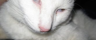
Unfortunately, a disease in which part of a cat's eyelid is covered with a white, opaque film is common. The film bears the name of the third century and is of great importance. It removes foreign particles and dust, and also distributes tear fluid. The third eyelid (conjunctival fold or nictitating membrane) is located at the inner corner of the eye. It is almost impossible to notice it in a healthy animal. If the cat is sick, the membrane becomes inflamed and turns white.
Conjunctivitis
As a rule, this disease is rarely registered as an independent disease. Most often, this is a signal of the presence of other problems: mechanical injuries, infectious diseases, parasitic worms in the eyeball, eyelash growth inside the eyelid (trochiasis), and even incorrectly selected dry food.
Symptoms of conjunctivitis:
- Photophobia;
- Swelling and redness;
- Frequent washing of the face;
- Decreased appetite;
- Discharge (which increases and the eye sticks together).
Main reasons
The reasons for the appearance of a white film on a cat’s eyes can be varied:
- eye diseases such as conjunctivitis;
- mechanical impact;
- getting foreign objects into the eyes;
- parasites (internal and external);
- neoplasms (adenomas);
- allergy.
Let's look at some of them in more detail.
Infection
The action of the main contagion is combined with the influence of secondary microflora:
- rhinotracheitis;
- calcivirosis;
- panleukopenia;
- chlamydia.
Congenital anomalies
It is believed that some cat breeds are genetically predisposed to eye diseases. At risk, for example, are Persian cats and other brachycephalic breeds.
Invasion
Eye inflammation is caused by the following pathogens:
- protozoa;
- toxoplasmosis;
- helminthiases.
Permanent pathologies
These include pathologies such as hepatitis, pancreatitis, diabetes mellitus and other chronic diseases. They weaken the immune system, making the cat defenseless against the microflora that causes conjunctiva.
Hypersensitivity
Plant pollen, perfumes and accessories can be irritants in the formation of conjunctivitis, which is accompanied by the appearance of a transparent film on the cat's eye.
Third eyelid on both eyes
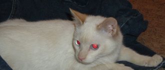
If the film is only on one of the eyes, then the reason for this is most likely a foreign body that caused a reaction in the eye. And if such a third eyelid appears on two eyes of a pet at once, then this is already a sign of the appearance of various serious diseases. Various eye problems in cats occur due to injury or infectious disease. Severe, severe injuries often appear in cats after fights, and problems with the cornea of the eye often arise because of this. Infected claw wounds are terrible in themselves, although it will be even worse if a dangerous infection gets into the wound, as this can lead to severe complications.
Most often, such wounds after a cat fight will cause the formation of acute keratitis and a creeping, terrible corneal ulcer.
Because of this, the cat's eye will first become cloudy and erosion will form on its surface with fairly pitted common edges. Further, your pet may develop white or greenish discharge; this is a very dangerous phenomenon that requires the intervention of a veterinarian who has good experience in this matter. It is required to carefully and as often as possible monitor the behavior of the animal, and if symptoms of the disease appear and if the cat behaves strangely, you must immediately go to a veterinary hospital. It is also recommended to feed your pet more and better, giving him vitamin B12 daily.
External manifestation
Third eyelid disease can be diagnosed not only by changes in the pet’s appearance, but also by changes in behavior. Most likely, the cat is hiding from bright lighting, rubbing its face with its paws, and especially its eyes, blinking and squinting frequently due to noticeable discomfort. You can also see profuse lacrimation and even the presence of pus. If treatment is not carried out in time and you do not consult a veterinarian, there is a high probability of vision loss.
You can also notice the third eyelid by the presence of the following inflammations:
- adenoma;
- cartilage malformations;
- protrusion of the nictitating membrane.
Adenoma
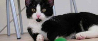
This is a pink tumor that is located near the bridge of the nose. It is benign and resembles a bean in shape. The main causes are injury or infection. It is easy to detect; the eye stops closing. If not treated promptly, serious complications can occur.
Cartilage malformations
Defective cartilage is deformed during the growth phase, as a result of which the nictitating film begins to become inflamed. It is one of the congenital anomalies.
Protrusion of the nictitating membrane
If the third eyelid appears in only one eye, the causes of the pathology may be as follows:
- foreign objects under the film;
- injuries to the facial nerves, cornea or cartilage;
- accretion of the membrane to the eyeball.
If the third eyelid appears in both eyes, the following reasons for its occurrence are distinguished:
- infectious conjunctivitis;
- helminthiases.
Own diseases of the third century
There are a number of specific diseases of the nictitating membrane.
Prolapse (loss) of the lacrimal gland
Lacrimal gland prolapse is rare but occurs in brachycephalic cats. This often occurs during a period of active growth of the cat, at the same time the size of its eyes rapidly increases. The ligament that holds the lacrimal gland of the nictitating membrane in its normal place - under the conjunctiva - is torn. The lacrimal gland emerges into the inner corner of the eye, becomes visible and looks like a small pink rounded formation. When displaced, the lacrimal gland is pinched, it swells and grows in size, and conjunctivitis develops.
Prolapse of the third eyelid lacrimal gland often occurs during rapid growth in cats.
This bothers the cat; when scratching with its paws, secondary flora is introduced, and the course of conjunctivitis becomes purulent. If the lacrimal gland is displaced significantly and for a long time, then its blood circulation begins to suffer and the production of tear fluid decreases. Its pronounced deficiency will lead, in the absence of measures taken, to the development of keratoconjunctivitis sicca. Also against this background, a crease (curvature) of the cartilage of the nictitating membrane may occur.
Only surgical treatment is applicable - the displaced lacrimal gland is immersed in the formed conjunctival pocket and sutured with sutures using atraumatic needles and thin absorbable threads (the sutures do not need to be removed later). The operation takes no more than half an hour; in the postoperative period, local and systemic antibacterial drugs are used, as well as an “Elizabethan” collar if the cat rubs its eyes with its paw.
Video: lacrimal gland prolapse
Kink (eversion) of the third eyelid cartilage
The manifestations of a hall of the third eyelid cartilage are similar to the prolapse of the nictitating membrane. Curvature of the cartilage occurs, and part of it is noticeable when examining the inner corner of the eye. In this case, the lacrimal gland can either shift or maintain its normal location. Treatment is also surgical - the curved and protruding part of the cartilage tissue is removed.
The third eyelid cartilage gap can only be corrected surgically
Third eyelid injury
Injury to the third eyelid usually occurs in fights. Initially, there is slight bleeding, secondary conjunctivitis develops, and there may be blepharospasm. Minor injuries heal on their own and without consequences for the function of the nictitating membrane, but in cases where its torn part becomes mobile or cartilage tissue is visualized, a surgical operation is performed to restore the size and full function of the nictitating membrane, as well as eliminate irritation of the conjunctiva by torn tissues and cartilage.
A rupture of the nictitating membrane usually occurs in fights between cats.
Neoplasms of the third century
Neoplasms of the third eyelid are also rare, but are dangerous due to the malignancy of most tumors in this location. A small formation is surgically removed and a histological examination is performed to clarify the nature of the tumor. With a more pronounced spread of the tumor process, the entire nictitating membrane must be removed. The identified type of tumor influences further treatment measures and the prognosis for the cat’s life. Therefore, in all cases of impaired mobility, changes in the structure, shape and color of the third eyelid, it is necessary to exclude the presence of a tumor.
Some veterinarians distinguish lymphoid hyperplasia of the third eyelid - the lymphoid tissue contained in the thickness of the third eyelid grows under the influence of an infectious process or constant irritation; When blinking, the overgrown follicles injure the cornea. The cat has discharge from the eye and blepharospasm. When examined on the surface of the third eyelid, overgrown follicles are identified as a rash or small volumetric formations. Often this causes a similar proliferation of lymphoid tissue on the inner surface of the upper and lower eyelids. Surgical treatment is curettage (scraping) of the overgrown lymphoid tissue, followed by the use of antibiotics and anti-inflammatory drugs.
Diagnostics
If you see that your cat's eyes are covered with a film, we advise you to contact a veterinarian as soon as possible. Timely treatment will help quickly restore vision and avoid re-infection.
A specialist doctor uses an ophthalmoscope to determine the cause of the underlying pathology of the eye disease and propose an effective course of treatment. In some cases, surgery may be required.
It is imperative to monitor the dynamics of the disease.
Keratitis in cats
Another one of the most common ophthalmological diseases in cats. Keratitis is an inflammation of the cornea. You can recognize it immediately by a clouded eye.
This disease can also occur due to other reasons: foreign body, conjunctivitis, eye burns (difficult to treat), allergies, infection, vitamin deficiency, inflammation of the lacrimal glands, genetic predisposition (most often artificially bred cats are prone to this disease - Siamese, Persians, British).
In addition to clouding of the cornea, you need to pay attention to a number of symptoms that will confirm your fears:
- Manifestation of blood vessels;
- Severe photophobia;
- Fluid accumulation inside the eye;
- The appearance of scars (indicates that the disease is at the last stage and further treatment will most likely not bring any results).
As you probably already understood, independent treatment is contraindicated, and first you need to find out the causes of keratitis. You can examine the eye for the presence of foreign bodies, assess the general condition, but only a qualified veterinarian can determine the severity of the disease and prescribe treatment.
Cloudiness of the eyeball indicates the possibility of more complex internal diseases.
Treatment
To treat this type of disease, veterinarians offer several methods:
- drops or ointments with an anesthetic effect - they help relieve inflammation;
- inclusion of vitamins B12 in the cat’s diet;
- balanced diet;
- washing the eyes using olive oil, warm tea, boric acid or just warm water.
The simplest method, but no less effective, is considered to be preventive eye rinsing. This procedure makes it possible to remove dust and foreign particles from the eyes.
How to wash your eyes
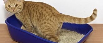
The rinsing procedure is required periodically when the eyes become watery, suppuration appears, or a film forms. Before starting, you should wash your hands thoroughly with soap to avoid further infection. It is better to rinse with an assistant: one person fixes the pet, the second acts, this way the process will go faster.
You need to use a special solution to wash your eyes. Rinsing is carried out as follows: thoroughly moisten a cotton swab in the solution and then carefully rinse both eyes of the animal. After completing the procedure, be sure to blot your eyes with a dry cotton pad.
Acute and chronic form
Keep in mind! In adult patients, dacryocystitis can develop in acute or chronic forms.
The two types of disease can be distinguished by symptoms, and in the acute form the following symptoms can be observed in the patient;
- increased body temperature;
- swelling in the area of the lacrimal sacs ;
- narrowing of the palpebral fissure;
- possible swelling of the eyelids ;
- pain in the orbit of the eye;
- manifestation of general symptoms of intoxication of the body.
The tumor , which can be easily felt in the area of the lacrimal sac, may initially be dense to the touch , but after a few days it begins to soften and the swelling subsides.
During this period, an abscess forms, which can spontaneously open, and due to the outflow of pus from it, swelling decreases.
In the chronic form, the patient does not experience pain , but at the same time he experiences strong constant lacrimation , and the swelling in the area of the lacrimal sac turns into a tumor, upon pressing on which pus begins to flow out of the lacrimal canaliculi.
Prevention measures
The most effective way of therapy is to prevent the problem.
To prevent inflammation of the third eyelid in a cat, use the following recommendations:
- Create a balanced cat diet from quality products.
- Be sure to add vitamins to your food.
- Periodically carry out eye rinsing procedures.
- Monitor your pet for changes in behavior and examine its eyes regularly.
- Visit a veterinarian at least once a year.
Remember, the sooner you detect eye disease in a cat, the greater the likelihood of its speedy cure. You should not self-medicate; you should immediately take the animal to the veterinarian.