Quite often, veterinary specialists are faced with diagnosing leukoma (eye sore) in patients. The pathological process is direct evidence of the onset of the inflammatory process in the transparent protective layer of the eyeball - the cornea.
Neglect of the disease and untimely assistance can lead to serious problems in the cat such as blindness or complete loss of the eyeball.
Leukoma or cataract is a clouding of the cornea of the eye with the formation of a scar. The development of leukoma is provoked by such negative factors as thermal and chemical burns, allergic reactions, and mechanical injuries. White connective tissue begins to grow after damage, impairs the cat's vision and can lead to complete blindness.
The cat's eyesore, like other animals, is divided into several types. It depends on the location of the white film. There are peripheral leukoma, which covers the edge of the eye (peripheral vision is impaired), as well as central (develops in the center) and total (affecting the entire visual surface).
The inflammatory process that develops with leukoma causes the scar to rise above the cornea.
General information
A cataract is any opacity of the cornea; Previously, this concept also included cataract - clouding of the lens - however, taking into account the fundamental etiopathogenetic and anatomical difference, currently these two diseases are separated and considered as independent.
As you know, a healthy cornea is transparent. Being the outermost, open part of the eye's optical tract, the cornea performs both protective and focusing functions. In fact, this is a natural concave-convex lens with a diameter of 1 cm, a thickness of 0.5-1 mm (thinner in the central part, thicker towards the points of diffuse transition into the sclera) and an optical power of 40 diopters.
Any damage, any changes in shape and refractive parameters, degeneration of the multilayer structure of the corneal tissue inevitably and automatically lead to a pronounced and, as a rule, uncorrectable decrease in vision. With all the variety of causes of cloudiness or complete opacity of the cornea, this condition can only be treated surgically, and it is necessary to take all necessary, available, and possible measures to protect the cornea and prevent the formation of a cataract.
Rehabilitation after therapy
During the recovery stage, the animal must receive high-quality and healthy nutrition.
Treatment usually takes a long time, on average about 3 months. After therapy and elimination of eye cloudiness, animals are often prescribed antiviral medications and deworming. In addition, it is important for the owner to monitor the animal’s diet. It is permissible to use only high-quality complementary foods recommended by the veterinarian. If surgery has been performed, immediately after it you should slightly limit your pet’s physical activity, and also regularly visit a veterinarian to reduce the likelihood of developing postoperative complications.
Causes of corneal opacities
Unlike the lens, which can become cloudy due to endocrine and hematological disorders, to some extent due to the aging of the body and other purely internal, endogenous reasons, the cornea almost never becomes cloudy “on its own.” In the vast majority of cases, the process, injury or other damaging factor that resulted in the occurrence and/or progressive development of the cataract is diagnosed and identified. The main reasons include:
- viral, bacterial, fungal infections, which often begin as “ordinary” conjunctivitis, spread to the cornea (keratoconjunctivitis, keratitis) and, with extensive ulceration, leave an opaque scar surface. As a rule, this is also accompanied by pronounced thinning of the corneal layer and the formation of a convex cataract (ectatic leukoma);
- mechanical wounds and eye damage, incl. trauma during ophthalmic surgery;
- chemical burn of the eye, especially with substances with an intensely alkaline pH reaction;
- intrauterine infectious-inflammatory process resulting in a “ready-made” congenital cataract in the fetus (statistically rare option).
Symptoms of the disease
Any visually noticeable change in the condition of the eyes signals the need to contact a veterinary clinic. What symptoms indicate the possible presence of cataracts in a cat? Let's look at the most common ones:
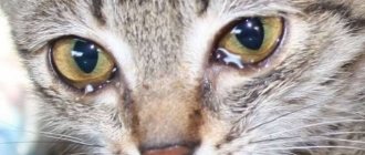
1. Covering the ocular surface with a whitish film with a grayish or yellow tint, without black dots. 2. Discharge from the affected area contains pus. 3. The animal is afraid of light and hides in the dark. 4. Deterioration of vision: the cat bumps into objects in a familiar room. 5. Redness and swelling. 6. Nodules may form. 7. Pain when trying to touch the eye.
Symptoms
Actually, the main and most severe symptom is a decrease in visual acuity in the affected eye. In all cases, the decrease reaches a deep degree, sometimes vision is limited only to residual light sensitivity or complete blindness occurs. With perforations and adhesions (“soldering”) of the cornea and iris, in addition to leukoma, a syndrome of increased intraocular pressure (secondary glaucoma) usually develops.
Depending on the nature of the lesion and its stereometric characteristics, for example, on the degree of convexity of the scar surface, secondary irritation of the conjunctiva of the eyelids may also develop, which creates favorable conditions for recurrent infection. Often, especially when the cornea is thinning and the layer of nerve endings is exposed, patients complain of photophobia, increased lacrimation, and pain.
Preventing eye problems in cats
To prevent the development of the disease, it is important to adhere to a number of simple rules:
provide the animal with healthy, nutritious nutrition;- regularly examine your eyes;
- protect your pet from contact with other animals;
- do not let outside unattended;
- carry out vaccinations in a timely manner.
It is also necessary to regularly visit the veterinarian as a preventative measure, which will allow the disease to be identified at the beginning of its development.
Treatment of corneal thorn
In cases of exposed nerve endings, severe hyperemia, lacrimation, and painful sensitivity to bright light, special contact lenses-protectors are sometimes prescribed. It should, however, be understood that this is not a treatment and serves only as a palliative method to alleviate symptoms.
Moreover, the countless “folk remedies” that quasi-medical websites vying with desperate patients cannot be considered a treatment: these “recipes” include honey (which for many people is a powerful allergen), aloe, and eyebright in various forms, and mother’s breast milk, and the bile of freshly caught pike, and many other substances, sometimes completely unexpected in this context.
As medicinal support, additional or symptomatic treatment, the ophthalmologist may prescribe an anti-inflammatory regimen, absorbable drugs; the use of physiotherapeutic procedures (for example, electrophoresis with hormone-containing agents) and the prescription of keratoprotectors (Balarpan, Korneregel, etc.) are reported.
However, conservative therapy in this case is just a palliative and does not radically solve the problem. The only effective method of eliminating a cataract has been and remains surgical treatment.
Our ophthalmology center successfully performs surgical treatment of all types of corneal opacities. Using various types of keratoplasty, our ophthalmic surgeons restore vision and remove cosmetic defects. You can find out prices for treatment and make an appointment by calling the phone number listed on the website.
Diagnostics: techniques and methods
During the initial examination, the veterinarian evaluates the clinical picture and conducts an external examination of the pet, especially the eye area.
The specialist also collects information about when the spot was first noticed, the quality of food, and studies the animal’s history.
Based on the data obtained, a diagnostic examination is prescribed, including the following manipulations:
- clinical and biochemical blood test;
- ophthalmoscopy;
- serological tests;
- determining the pressure in the eye (to assess how likely it is to develop glaucoma or cataracts);
- Seidel test (fluorescein test);
cytological examination of damaged tissue taken from the conjunctiva;- biopsy;
- microscopic examination of scraping;
- gonioscopy.
If for some reason it is not possible to conduct a standard diagnostic examination, the doctor may prescribe an ultrasound examination, which will help identify the true causes of clouding of the eyes.
If concomitant diseases of viral and infectious origin are suspected, additional diagnostic methods may be prescribed.
Operations for corneal opacities
Various techniques and protocols are successfully practiced, selected “according to indications” depending on the specific clinical situation. Typically, penetrating or partial keratoplasty is used - respectively, complete (over the entire thickness and area) or partial, more gentle replacement of the affected area or layer of the cornea with a donor keratobioimplant. Such operations are well established and, as a rule, effective, especially if all instructions for the rehabilitation period are followed, which in some cases can last up to a year or more.
According to statistics, the most successful, in terms of short-term and long-term prognosis, are operations for keratoplasty of cataracts caused by infectious ulcerations/scarring. A necessary condition is sanitation and prevention of recurrence of infection.
As for the prevention of the cataract itself, the first and main rule should be to contact a qualified ophthalmologist for any signs of conjunctivitis or other inflammatory process (before the cornea is involved in it), for any injuries and burns of the eye. In a very significant proportion of cases, the cataract could have been prevented without delaying the situation and without bringing the situation to ophthalmic surgery.
Types of pathology
An eyesore is classified according to several criteria.
By location type
Highlight:
- peripheral;
- total;
- central.
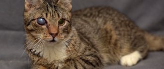
In the first case, the pupil is not affected; the spot is mainly localized on the side of the eyeball, has a white tint and black inclusions.
With the total type, the cataract completely covers the entire eye.
In the central form, the center of the visual organ is affected, and complete occlusion of the pupil is often noted.
According to the pathological process
Leukoma can be congenital or acquired . The first type of disease is rarely diagnosed and occurs against the background of negative changes in the cat’s body that occurred during the period of intrauterine development.
The second form is more common and can be caused by many factors.
What to pay attention to
The formation of the film is preceded by the appearance of signs that cannot be ignored.
Tearing, white veil
Increased lacrimation can develop from infections, after injuries, or from a foreign object.
A white veil on the organ of vision appears due to weight loss, dehydration, and oncological processes. This is the third eyelid, which droops with adenoma, exophthalmos, enophthalmos .
Signs of pathology
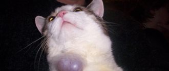
When a white veil covers the floor of the eye, they speak of prolapse of the third eyelid. In a normal state, its movement is invisible to prying eyes. The eyelid cleans the surface of the apple and produces a third of the tear fluid.
When the gland protrudes, the following symptoms are observed:
formation of a reddish clot in the inner corner of the eye;- profuse lacrimation;
- redness of the edge of the apple;
- development of conjunctivitis.
If the condition is advanced, pus is released from the corners of the eyes, the mucous membrane swells, the animal squints, and the eyelids become swollen.
Keratitis
Keratitis is a lesion of the cornea of the eye. It can be caused by either trauma, including foreign body impact, or infection. The main symptoms of keratitis are:
- photophobia;
- profuse lacrimation;
- corneal clouding;
- accumulation of discharge in the corner of the eye.
When treating keratitis, it is important to exclude the possibility of its relapse : eliminate the cause of the disease. Effective drug intervention is impossible without the help of a veterinarian. Only a specialist can correctly determine the cause of the disease and prescribe the correct therapy.
How to help your pet?
When an illness is identified, it is better to immediately identify its cause by seeking the help of an experienced veterinarian.
First, the veterinarian will examine the affected area using an ophthalmoscope and take smears from tear secretions, which will help exclude or confirm the presence of an infectious disease.
If the root of the problem is a disease, the specialist will prescribe anesthetic drops for sore eyes and special ointments. Of course, it is very important to stop the disease itself.
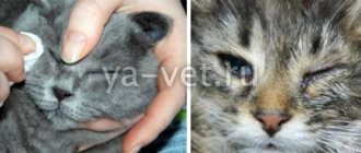
At the same time, the cat's eyes will need to be washed at home to rid them of dirt. If you notice that a kitten or an adult suffers from lacrimation, suppuration and constantly rubs its eyes, then use the following ingredients as a wash:
- boric acid;
- olive oil;
- warm clean water.
Secure the pet well; it is better to carry out the procedure in pairs, wrapping the mustache in a towel. Then gently rinse her eyes using a generously moistened cotton pad or pipette. Afterwards, wipe your pet’s eyes with a clean swab from the outer corner of the eye to the inner.
For suppuration and tearfulness, Lacrimin drops are effective. If creatitis is detected in a tailed creature, then you cannot do without medical help - follow all the doctor’s instructions, this will preserve your pet’s vision.
For cataracts (when, in addition to the formation of a film, the lens becomes cloudy), blockage of the ocular ducts and strabismus, surgical intervention will most likely be required.
It is undesirable for the cat to rub its eyes during therapy, so it is better to use a special veterinary collar.
Blepharitis
Inflammation of the eyelids, which is quite common in furry cats, is divided into three types:
- Scaly blepharitis (also called simple);
- Ulcerative blepharitis;
- Meibomian blepharitis.
Symptoms differ for each type, but there are common ones that are always noticeable. Namely:
- Itching;
- Edema;
- Redness;
- Tearing.
With scaly blepharitis, you can notice a clear thickening of the eyelids, after which gray scales appear there. Over time, the eyelashes will begin to fall out, and pus will be released in place of the scales.
If treatment is not started, scaly blepharitis can quickly turn into ulcerative blepharitis. With it, crusts of pus dry out, fall off, and in their place sores open. The inflammation gets worse. Scarring may cause the skin to tighten.
Meibomian blepharitis is similar to a stye. The eyelid swells and turns red. Pus begins to ooze out profusely.