Signs Diagnosis Treatment The diaphragm is the muscular partition between the chest and abdominal cavity. Performs an important respiratory and barrier function. A diaphragmatic hernia occurs when the integrity of the diaphragm is disrupted or ruptured. In this case, the abdominal organs are displaced into the chest. Most often, a diaphragmatic hernia occurs due to trauma and is combined with multiple injuries (a cat gets hit by a car, falls from a window).
There are 2 types of diaphragmatic hernia:
- traumatic – rupture of the diaphragm due to a strong blow or injury;
- congenital - a cat is born with a defect (the most common is peritoneo-pericardial diaphragmatic hernia, in this case the abdominal organs are displaced into the pericardial cavity through a hole in the diaphragm).
Why does it happen?
Mostly in cats, a diaphragmatic hernia is observed due to traumatic factors. The injuries can be open, resulting in damage to the chest and abdominal cavity, and the diaphragm is also injured. If we talk about closed injuries of a traumatic nature, they are observed during a fall from a height, road traffic accidents, and also in a situation where intra-abdominal pressure increases sharply and greatly.
Congenital diaphragmatic hernia in kittens is also not uncommon. This pathology is usually divided into 4 types:
- Pleuroperitoneal. Rarely diagnosed. In most situations, pets with this diaphragm defect die during childbirth or a few hours after birth.
- Pericardio-pleuroperitoneal. A common type of disease. Mostly found in Persian felines.
- Sliding. It develops due to weakening of the diaphragmatic ligament, which causes the stomach to move upward. No strangulated hernias are observed.
- Paraesophageal. This type of pathology is characterized by fixation of the cardiac region and displacement of the digestive organs into the chest cavity through the esophageal opening, which is excessively dilated. Hernias can be strangulated and cause severe pain.
Peritoneo-pericardial diaphragmatic hernia is a congenital defect, as a result of which a connection between the abdominal cavity and the cardiac membrane is formed in a cat during intrauterine development due to a violation of the integrity of the diaphragm.
What symptoms indicate pathology?
Against the background of this pathology, the animal develops shortness of breath, which is difficult for the owner to notice.
It is important for pet owners to monitor the general condition of the kitten. As the disease progresses, it becomes apathetic and does not make contact with the owner. If the pathology has not been identified and appropriate treatment has not been carried out, breathing problems develop. The animal may choke and experience shortness of breath. Since the body does not receive the required amount of oxygen, changes from the pink tint of the mucous membranes to blue are observed. If the abdominal organs put too much pressure on the blood vessels, there is a risk of pulmonary edema.
With pericardial-diaphragmatic disease, cats may not have any symptoms. This is often due to the fact that abdominal organs slide freely into the pericardium. In such a situation, a hernia is often diagnosed when an x-ray is routinely performed. However, there are pets in whom the pathology provokes loss of appetite, constant vomiting, diarrhea and weight loss. This symptomatology is caused by strangulation of the stomach. Some pets suffer from compression of the heart and lungs.
Types of hernias in kittens
Pathology is classified depending on the location of its occurrence. The most common ones include:
- umbilical hernia . The pouch appears in the middle of the young animal's abdomen, in the navel area. This is the most common location of the disease;
- inguinal _ Forms in the lower abdomen, between the last nipples and the genital area;
- perineal _ Located under the anus in males or in the vaginal area in females;
- diaphragmatic _ The most dangerous type of disease. A hernial protrusion forms inside the body in the diaphragm. This is a muscle-connective tissue formation that separates the abdominal cavity from the thoracic cavity and plays an important role in the breathing process.
In very rare cases, kittens may develop intervertebral or pericardial-peritoneal hernias . Such pathologies are typical for older pets, but practically never occur in young ones.
What to do?
Diagnostic measures
Sometimes it is advisable to examine an animal using an ultrasound method in order to accurately diagnose it.
If the owner notices breathing problems in a cat or other undesirable symptoms, it is important not to delay a visit to the veterinary clinic. First of all, the veterinarian interviews the owner and clarifies how long ago the cat’s condition worsened. Then the cat is examined and x-rayed. A routine examination or a technique during which a contrast agent is used is prescribed. If part of the digestive organs is localized in the chest cavity, the preliminary diagnosis of diaphragmatic hernia is confirmed. To detect a diaphragmatic rupture, the cat is prescribed a peritoneogram. Sometimes an ultrasound examination is required.
How is the treatment carried out?
Only surgical intervention can help cope with diaphragmatic hernia in cats. The operation involves opening the chest by a veterinarian. After this, he returns the organs that have moved back to the peritoneum. Then the doctor sews up the diaphragm, but first resorts to plastic surgery. The surgeon places a special mesh on the muscle-tendon plate. In a situation where internal organs are strangulated, their resection is sometimes required.
If after the intervention the animal may develop a bacterial infection, then appropriate medications are prescribed.
12 hours before surgical treatment of a diaphragmatic hernia, the cat is prohibited from giving food. It is not recommended to give water to your pet 2-3 hours before surgery. In most situations, surgery is scheduled in the morning. By evening the animal can return home. The rehabilitation period takes about 14 days on average. After this time, the stitches are often removed. For 2 weeks, the cat's owner will need to give her complete rest and monitor the operated area. Sutures must be treated with medications prescribed by a veterinarian. Sometimes antibacterial treatment is prescribed to avoid infection of the operated area.
Preventive actions
To prevent the occurrence of a diaphragmatic hernia in a cat, veterinarians from the Pride clinic recommend that pet owners be attentive to changes in the cat’s condition. If any problems with digestion or bowel movements are detected, it is important not to delay a visit to the veterinary clinic. Timely diagnosis allows us to identify the possible presence of a congenital hernia and carry out appropriate treatment. If your pet is kept in an apartment on the upper floors, you should not let him sleep on the windowsill or walk along the ledge. Once again, it is not recommended to open the balcony and windows unless necessary. Such precautions are due to the fact that many situations have been recorded when cats, trying to jump onto the windowsill, did not calculate the distance, missed and fell.
Veterinary clinic of Dr. Shubin
Description and reasons
Diaphragmatic hernia is a violation of the integrity of the diaphragm, accompanied by displacement of the abdominal organs into the chest cavity. The diaphragm is a muscle-tendon plate that separates the abdominal cavity from the thoracic cavity. Mammals are the only ones in which complete muscular separation of these cavities is determined (± crocodiles). The formation of the diaphragm during evolution has dramatically improved lung function. The esophagus, posterior vena cava and aorta pass through this plate.
Drawing. Diaphragm of a dog, view from the abdominal cavity. Source. Guide to the dissection of the dog, Seventh Edition.
Diaphragmatic hernia can be congenital or acquired. The congenital form of diaphragmatic hernia is rarely diagnosed in a veterinary clinic, because Death with this type of hernia in an animal occurs either at birth or in the neonatal period (immediately after birth). Acquired form of diaphragmatic hernia - develops secondary to any mechanical impact; It is with this disease that we have to work and further we will talk about it. So, a traumatic hernia develops as a result of any impact of mechanical factors on the body, which is often noted in an accident, a fall from a height, and bites by other, larger animals.
Depending on the time of occurrence, diaphragmatic hernia is divided into acute and chronic. The bottom line is that the development of this type of hernia does not always lead to the death of the animal (only 15% of animals die before diagnosis), and cases of diagnosis in animals in a period remote from the injury are quite common.
The movement of abdominal organs through the diaphragm leads to compression of the lungs, which is accompanied by visible manifestations of respiratory failure. At the beginning of diaphragm rupture, these signs are most pronounced, then they may decrease somewhat. However, it is difficult to recognize the presence of a hernia only by external signs of difficulty breathing. Thus, during routine sterilization of cats, there are frequent cases of difficulty breathing and detection of a diaphragmatic hernia in an emergency (radiographic examination already under anesthesia).
Clinical signs and diagnosis
Diaphragmatic hernia can develop in both dogs and cats. In many animals (15%–25%), a hernia is diagnosed within the first week after any traumatic exposure. Due to the fact that this type of hernia develops against the background of various injuries, various concomitant lesions may be detected in the animal (eg severe blood loss, shock, bone fractures). In the chronic course of a diaphragmatic hernia, signs of the disease can be expressed in various types of respiratory disorders (eg shortness of breath, exercise intolerance) or gastrointestinal tract (eg lack of appetite, vomiting, diarrhea, weight loss, pain after eating). As mentioned above, many animals with a chronic form of diaphragmatic hernia do not exhibit dyspnea at the time of examination.
When examining the animal, the veterinarian may identify some signs that suggest the presence of a hernia (eg breathing problems, decreased abdominal volume), but these only serve as a reason for further diagnosis (usually a radiographic examination).
The final diagnosis of a diaphragmatic hernia is usually determined by radiographic examination (X-ray); in rare cases, the use of contrast and ultrasound diagnostics (ultrasound) may be used. Diagnosing most cases of clinically significant forms of diaphragmatic hernia using x-rays usually does not cause difficulties.
Preoperative preparation
An operation to correct a diaphragmatic hernia may require some preparation; this is aimed at reducing the probable anesthetic mortality of the animal and making certain procedures easier to carry out. In most cases, an intravenous catheter is installed and infusion therapy is administered. In case of severe breathing difficulties, the animal can be provided with oxygen, and its placement is carried out taking into account the facilitation of respiratory movements. If concomitant diseases are suspected, the veterinarian may recommend various examination methods (eg blood tests, ultrasound, x-rays). Immediately before the operation, the animal is given an antibiotic.
Anesthesia
Carrying out anesthesia in patients with diaphragmatic hernia is associated with a high risk of complications, including fatal ones. Total intravenous anesthesia (injection of drugs into a vein) is used as the basis of anesthesia, because artificial ventilation is used intraoperatively and this greatly complicates the administration of inhalation anesthesia. An animal with a diaphragmatic hernia is subject to mandatory intubation - a special tube is inserted into the trachea under anesthesia, through which the breathing function is maintained.
Anesthetic mortality during surgical correction of diaphragmatic hernia in our clinic tends to zero, but be that as it may, employees warn every animal owner that any anesthesia can lead to the death of the animal.
Drawing. A ventilator used in a veterinary clinic to maintain breathing in animals during the correction of a diaphragmatic hernia. This device provides excellent intraoperative ventilation and reduces the risk of lung injury due to excess pressure of the inhaled mixture.
Surgical correction
Diaphragmatic hernia is a primarily surgical disease; its correction is achievable only by using various methods to restore its integrity. Conservative treatment (eg intravenous and antibacterial therapy) is aimed only at facilitating surgical intervention. The doctor may slightly delay the operation for drug correction of various complications, however, the operation should be performed immediately after the animal has been stabilized, this improves the prognosis for a favorable outcome.
A patient with a diaphragmatic hernia is placed in a special groove on the back, the incision is usually made along the midline of the abdominal wall from the xiphoid process of the chest half or two-thirds of its length towards the tail. The abdominal organs are shifted to their proper physiological position, which frees up the chest cavity. After achieving the correct anatomical location of the internal organs, the defect of the diaphragm itself is assessed and approximating sutures are placed on its edges. After which, excess air is removed from the chest cavity. In chronic cases of diaphragmatic hernia, various defects and adhesions between internal organs may develop, and they are also subjected to various types of surgical procedures. When correcting a diaphragmatic hernia, non-absorbable sutures (nylon, etc.) are often used.
Postoperative care
The immediate postoperative period is very important in terms of the animal’s recovery; staff “takes” the animal out of the operating room only after spontaneous breathing has been restored and in a stable condition. After the operation, antibiotics and painkillers are administered for three to four days. The optimal way of postoperative care is to keep the animal in a 24-hour hospital, but at the moment we cannot provide this type of service.
Sutures on the abdominal wall are subject to mandatory protection for a period of 14 days; for this, either a protective blanket (optimally) or an Elizabethan collar is used. Protecting the wound prevents the animal itself from affecting the stitches, which reduces the risks of various complications. During this period, the wound itself is monitored for the development of swelling, redness, etc., at the end of this period, the stitches are removed in a veterinary clinic and there is no need for protection. If there is a change in the general condition of the animal, the veterinarian must be immediately informed for a competent assessment and correction of any complications.
Possible complications and prognosis
Postoperative prognoses for animals with diaphragmatic hernia have improved significantly over the past few decades. Thus, in the 1980s, only 52% of cats and dogs underwent surgical correction; recently, the survival rate for diaphragmatic hernia was 82%-89%. Survival data differ slightly between cats and dogs, and between acute and chronic forms of diaphragmatic hernia. However, there continues to be a generally accepted opinion that survival rate for chronic hernia is somewhat lower than for acute hernia. The main reasons for the unfavorable outcome of hernia correction surgery are considered to be deficiencies in anesthesia and artificial ventilation.
Various complications develop in half of patients after surgery to correct diaphragmatic hernia. Fatal complications can develop from both the thoracic and abdominal organs. Also, complications can develop immediately after surgery or at a somewhat distant period of time. Soon after surgery (the first day), death may develop for the following reasons: accumulation of blood, air and fluid in the chest cavity; pulmonary edema; shock, heart rhythm disturbance. In the long term, death may occur secondary to rupture, torsion, or obstruction of the gastrointestinal tract or diseases unrelated to hernias. Recurrences of a hernia after proper correction are quite rare, but they can still amount to 4%-5%. Rarer complications may also occur and should be discussed with your veterinarian.
Conclusion
Be that as it may, with surgical correction of diaphragmatic hernia, a fairly high recovery rate is observed in cats and dogs. The staff of the veterinary clinic have accumulated enough successful operations, and in our opinion, the only way to restore the health of the animal is to suturing the diaphragm defect.
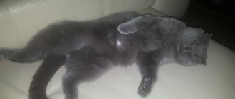
Screening image of a kitten admitted to the veterinary clinic with severe shortness of breath; the x-ray is not of the best quality, but it clearly shows signs of a diaphragmatic hernia. Due to the serious condition of the animal, the veterinary clinic staff did not conduct a detailed X-ray examination, but decided to undergo surgery.
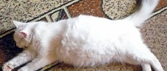
The same kitten the day after surgery in the veterinary clinic. The veterinary clinic staff, under anesthesia, returned the organs to their normal position and sutured the diaphragm defect. While in the hospital at the veterinary clinic, a “butt with a handle” was stuck to the kitten due to its catastrophic thinness, long tail and some character traits.
The same kitten, 14 days after surgery, allows doctors to remove the stitches.
He looks serious and looks confidently into the future.
July 2016
Veterinary clinic of Dr. Shubin, Balakovo.
Main manifestations of a hernia
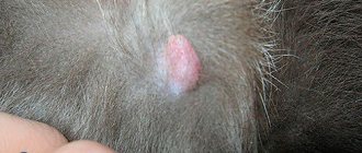
At the initial stages of pathology formation, it is rarely diagnosed, since it does not cause discomfort to the animal and practically does not manifest itself externally. Characteristic symptoms appear much later, when the formation reaches a large size (looks like a soft/elastic spherical protrusion) or an organ trapped in the hernial sac is pinched.
In this case, the following symptoms are observed:
- severe pain in the abdomen. The animal will meow loudly, cower and arch its back;
- increased body temperature;
- refusal of food;
- constipation and problems with urination;
- nausea and vomiting;
- inactivity, apathy;
- Possible problems with breathing and coordination of movements.
Another important symptom of strangulation is the inability to reduce the hernia. In this case, the hernial sac becomes hard to the touch and extremely painful. It is important to remember that strangulation is an extremely serious complication and requires immediate surgical intervention.
Hiatal hernia (HH) - symptoms and treatment
To diagnose hiatal hernia, in addition to detailed questioning of the patient, almost all research methods used in gastroenterology are used. Mandatory diagnostic methods include:
- clinical and radiological examination;
- fibroesophagogastroduodenoscopy (FEGDS);
- esophagotonometry;
- pH-metry of the esophagus and stomach;
- Ultrasound of the abdominal cavity.[12][158]
The leading instrumental methods are considered to be X-ray diagnostics and FEGDS.[8][16]
X-ray diagnostics
Thanks to the X-ray diagnostic method, fundamental research has been carried out on hiatal hernias, classifications have been developed, various forms of this pathology have been studied, and a number of indications and contraindications for various types of treatment of hiatal hernias have been developed.
The modern full name is “Polypositional X-ray diagnostic examination of the esophagus, stomach and duodenum using a liquid suspension of barium sulfate on a troscope.”
This X-ray examination allows you to reliably diagnose various forms of hiatal hernia, including “small” esophageal hernias, identify cardia insufficiency, gastroesophageal reflux, reflux esophagitis, and exclude cardia insufficiency associated with impaired passage of food in the underlying sections of the gastrointestinal tract.
Endoscopic esophagogastroduodenoscopy
In the middle of the 20th century, the latest technologies in endoscopy were developed and widely introduced into clinical practice. They have made it possible to significantly expand the possibilities for diagnosing gastroenterological diseases.
The peculiarity of endoscopic esophagogastroduodenoscopy is:
- the use of flexible fiber optics and the creation of endoscopic devices - fiber gastroscopes;
- high resolution of these devices with the ability to conduct research while visualizing images on the monitor;
- minimally invasive endoscopic diagnostics;
- minimal percentage of complications that arise;
- no need for special preparation of the patient before the start of endoscopic examinations;
- outpatient nature of endoscopic diagnostic methods.
All this allows us to recommend this diagnostic method not only to patients, but also to the population as a whole for medical examination and detection of the disease in the early stages.
Of course, endoscopic diagnosis of hiatal hernia is not an easy procedure, but FEGDS doctors consider it as a screening method indicated for all patients, including people with minimal symptoms of gastroesophageal reflux, dyspepsia or dysphagia (digestive or swallowing disorders), as well as anyone who suffers from digestive diseases. tract.
The main direct and indirect symptoms of hiatal hernia, usually manifested during FEGDS, include:
- reduced distance from the anterior incisors to the cardia;
- reduced length of the abdominal esophagus;
- hernial cavity;
- “second entrance” to the stomach;
- gaping (opening) of the cardia or its incomplete closure;
- prolapse (protrusion) of the gastric mucosa into the esophagus;
- reflux (reverse flow) of stomach contents into the esophagus;
- segmental dilation (expansion) of the esophagus in the area of the ninth segment;
- absent, poorly visualized or blurred Z-line;
- flattened fold of the cardioesophageal junction detected during inversion examination of the cardia;
- a smoothed angle of His, also detected during inversion examination of the cardia.
Most of the listed endoscopic symptoms of the hiatal hernia can be detected through video monitoring during FEGDS, which helps to establish an accurate diagnosis.