To understand why tumors appear in cats after sterilization, you need to understand what operations to “disable” the reproductive function in females are. In modern veterinary clinics such procedures are carried out in one of the following ways:
- laparoscopic;
- classical.
Laparoscopic .
Through small (up to 1 cm) incisions on the abdomen, a laparoscope (tube with a video camera) and a manipulator are inserted into the abdominal cavity. Carbon dioxide is supplied to raise the abdominal wall so that the surgeon can see all the organs. Directly in the abdominal cavity, the ovaries and uterus are removed and removed through incisions, bleeding vessels and tissues are cauterized with a laser or electric current. The cat requires virtually no post-operative care. If the incisions are smaller than 0.5 cm, they are not sutured, but sealed with medical glue. Classic method . The incision is made along the linea alba of the cat's abdomen. The aponeurosis (wide tendon plate) of the abdominal wall is dissected, the muscles are not affected. The surgeon removes the ovaries and uterus, ligates the vessels and cuts off the organs. The peritoneum is sutured with special threads, which dissolve on their own after a certain time. Such seams do not require any special care; during this period the main thing is to ensure that the cat cannot reach them.
During the rehabilitation period after sterilization, lymphatic fluid may accumulate in the cavity formed under the external suture, forming swelling visible to the naked eye. This symptom usually appears 2–5 days after surgery. The “tumor” does not cause discomfort to the cat, does not require treatment and resolves on its own.
Post-operative swelling is normal and should not cause any alarm to the pet owner. If the appearance of swelling near surgical sutures is observed after laparoscopy or is accompanied by suspicious symptoms, consultation with a veterinarian will be required.
Alarming symptoms
What signs of deterioration in health in an animal that has recently undergone sterilization should alert a caring owner? First of all, the cat owner should suspect something is wrong if, more than 2 days after the operation, his pet still shows signs of abdominal pain:
- screams a lot and loudly;
- refuses food;
- if he lies, then only on his side or on his back.
Another alarming sign is a temperature that does not subside for a long time. Normally, surgery provokes an immune response in the cat’s body, as a result of which the thermometer consistently shows a value greater than 39°C for 3 days after the operation, which is completely normal.
However, if the tailed patient’s temperature does not decrease either on the fourth or fifth day after sterilization, this is already a clear sign that an inflammatory process is underway in the body, and the lump that has formed on the pet’s belly is its direct consequence.
After sterilization, the following symptoms may appear:
- allergic reactions (rash, swelling of the mucous membranes);
- lethargy, drowsiness;
- arrhythmia;
- loose stools or constipation;
- urinary incontinence;
- enlargement of the mammary glands;
- wheezing breathing;
- bleeding of sutures.
Sometimes more than one of the above symptoms may occur. As mentioned earlier, signs of the inflammatory process are increased body temperature and redness of the skin. If you do not contact a veterinarian in time, complications can lead to the death of the animal.
Important! Post-surgery lumps are often confused with a mammary tumor in a cat. There is a difference between them. In most cases, the lump goes away on its own. The tumor only grows over time, and similar tumors form next to it.
Reasons for appearance
There are many reasons for the appearance of lumps on a cat’s belly after sterilization. Let's look at the most common of them:
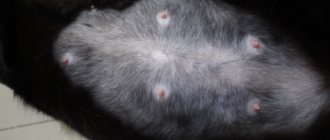
- Postoperative swelling. This is a safe and harmless reason for the formation of a lump after surgery. It occurs for physiological reasons due to the healing of tissue damaged as a result of the incision. It is worth noting that despite the possible impressive size (up to 5 centimeters in diameter), the formation itself does not cause the animal any discomfort. The swelling subsides on its own within 3–5 days after surgery.
- Hernia. The formation of a postoperative hernia is a more dangerous phenomenon that can lead to significant health problems for the animal. The cause of its occurrence is the divergence of the suture of the abdominal wall. This is due to the fact that in the process of making the incision, the veterinarian has to cut through several layers of tissue - the skin, the subcutaneous fat layer, the muscles of the abdominal wall and, in fact, the peritoneum itself. After the operation is completed, tissue stitching occurs in the reverse order. Sometimes it happens that the suture on the peritoneum is made incorrectly, as a result of which it diverges and a small gap is formed into which the cat’s internal organs fall out. They protrude, which on the outside looks like a lump or lump. Postoperative hernia is a complex pathology, the treatment of which involves repeated surgery.
- Ligature fistulas. They are formed as a result of inflammation and suppuration of non-absorbable threads that were used to make a surgical suture. At first they may look like small bumps on your cat's belly. The bumps very quickly develop into pustules, which are foci of infection. The disease requires repeated surgical intervention in rare cases; usually treatment is conservative, and it is aimed at eliminating the infectious process.
- Accumulation of lymph. If the lymphatic vessel is poorly sutured, the lymph may leak and accumulate, forming a lump. Over time, it resolves on its own or is removed with the help of drainage.
My animals had just an accumulation of lymph. The accumulated fluid was pumped out of the dog with a syringe. In cats, the cones resolved on their own within 2 weeks.
When to see a doctor
During the recovery period after sterilization, it is recommended to show the cat to a veterinarian. How painlessly and quickly she recovers largely depends on this.
Of course, lumps, which are postoperative edema, are quite physiological. In this case, contacting a veterinarian is not required. However, if the lump has a large diameter, a rash, redness appears around it, and ichor or pus is released, then this indicates the seriousness of the pathology and requires prompt seeking veterinary help.
Diagnostic methods
Diagnosis of a simple unbranched single-channel fistula is not difficult - it is enough to feel a cord in the local seal, from which contents can leak when pressed.
All external exits of the fistula are examined with a button probe, this is how the localization of the passages is determined. The probe is inserted from the side of the skin, carefully pushing it all the way; if a rectal fistula is being examined, then the passage of the probe inside the rectum is determined with the index finger.
Next, a test is carried out with a dye - methylene blue, which is injected into the outer hole with a syringe. In case of rectal fistula, a cotton swab is inserted into the intestine before the test, and the exact location of the internal opening is determined by the dye marks on it.
For any fistula, the paint output can be recorded during endoscopy: anoscopy, rectoscopy, colonoscopy, cystoscopy, colposcopy, and so on. Endoscopic examination is one of the leading ones and is performed repeatedly in the process of diagnosis and treatment.
In some cases, fistulography is performed - an x-ray of the anatomical area before and after the injection of a contrast agent into the fistula. The procedure is not required only for simple and short rectal fistulas without exacerbation of inflammation.
Imaging methods - CT and MRI also make it possible to clarify the localization of tracts and leaks, branching and the root cause of the disease.
When the rectum is involved, ultrasonography (ultrasound) with a special rectal sensor is informative, when a computer program allows you to see the pathology in a three-dimensional image. When planning the operation, the function of the anal sphincter is additionally determined.
| Examination stage | Techniques | What are they used for? |
| Techniques used in the doctor's office during the initial examination |
|
|
| Lab tests |
|
|
| Instrumental studies |
|
|
What to do if a lump forms on your stomach
As mentioned above, if the swelling does not subside for several days, increases in size, festeres, or the pet feels unwell, be sure to contact a veterinarian to avoid serious complications. The veterinarian, having established the cause, will prescribe symptomatic treatment aimed at normalizing the general condition of the operated animal.
When a postoperative hernia forms, if there is no pinching, the problem is solved by repeated sutures or reduction of the hernial protrusion. If the process is started, surgery may be required.
If the tumor is malignant or benign, the veterinarian, after tests, decides on the choice of treatment. The animal is prescribed maintenance therapy or surgery.
In any case, the choice of treatment methods for the appearance of a lump on the abdomen, above or below the suture, depends on the root cause, the general condition of the cat, and diagnostic results.
Important! Do not self-medicate so as not to harm your mustachioed pet. All manipulations and therapy should be prescribed by a veterinarian.
Signs of a strangulated hernia
If the female shows anxiety when pressing on the bump, then it is necessary to immediately show her to a veterinarian, since pain may indicate a strangulated hernia.
Without timely surgical intervention, this leads to prolonged compression of blood vessels and nerves, causing the areas of the compressed organ to die.
Other symptoms of a strangulated hernia include:
- loss of appetite or complete refusal to feed;
- constipation;
- vomit;
- redness and increased local temperature in the area of protrusion.
Important! Infringement of the bladder greatly complicates the act of urination and can lead to rupture of the organ. In this case, the only way to save the animal is immediate surgical intervention.
Preventing the appearance of a lump on the stomach
In order for the postoperative period to pass calmly and without the development of complications such as the appearance of a lump on the abdomen, you should carefully follow all the veterinarian’s prescriptions.
Make sure that for the first two days the cat does not jump from a height or show excessive activity. Coming off of anesthesia, reflexes are dulled, the body is weakened, and behavior is unpredictable. Therefore, do not leave your pet uncontrolled.
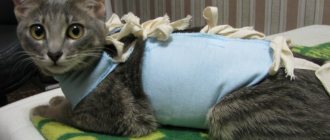
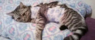
The seam should be regularly treated with disinfectants until it is completely healed. To prevent the cat from licking it and tearing out the threads, a blanket is put on it. Some animals manage to get out of this “clothing”; for such cats it makes sense to use a protective collar.
As soon as your cat shows interest in food, feed her small portions. Food should be nutritious and easily digestible. You can give special foods intended for feeding during the postoperative period or formulations for sterilized, neutered animals.
Proper prevention and systematic proper care will help the cat return to normal faster after sterilization. If there are no complications and you follow all the veterinarian’s recommendations, the rehabilitation period will take no more than five weeks.
After sterilization, a cat has a lump under the seam: what to do
In any case, complications are reported to the veterinary surgeon. If postoperative swelling has developed, the swollen surface is treated with an antiseptic spray that forms a protective film. The condition of the swelling is monitored, and re-treatment is carried out if the need arises.
If a significant amount of lymph is released, the veterinarian installs drainage or prescribes a wound-healing powder. The abscesses are opened, the wounds are treated with antimicrobial external agents.
If a hernia has formed, a repeat operation is performed.
Consequences of sterilization
A high temperature – above 39 degrees – may indicate the onset of a complication with the pet’s health after surgery. This temperature within 3 days after surgery is considered normal, but after the fourth day it is necessary to urgently seek help from a veterinarian. When the animal's temperature drops, you need to do everything to keep the pet warm: move it to another place, cover it with a blanket, rug, put a heating pad on it. If your cat still does not show signs of active behavior, you should seek help from a veterinarian.
Increased drowsiness, lethargy in behavior
The recovery process after anesthesia can last from 12 to 18 hours. During this time period, the cat may exhibit unusual behavior such as lethargy, vomiting, nausea, or lack of appetite. Some individuals may remain in this state until the sutures are removed. The operating veterinarian must answer any questions the owner has about the care of the cat and its behavior after surgery.
One way or another, the cat needs rest after a difficult operation. It is recommended to leave her in a calm, quiet place and limit access to her by children and other pets. During this period, the cat needs to get enough sleep and restore its own strength. Despite all the friendliness and affection of the purr, during the recovery period after surgery, she can behave aggressively. If the cat has obvious prolonged lethargy, lack of appetite, constant vomiting, or swelling of the suture, it is necessary to immediately show it to a veterinarian.
Sometimes owners notice the cat trembling at this time. The fact is that during this period the cat’s temperature drops significantly, so it is important to provide her with a warm place and good conditions for warming it up. In this case, the trembling should stop.
Lack of appetite
During the period after surgery, the question arises about the cat's diet?
| For the first day, the cat may not touch food or water at all, and this should not cause a feeling of excitement. If the cat does not drink or eat for more than 3 days, then it is time to consult a veterinarian, since such ignoring of food is a sign of a complication. |
The way out of this situation, according to most veterinarians, is intravenous nutrition or liquid food in the form of broth, which is fed to the pet from a pipette or syringe. During the recovery period after sterilization, a cat may change its taste preferences quite often, for example, not eating food that it previously really liked.
This shouldn't be a cause for concern either. It is necessary to include vitamins in your diet that your doctor recommends. There are separate recommendations for feeding a cat during the recovery period after sterilization. Such rules should include:
- assessing the volume of water consumed and ensuring unlimited access to it;
- constant vomiting is a sign of “coming out” of anesthesia, so it is entirely possible to completely stop feeding;
- Do not give the animal too much food;
- It is better to choose a semi-liquid diet that will not cause constipation.
Methods for treating fistulas
Fistulas rarely close on their own; this can only be hoped for by creating favorable conditions, for example, limiting and partially controlling the movement of feces through the rectum using cleansing enemas. In the vast majority of cases, conservative therapy is ineffective; the only radical treatment is surgical, that is, excision of the pathological area, including reconstruction of the missing tissue.
Technically simple surgical intervention, including endoscopic, and a hundred surgical modifications cannot cure about half of the patients who suffer from relapses. It is especially difficult to achieve success with intestinal and urinary fistulas, since they are always contaminated with microflora. In some cases, it is necessary to resort to the formation of an intestinal stoma, temporarily stopping the movement of feces through the pathologically changed area of the intestine for several months.
In isolated cases, they resort to “old-fashioned methods” of treatment with scraping the mucous membrane of the tract, burning it with chemical reagents and enzymes, achieving sticking of the walls. Better results - in approximately 50% - are achieved by introducing fibrin glue into the fistula tract, which glues the walls together.
Tampons made of biomaterials act similarly to glue, sealing the internal opening; emptying of the passage can cause the walls to stick together and close the fistula.
Until now, the role of antibiotics in the treatment of fistulas caused by inflammation has not been determined, since drugs are not able to penetrate into the infiltrate due to massive scar changes. However, with fistula tracts due to Crohn's disease, specific drug therapy is mandatory and not unsuccessful.
Surgery for fistulas using the LIFT method
Prevention of fistulas
Not all diseases can be prevented, especially fistulas, which complicate the course of purulent paraproctitis. However, it is possible to adequately treat the diseases that lead to paraproctitis - hemorrhoids and fissures, and this is precisely what will prevent the formation of fistulas.
Complicated childbirth cannot be prevented, but high-quality and timely obstetric care, attentive attention to the woman and a thorough postpartum examination are available preventive measures.
The high incidence of post-radiation tissue damage and fibrosis progressing over time forced oncologists to abandon high doses of radiation therapy and even change approaches to the treatment of malignant tumors of the genital area.
Particular importance is attached to the correct choice of method of surgical treatment of diseases of the hollow organs and adequate management of the postoperative period.
In our clinic, a complicated course of the disease is very rare, because we not only know about prevention methods, but also actively use them.
| Price : | |
| Primary consultation with a surgeon | 5,100 rub. |
| Repeated consultation with a surgeon | 4,600 rub. |
| Consultation with a surgeon Ph.D. | 6,900 rub. |
| Primary consultation with an oncologist | 5,100 rub. |
| Repeated consultation with an oncologist | 4,600 rub. |
| Consultation with oncologist Ph.D. | 6,900 rub. |
| Consultation with an oncologist, MD. | 10,500 rub. |
| Fistulography | RUB 41,800 |
Book a consultation 24 hours a day
+7+7+78
Bibliography:
- Shelygin Yu.A., Blagodarny L.A. /Handbook of coloproctology// -M.: Litterra; 2012.
- Becker A., Koltun L., Sayfan J. /Simple clinical examination predicts complexity of perianal fistula// Colorectal Dis.; 2006; 8.
- Gaertner WB, Hagerman GF, Finne CO, et al. /Fistula-associated anal adenocarcinoma: good results with aggressive therapy// Dis Colon Rectum; 2008; 51.
- Genadry RR, Creanga AA, Roenneburg ML, Wheeless CR /Complex obstetric fistulas // Int. J. Gynaecol. Obstet.; 2007; Vol. 99; Suppl. 1.
- Ommer A., Herold A., Berg E. / S3-Leitlinie: Rektovaginale Fisteln (ohne M. Crohn) // Coloproctology; 2012; Vol. 34.
- Sahni VA, Ahmad R., Burling D. / Which method is best for imaging of perianal fistula?// Abdom Imaging.; 2008; 33.
- Zoulek E., Karp DR, Davila GW/ Rectovaginal fistula as a complication to a Bartholin gland excision // Obstet. Gynecol.; 2011; Vol. 118; N 2.
Diarrhea, constipation
As a result of sterilization, the cat's stool may change and constipation may occur. After surgery, some animals may experience difficulty going to the toilet; constipation and diarrhea develop, accompanied by fairly severe pain attacks. The following signs may indicate constipation:
- lack of appetite;
- plaintive cries;
- failure to go to the toilet;
- trembling in the tail.
If constipation occurs, it is necessary to ensure that the cat takes laxatives prescribed by the veterinarian and adjust its diet. Among the household ways to relieve constipation, one can highlight the use of a tablespoon of Vaseline.
Diarrhea occurs as a result of the use of narcotic drugs that have a toxic effect on the body during surgery. When diarrhea occurs, the cat poops liquid feces, and the owner is advised to introduce restrictions on drinking and eating, and give the cat the drug prescribed by the veterinarian. As a household way to prevent diarrhea, you can prepare rice water for your pet.