Etiology
The causative agent of the pathology is a fungus of the genus Achorion, which has selectivity. In birds, symptoms are caused by A. gallinae, in rats and mice by A. quinckeanum, but cats, dogs, horses, and sheep can become infected. Favus in humans is caused by A. Schonleini, and it can sometimes cause pathology in primates, dogs, cats, calves, and mice.
The mycelium of the microorganism is thin. The cells are double-circuit, rectangular. The spores are round or multifaceted and are arranged in chains or groups. The optimal temperature for growth is 28-30 ᵒC. The fungus prefers wort agar and Sabouraud. Growth appears as white, smooth colonies. As it ages, the growth becomes wrinkled, mealy and changes color to all shades of pink, with a purple underside.
The bioassay is successful on white mice, guinea pigs, and rabbits.
Most often, the pathogen is recorded in young chickens, less often in turkeys, ducks and wild birds. The source of the disease is sick animals. Infection occurs through contact, through damage to the skin, through airborne droplets, through care items infected with the secretions of a sick bird. Carriers of fungal spores can be ticks, mice and rats. Soil is a natural reservoir of the favus pathogen.
The disease can be widespread or sporadic. When the disease occurs, the lesions become stationary.
The fungus begins its development in the peri-root zone of the feather and hair, forming a mycelial shield. The site of settling becomes inflamed, feathers become deformed, fall out, epidermal layers, sweat and sebaceous glands atrophy. If the fungus penetrates the blood, it can grow in parenchymal organs, where it forms favus nodules.
Animals that have recovered from the disease acquire lasting immunity.
Are cat dermatological diseases dangerous for humans?
Cat skin pathogens pose a danger to humans when the following factors are combined:
- neglect of hygiene standards;
- childhood or old age;
- diseases accompanied by weakened immunity.
The greatest danger is caused by cats suffering not from scab, but from trichophytosis or microsporia. However, most often when one type of fungal infection occurs, pathogens of other diseases join it, so the owner of a sick cat must be extremely careful.
Be sure to read:
Dermatomycosis (ringworm) in cats: symptoms, treatment, veterinary advice
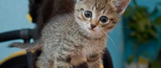
Symptoms
The incubation period is up to 4 months.
In dogs, cats and rabbits, the base of the claws, head, ears, and abdominal wall are affected. In sheep, the fungus most often localizes to the skin around the lips and nose, as well as the inside of the ears.
Rodents are very sensitive to the fungus. The scalp and body are initially affected, then the deeper layers, muscles and bones are affected, and the mice lose their vision.
The crusts (shields) that “grow” under the action of the fungus are easily removed in the initial stages, revealing underneath the wet surfaces with a depression in the center. Old processes do not allow the scutula to be removed and they continue to grow and occupy larger and larger spaces.
In birds, the incubation period is 3-4 months. The process begins with areas devoid of feathers (head, comb, earrings). These places are covered with white nodules, which, as they increase, merge, become dirty yellow, then gray. Within 2-3 months, the comb becomes completely covered with extensive crusts extending to the feather areas. During the process of generalization, feathers fall out and the skin becomes covered with scales.
With a protracted nature, the mucous membrane of the oral cavity, goiter, and intestines is affected. The bird refuses to eat food and develops diarrhea. Pathology can also affect the upper respiratory tract.
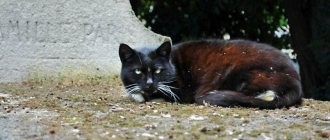
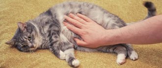
Scab in cats: description of the pathology
Scab or Favus is a fungal disease that affects the skin and coat. Reports that scab-causing fungi can attack internal organs are questionable. However, since Favus develops against the background of a weakened immune system, the development of septic processes under the influence of other microorganisms cannot be ruled out.
Infection occurs by contact or oral-fecal route. The reservoir of infection is rodents and stray cats. The incubation period depends on the condition of the pet at the time of infection. In weakened animals, pathological signs may appear after a few days, in stronger ones - after several months.
Treatment
Build-ups should be softened. To do this, use a soap-creolin emulsion or ointment. The affected areas are lubricated with a solution of formaldehyde 5-6%, potassium permanganate 1-2%, salicylic alcohol 4-5%. Treatments are carried out daily.
Small animals use zoomicol spray.
We will use a preparation prepared on the basis of a tincture of iodine 5% and a solution of salicylic acid 10%, or crystalline salicylic acid is added to the tincture of iodine to prepare a 10% concentration.
In the period between treatments, it is recommended to use ASD-f3. The affected areas are repeatedly lubricated with a swab.
To quickly restore the functionality of the skin and hair growth, it is recommended to take vitamin preparations “Trivit” and “Tetravit”.
Favus (scab)
Favus (scab, trichophytosis, trichophytosis) is an infectious disease of birds, less commonly mammals, as well as humans, characterized by damage to the skin, feathers, hair and claws. Sometimes the pathological process occurs on internal organs.
Historical reference . The etiological role of the fungus was established by German scientists L. Schönlein in 1839 and independently in 1841 by D. Grüby. Favus in birds and children was described in 1881 by P. Meunien. In the USA, this disease was described by B. Beach and J. Chalpin in 1918, calling it white comb. Favus is registered in France, Great Britain, Turkey, USA, New Zealand, Australia and other countries. The disease also occurs in Russia.
Etiology . The causative agent of Favus is fungi of the genus Achorion. In birds, favus is caused by Achorion gallinae Sabourand; in rats and mice – Achorion quinckeanum. Cats, dogs, sheep, horses, as well as humans can become infected; Achorion Schonleini is the causative agent of human favus. Sporadic cases of favus caused by this fungus are known in dogs, cats, mice, monkeys and calves. In pathological material, the mycelium of achorions is thin, rarely septate, consisting of rectangular cells with a double-circuit shell. The spores are round or multifaceted, arranged in chains or groups. Their sizes range from 4 to 8 microns. Air bubbles are detected in the form of long black strands. The elements of the mushroom are located along the length of the hair. Fungi grow on Sabouraud agar, wort agar and others, at temperatures up to 30°C, and gallinae grow on Sabouraud agar in the form of smooth, white, velvety colonies, which with age become folded, mealy, pink-red or crimson in color. A. quinckeanum on wort - agar - forms flat, velvety, folded with age, sometimes with depressions in the center, mealy colonies. The reverse side is purple. On potatoes, colonies are wrinkled, gray-yellow; the periphery of the colonies is radiant. Young colonies of A. Schonleini on wort agar are smooth, yellow-white, then rise above the surface of the medium, becoming wrinkled, with a crumbly consistency. Among laboratory animals, white mice, guinea pigs, and rabbits are susceptible.
Epizootological data . Under natural conditions, chickens and, less commonly, turkeys, ducks and wild birds get sick. Young birds are more susceptible during the period of comb and earring development. Roosters get sick more often. In mammals, favus is rarely recorded. The person is susceptible. The source of the infectious agent is a sick animal. The pathogen enters the body of birds and mammals through damaged skin, through airborne droplets when sick and healthy animals are kept together. The transmission factors for the fungus are various objects infected with the secretions of sick animals. An enteral route of infection is possible. Unsatisfactory conditions of keeping and feeding animals contribute to a wider distribution, duration of course and stationarity of favus. Favus is transmitted by ticks in birds, and mice and rats in mammals. The natural reservoir of achorions is probably the soil. Favus can be sporadic or widespread.
Pathogenesis . The fungus, developing near the roots of feathers or hair, gradually penetrates into their vagina, causing an inflammatory process. The mycelium of the achorions forms a plexus on the skin and at the mouth of the hair follicle - the favous shield. Underneath it, all layers of skin atrophy. The sebaceous and sweat glands are usually destroyed. If the fungus gets into the feather pouches, the feathers become deformed and fall out. When the fungus penetrates the blood, a generalized form of favus develops. In this case, internal organs are also affected.
Immunity Research has established that the body of an animal and a person who has suffered a favus infection becomes immune, both in relation to repeated infection (reinfection) with live mushrooms, and in relation to their poisonous products (endotoxin, favin).
Clinical signs . The incubation period ranges from several days to 3-4 months. With favus, it is customary to distinguish between scutulatory (generalized) and visceral forms.
In mammals, the disease primarily affects cats, dogs, and rabbits, which appear to be most often infected by rats and mice. In rats and mice, favus often occurs en masse.
A characteristic manifestation of the disease is the scuticular form.
In rabbits, dogs and cats, the favorite location of the fungus is the skin around the base of the claws of the paws and head; In cats, the skin of the ears is also affected. In some animals, skin lesions can be found on the hairless part of the abdominal wall, on the surface of the chest; In sheep, favus is most often recorded around the lips, nose and on the inner surface of the auricle. In mice, lesions are localized on the scalp, then the process can invade muscle and bone tissue, the fungus penetrates into the brain, and mice lose vision; in rats - on the face, paws, near the claws and on the ears.
The disease begins with the formation of round spots gradually increasing to the size of a lentil, covered with grayish-yellow crusts, the skin around the lesions is hyperemic, the epidermis swells, rises along the periphery, and the scab takes the shape of a saucer or shield (Scutulum) with a depression in the center.
If the scutula (scutellum) is separated, a moist surface with a depression in the center is exposed. During deep disease processes, the crusts do not separate; they enlarge and, merging with nearby ones, form thick layers; there is a mouse smell, indicating putrefactive processes. The hair follicles are destroyed or atrophied; therefore, hair in place of the scutes does not grow, and the sebaceous and sweat glands are destroyed. With a deep scuticular form of the favus, the skin atrophies and scars form on it.
In birds (chickens, turkeys, ducks), favus can occur at any time of the year. The incubation period of the disease lasts 3-4 months. The onset of the disease is indicated by the appearance of white spots on the featherless areas of the scalp, comb, earrings and near the beak. As the process develops, small nodules appear in these areas, which, increasing in size, turn into yellowish-gray and grayish-white crusts-scutules. Usually the lesions merge, and later (after 2-3 months) the ridge and earrings are covered with a solid, gray-white, dry, thick, cracking scab. Closely related to the skin.
The lesion can spread from the crest to the back of the head, neck, and sometimes appears near the cloaca. In these cases, the fungus develops near the roots of the feathers, penetrates into their sheaths and causes inflammation, accompanied by the accumulation of grayish-white scales or thick crusts on the skin around the feather shafts. The fungus can grow into the bags and shells of feathers; in such cases, the feathers fall out, and in their place are bald, reddened, and sometimes flaky, painful lesions. The edges of fallen feathers are covered with a white sheath; in scraping this sheath, threads of mycelium and chlamydospores are visible under a microscope. With the scuticular form, birds emit a mouse-like odor.
Scab can also occur in a generalized form, involving a large surface of the feathered part of the head, body and damage to the nasopharynx, goiter, and small intestine. There may also be a visceral form, accompanied, in addition to the indicated lesions, by anemia, jaundice and prolonged diarrhea. This form of the disease leads to complete exhaustion and death of the bird.
Pathological changes . The corpses of dead animals are emaciated and emit a pungent mouse-like (not moldy) odor. Some areas of the skin are bald and have scabs. On the mucous membrane of the upper respiratory tract, and also often in the lungs, goiter, and small intestines, nodules, ulcers or ring-shaped overlays are found.
The diagnosis is made on the basis of typical favous overlays on the comb and earrings. Clinical diagnosis is confirmed by microscopic examination of pathological material and isolation of a pure culture of the pathogen. In scrapings, large, round and multifaceted spores (aleirospores) are found, arranged in groups, sometimes in chains.
Differential diagnosis . Favus must be distinguished from microsporia and trichophytosis based on clinical signs and mycological studies. At the same time, favus must be differentiated from scabies, dermatitis and other skin diseases.
Treatment . The cuticles (crusts) are softened with soap-creolin emulsion or 3-5% creolin ointment. Then the affected areas are treated with a 2-6% solution of formaldehyde, 1-2% solution of potassium permanganate, 5% solution of salicylic alcohol. The treatment is repeated every 3-4 days until complete cure. Hair, scales, crusts, affected feathers, and fluff are burned. Places where animals are processed are disinfected.
Prevention and control measures . A very important measure in the prevention of favus on the farm is the organization of adequate feeding and compliance by animal owners with the rules of keeping and care, and additionally the creation of an optimal microclimate for birds. Veterinary and sanitary measures should include mechanical cleaning and disinfection of places where animals are kept, systematic preventive treatment of animal skin with a 2-3% creolin emulsion or other substances, and rodent control.
To prevent the introduction of the disease, animals newly entering the farm are kept in quarantine and checked for the presence of skin diseases.
When a disease is detected, the farm (part of it) is declared unfavorable according to the favus. Sick animals are isolated and treated, healthy livestock are carefully examined every 5-6 days for timely detection of sick animals. Animals suffering from the generalized form of favus are destroyed.
A group of sick animals is assigned specific service personnel who are well acquainted with the rules of personal prevention. On-farm regrouping of animals, transferring them to other premises and changing pastures is prohibited.
Until the restrictions are lifted, the introduction and removal of animals from the disadvantaged group is prohibited.
The premises, after the isolation of sick and suspected animals, are cleaned and disinfected every 10 days until restrictions are lifted with a 20% slurry of freshly slaked lime, an alkaline solution of formaldehyde containing 2% formaldehyde and 1% caustic soda, a hot 10% solution of sulfur - carbolic mixture (treated twice after 1 hour), hot formalin - kerosene emulsion (10 parts each of 40% formaldehyde and kerosene, 5 parts creolin and 75 parts water), 5% creolin emulsion.
The surface of the soil infected with fungi (walking areas) is cleaned of debris, manure and other contaminants, the grass is removed, then irrigated with a suspension of bleach containing 5% active chlorine, or a 4% formaldehyde solution. Manure is disinfected biothermally.
The farm is considered healthy 21 days after the last sick bird has recovered and the final disinfection has been carried out.
Prevention
Preventive measures are based on the organization of balanced, nutritious feeding and compliance with veterinary and sanitary conditions for keeping animals and birds.
When a disease occurs, the farm is declared unsafe. It is necessary to provide mechanical cleaning of the places of detention and treatment with a solution of creolin, caustic soda, formaldehyde, and a sulfur-carbolic mixture every 10 days. The soil and walking yards are cleaned and irrigated with bleach with at least 5% active chlorine or a 4% formaldehyde solution.
Patients are isolated and subjected to therapeutic procedures. In the generalized form, slaughter is carried out with the disposal of carcasses in a biothermal pit. It is prohibited to regroup, change pastures, or remove animals from the farm territory.
Quarantine measures are lifted 21 days after the appearance of the last clinical case and final disinfection.
When forming a herd, quarantine measures should be carried out with mandatory examination of the skin and only from prosperous farms.
Share:
Miliary dermatitis in cats: treatment at home
Scab is often identified with miliary dermatitis.
Be sure to read:
Feline lichen in humans: what it is, how you can get infected, symptoms, treatment, how not to get infected
However, the latter concept is broader, since skin diseases, as a rule, develop with the combined action of the following pathogenic factors:
- atopic dermatitis;
- food allergies;
- bacterial infections;
- interaction of fungi: trichophytosis, microsporia, malassezia and parasites - fleas, ticks;
- dry eczema;
- flea dermatitis;
- hormonal imbalances;
- side effects of medications;
- stress;
- unidentified causes.
Miliary dermatitis does not require hospitalization and can be treated at home with remedies prescribed by a doctor. Making independent decisions is dangerous due to the deterioration of the pet’s condition and the emergence of new diseases.
Any medical techniques are useless without providing the animal with comfortable housing and adequate feeding.
Drug treatment is developing in the following areas:
- Application of external agents.
- Internal and parenteral antifungal drugs.
- Allergy remedies.
- General strengthening medications.
External agents are used for local lesions. Before treating the affected areas, the surface is sanitized: the hair is cut off, softened and the crusts are removed.
The following external remedies are popular:
- Ointments: Clotrimazole, Mycozon, Yam.
- Solutions: Fungin, Thermikon, Imaverol, Fukoricin.
Internal and parenteral agents are prescribed when external agents are ineffective.
The following drugs are in demand:
- Internal: Griseofulvin, Intraconazole, Irunin.
- Injection - Dermikotsid - suspension for parenteral administration.
If hypersensitivity to external stimuli is diagnosed, the veterinarian prescribes desensitizing drugs. If skin irritation occurs in response to the consumption of food components, switch to a hypoallergenic diet prescribed by a veterinarian.
To combat infections, as well as to speed up the healing of skin areas, immunomodulators are used: Gamavit, Fosprenil, Maxigan, Anandin, multivitamin preparations.
How to treat
For proper treatment, specific therapy must be carried out to confirm scabies. If the doctor was unable to identify the mite and scabies, a trial treatment will be prescribed. In addition, preventive treatment is prescribed to all family members and people in contact with the patient.
The doctor will prescribe medications that kill ticks. Most often they are:
- aerosols
- ointments
- creams
- suspensions
- emulsions
The prescribed drugs will be based on the following substances:
- benzyl benzoate
- permitrina
- sulfur
- piperonyl butoxide
- esbiola
Sometimes treatment includes washing with soap or treating the skin with emollient ointments before using the drug.
Only a doctor can prescribe a medicine and treatment method. It is based on the following criteria:
- clinical picture
- complications
- person's age
- pregnancy
Self-medication can cause complications, apathetic forms of infection or a protracted course of the disease.
Division into types depending on the pathogen
Symptoms and treatment for mange in cats depend on the type of mite. According to this criterion, 4 types of disease are distinguished.
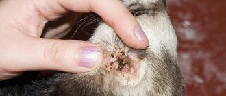
Otodectosis
The causative agent is the tiny ear mite Otodectes cynotis. The maximum size of its body is only 0.75 mm. It affects the outer and inner parts of the ear. Distinctive signs of otodectosis are severe ear itching, the appearance of a nauseating smell of rot and dark grains inside the ears. An infected cat constantly shakes its head and rubs its ears against any objects.
Demodicosis
Demodex canis is a cigar-shaped parasite that is light gray in color. The length of its body is 0.2-0.3 mm. Unlike other pathogens, it lives in the sebaceous glands and hair follicles from the moment the animal is born. The development of demodicosis occurs with a sharp drop in immunity.
According to the degree of damage, there are 2 forms: localized and generalized. In the first case, the diameter of the lesions is no more than 2.5 cm, in the second, the disease affects most of the body. The localized form often goes away on its own and responds well to drug therapy. The chance of recovery from chronic demodicosis depends on the complications that arise. When a secondary infection occurs, they drop to 50%.
Depending on the symptoms, demodicosis can be scaly or pustular. In the first case, redness and cracking of the skin is noted, in the second - the appearance of suppuration and ulcers. The danger of generalized demodicosis is in the combination of scaly and pustular forms. The disease is accompanied by severe complications and is difficult to treat.
Sarcoptic mange
Sarcoptes scabiei is a broad-oval tick that is white-yellow or completely white. The length of the smallest representatives is 0.14 mm. They affect the entire surface of the body and cause sarcoptic mange. You can suspect their appearance by thin light gray lines with a small bubble at the end that appear on the affected areas.
Notoedrosis
Notoedres is a round, dirty gray parasite. The length of its body is 0.5-0.45 mm. Most often it affects the outer side of the ears. In this case, dark gray discharge with a foul odor appears from the ears. Due to liquid secretions, the fur sticks together and takes on an unkempt appearance. The disease is called pruritic scabies, or notoedrosis.
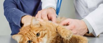
Diagnostics
To determine the clinical picture, you need to do the following:
- Explain your symptoms to your doctor in detail. For example, itching only bothers you at certain times
- get tested
- examine the patient's body
- determine the source of infection
- find traces of the tick itself or its larvae
Typically, the disease is determined by the following criteria:
- purulent blisters
- dry crust
- redness on the buttocks
- scabies (the most important criterion)
To determine the itch, the lesion can be stained with iodine, oil containing minerals, or simply pressed on the epidermis with a glass slide. This will block blood from reaching the affected area and allow you to clearly see the affected area. It can also be detected using dermatoscopy (during the procedure the tick itself is detected).
Ticks can be detected using lactic acid. One drop (40%) should be applied to any lesion and wait 5 minutes. The loosened skin can then be scraped off with a sharp spoon until capillary bleeding appears. The resulting epidermis must be applied to a glass slide and looked at under a microscope. The method will allow you to identify most parasites and the products of their activity.
Pathogens
The causative agents of scabies in animals can be:
- sarcoptes (Sarcoptes);
- notoedris (Notoedres);
- Cheyletiella;
- demodex (Demodex);
- Otodectes.
Scabies mites are microscopic arachnids. These are permanent parasites that settle on the skin or inside the skin, including in the deep layers. Dogs are most often affected by Sarcoptis and Demodex, and cats by Notoedris and Cheyletella. Otodectis, the causative agent of ear scabies, is dangerous for both species of animals.
By parasitizing a cat or dog, microscopic arachnids go through all stages of development from egg to adult.
Medicines prescribed for scabies
Today, veterinary pharmacies have a significant selection of drugs, but using them without a doctor’s recommendation and a preliminary examination of the animal is extremely irrational.
Drops "Bars".
Produced for cats and dogs to combat arachnoenthomosis. Active ingredients: fipronil, diflubenzuron and dicarboximide.
Drops "Bars Forte".
There are different packaging options for kittens and adult cats, puppies and adult dogs. "Bars Forte" for puppies and "Bars Forte" for kittens are convenient to use on animals with low weight.
Spray "Bars".
An insectoacaricide with fipronil is aimed at the larval and adult stages of parasites: fleas, lice, lice, microscopic and free-living mites.
Spray "Bars Forte".
Insectoacaricidal agent for treating animals (cats, dogs) and their habitats. The active ingredient is fipronil. The drug also helps to normalize metabolic processes in the skin and hair follicles of the animal.
Ear drops "Bars".
Used for the treatment and prevention of otodectosis in dogs and cats. The active substance, diazinon, acts against larvae and adults of scabies mites, including Otodectis.
"Dironet spot-on."
Drops on the withers (praziquantel and ivermectin) are prescribed to dogs and cats for arachnoenthomosis and helminthiasis.
"Amit forte"
Due to fipronil and diflubenzuron, the drug effectively affects ticks and is used to treat cats and dogs with tick-borne scabies.
Attention! Do not lick the drug! Before use, consult a specialist! The products are not intended for use in humans!