When pyogenic bacteria invade the organ of vision, panophthalmitis occurs in kittens and adult cats. The disease is accompanied by the release of pus, profuse lacrimation, increased reaction to light, and protrusion of the eyeball. At the first symptoms of infection in a pet, the owner should contact a veterinarian, who will diagnose, prescribe medications and perform surgery.
According to the observation of veterinary ophthalmologist A. Shilkin, most eye injuries occur in the summer, when cats walk outside and injure their mucous membranes on surrounding objects or during fights with relatives.
Description
This occurs when an infection enters the eyeball.
It usually penetrates through wounds, burns, and during cat fights. Defense mechanisms are launched in the pet’s body and E-lymphocytes begin to be produced. However, the eye tissues are very soft, vulnerable, and inflammation spreads quickly and a purulent process develops. It gradually affects all tissues - eyelids, nerves, muscles, etc. The disease is accompanied by severe tearing and protrusion of the eye. Treatment necessarily includes antibiotics to clear the infection. Sometimes surgery is required.
Panophthalmitis is divided into two types. Exogenous develops as a result of injury and subsequent infection of tissues. Inflammation can spread from neighboring organs after ulcerative keratitis. Endogenous appears as a result of any infectious diseases (erysipelas, pyaemia, purulent processes in the ears and nose).
Diagnostic measures
To identify panophthalmitis, the veterinarian conducts a visual examination, finds out the cause of injury to the organ of vision or the presence of other foci of inflammation in the animal’s body. To make a final diagnosis, it is recommended to undergo procedures such as:
When examining the animal, an ophthalmoscopy must be performed.
- PCR (Polymerase chain reaction) analysis to determine the pathogen;
- cytological examination;
- ophthalmoscopy;
- Ultrasound of the organ of vision;
- measurement of intraocular pressure;
- biomicroscopy;
- Seidel test;
- test using fluorescein dye;
- Schirmer's study.
Symptoms
The disease develops rapidly - within a few days. The incubation period for panophthalmitis is 2-3 days after infection enters the eye.
Symptoms of the disease:
- purulent discharge sticking together the eyelids;
- temperature increase;
- blepharospasm;
- increased lacrimation;
- loss of appetite or complete refusal to eat;
- wrinkling and atrophy of the eyes;
- exophthalmos (protrusion of the eyeball);
- soreness in the eyes;
- clouding of the lens and cornea;
- redness and swelling of the eyelids;
- conjunctivitis;
- intolerance to light.
The cat does not strive to play, becomes lethargic, and constantly rubs its eyes. Vision deteriorates and the pet may go blind. Due to the sharp pain, the cat suffers, becomes restless, and cannot find a place for itself. The purulent process does not develop immediately, but after a few days. Exudate accumulates between the retina and the lens. The mobility of the eye is lost, the eyeball is displaced, and the pet develops a fever.
Causes of the disease
Panophthalmitis can be caused by both exogenous and endogenous penetration of pathogenic microflora into the eyeball. As a rule, the causative agents of the disease are streptococci, pneumococci, staphylococci, Pseudomonas aeruginosa or Escherichia coli, as well as Mycobacterium tuberculosis.
Risk factors for the development of panophthalmitis are often penetrating eye injuries (foreign bodies, mechanical damage, burns). In this case, a purulent infection penetrates into the eyeball through the formed wound canal. Panophthalmitis is often a consequence of bacterial keratitis, perforated purulent corneal ulcer, blenorrhea, severe uveitis, eyelid abscess, endophthalmitis, orbital phlegmon.
Endogenous infection of ocular structures is less common and is often caused by metastatic introduction of bacterial microflora from distantly localized purulent foci. Panophthalmitis can develop against the background of pneumonia, tuberculosis, furunculosis, as well as postoperative or postpartum sepsis, sinusitis, meningitis, typhus.
Treatment
Treatment should begin immediately after the first symptoms appear. Otherwise, sepsis or bacterial meningitis may develop, and this leads to the death of the pet. First, drug treatment is prescribed; in advanced cases, surgery is required when the eye is completely removed. You cannot provide first aid yourself; this may worsen the cat’s condition. Treatment is carried out only in a hospital setting, in a veterinary clinic.
Basic treatment
In the initial stage of the disease, a novocaine blockade is performed to reduce pain. The painkiller is injected behind the apple. To enhance the effect, sulfacyl or syntomycin ointment is added.
To eliminate the infection, the cat is given orally (or injected) with broad-spectrum antibiotics:
- "Amoxiclav";
- "Biosol";
- "Neomycin";
- "Neopen";
- "Ampicillin";
- "Tetracycline";
- "Gentamicin";
- "Bicilin";
- "Streptomycin".
The duration of antibacterial therapy depends on the stage of the disease and the pathogen. The minimum treatment time with antibiotics is a week. Most often, medications are administered by injection; it is difficult to force your pet to swallow pills. To avoid addiction, after 14 days the antibiotics are changed to another drug.
Sulfonamides (Biseptol, Streptotsid, Etazol sodium), and ophthalmic drugs (Levomekol drops, Dekta-2, Tetracycline ointment, Gentapharm, Anandin) can also be prescribed.
Surgery
Three types of operations can be used as a surgical intervention. During evisceration, the structures of the apple are removed - the lens, retina, uveal tract, vitreous body. During enucleation, the eyeball is excised and some parts of the visual apparatus are partially removed. During exenteration, the organ is completely removed. The affected tissues are also excised - nerves, muscles, eyelids, blood vessels, lacrimal canal. The eye socket becomes empty, the skin around it is sewn together, and sutures are placed.
They are treated with antiseptics and removed after two days. After surgery, drainage of the cavity may be required. When the eye is completely removed, a blood clot remains in the empty socket, which resolves within a month. Despite the absence of one eye, this does not prevent the cat from living a full life.
Diagnosis of panophthalmitis
Of primary importance in the diagnosis of panfotalmitis is the mention in the anamnesis of a penetrating injury to the eye or infectious foci localized in the body. Clinical signs of panophthalmitis (changes in the structure of the conjunctiva and iris, accumulation of purulent exudate in the anterior chamber and vitreous body) are detected by an ophthalmologist during examination (diaphanoscopy, biomicroscopy). If it is possible to examine the fundus of the eye (ophthalmoscopy), dilated retinal veins are detected. When performing an ultrasound scan, lesions are detected in the deep structures of the eye.
To determine the pathogenic microflora that caused panophthalmitis, a corneal paracentesis (puncture) is performed to obtain a sample of purulent exudate necessary for bacteriological culture. Differential diagnosis of panophthalmitis is carried out with endophthalmitis, the course of which is not so rapid.
Prevention
To prevent panophthalmitis, it is recommended to vaccinate your cat annually against blood-sucking insects, treat it with special means, and treat diseases in a timely manner. It is better not to walk your pet and avoid contact with other animals or aggressive cats. Even with a minor injury to your pet, you should immediately take it to the veterinarian.
Panophthalmitis is rare in domestic cats. However, if the first signs of the disease appear, you should immediately contact a veterinarian. This will help not only preserve the eyes and vision, but also the life of the pet.
Eye wash
The washing procedure itself does not require special skills and knowledge, but you should consult a veterinarian about the product. Otherwise, the condition will only worsen, and this will lead to the progression of the disease. Often use regular saline solution, boric acid or herbal decoctions.
Washing the cat's eyes
Healthy! You can buy ready-made solutions in pharmacies.
What are there
There are a lot of descriptions of eye diseases in cats. The disease can manifest itself as ordinary inflammation or suppuration, or a serious violation of the integrity of the lens.
It is very important to notice and recognize the disease as soon as possible in order to begin its treatment.
Due to the special structure of the cat’s eyeball, any damage and infection develops very quickly, affecting more and more healthy areas.
Photo: eye diseases are divided into inflammatory and non-inflammatory
Veterinarians distinguish two types of eye diseases: inflammatory and non-inflammatory. The first group includes the following ailments: conjunctivitis, keratitis, iritis, blepharitis and others.
The second includes various bruises and scratches, foreign particles entering the eye, eversion of the eyelid, prolapse or protrusion of the eyeball, glaucoma and cataracts.
Diseases are also divided into primary (main) and secondary. Veterinarians determine this by how the disease develops.
If the eye disease is the main one, then it is classified as primary. If the damage or infection is the result of another illness, then it is considered secondary.
Conjunctivitis
Conjunctivitis is considered the most common eye disease in cats. It can be caused by a number of reasons, including foreign bodies, injuries, lack of vitamins, and infections.
In kittens with weak immune systems, inflammation of the nose quickly spreads to the eyes, causing conjunctivitis.
Due to many reasons, the following types of this disease are distinguished:
- ulcerative;
- catarrhal;
- follicular;
- purulent.
It is very important to determine the source of infection in order to begin proper treatment. As a rule, special ointments are used to eliminate conjunctivitis.
Veterinarians warn that you should not apply the product with your finger (there may be bacteria and dirt on your hands, which will only worsen the situation).
Photo: conjunctivitis at the initial stage can be cured at home
Do not delay treatment under any circumstances! At first glance, a harmless disease quickly develops into a very serious one.
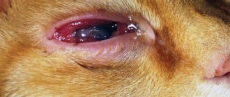
Keratitis
Keratitis is serious damage to the cornea of the eye. This disease can be caused by either injury or infection.
It is not so easy to notice that with a pet: the cat will try to close or squint the sore eye.
In addition, with keratitis, profuse lacrimation is observed. The eye instinctively tries to get rid of the aching lesion.
Photo: keratitis is caused by inflammation or injury
If treatment is delayed, the cornea will begin to noticeably become cloudy. The animal will develop photophobia.
Determining the cause of the disease is quite simple, even at home. Due to the injury, only one eye is affected. Infections affect both corneas.
Fungal diseases are a little more complicated: they first attack one eye and only after some time they move to the second.
It should be noted that wounds or foreign particles trapped under the eyelid provoke rapid clouding of the lens. Infections severely inflame the surface of the visual organ, causing severe suppuration.
Third eyelid
Cats have a special protective device that protects their eyes from foreign bodies getting into them. The third eyelid is a whitish, thin film of skin.
Its loss can be recognized by a number of signs:
- severe lacrimation;
- redness of the skin and eyes;
- discharge in the form of mucus and pus;
- swelling in the corners of the eyes;
- involuntary muscle spasms (eyelids visibly tremble).
Photo: prolapse of the third eyelid cannot be treated on your own. Prolapse of the nictitating membrane is a very dangerous disease that can quickly develop into others.
It can be caused by a number of problems that have already been observed in the cat:
- allergy;
- injury;
- conjunctivitis in an advanced stage;
- diseases of internal organs (usually the gastrointestinal tract);
- fungus;
- internal and external parasites.
Under no circumstances try to self-medicate. Wrong actions can only harm the animal (even completely deprive it of its vision). At the first suspicion of prolapse of the nictitating membrane, you should contact your veterinarian.
Blepharitis
Blepharitis is an inflammation of the eyelid in cats. According to stages and complexity, this disease is divided into: simple, scaly, ulcerative, meibomian. The main signs of blepharitis are redness and swelling of the eyelid.
With proper treatment, this disease can be quite easily eliminated. The problem is that simple blepharitis very quickly develops into ulcerative, and then scaly.
Then the eyelashes begin to fall out, and if nothing is done, the disease will spread to the lacrimal sac, and then to the eye, which, if left untreated, can lead to loss of vision.
Photo: blepharitis affects a cat's eyelids
The pet itself can significantly worsen its situation. A cat, trying to scratch a sore spot, can injure the eye with its claws.
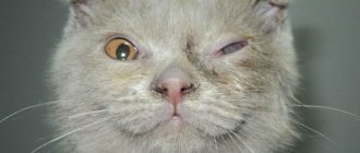
Panophthalmitis
Panophthalmitis is a very rare but very dangerous disease of domestic animals. It is characterized by simultaneous damage to all capillaries and tissues of the eye, which makes it extremely difficult to treat.
The symptoms of panophthalmitis in dogs and cats are similar: the eyeball initially noticeably increases in size, then redness and pus are detected. Very often complications appear in the form of other ailments.
Photo: panophthalmitis is a very dangerous viral disease
Unfortunately, it is almost impossible to save an eye with panophthalmitis. There is a high risk of infection spreading to the cerebral cortex, which usually leads to the death of the cat. To save the animal, the entire diseased organ is removed and the eyelids are sutured.
If you notice that your cat's eye is not in normal condition, you should not try to cure it on your own.
Careless actions (for example, wiping tears or pus with your hands, processing with human means) can only contribute to the fact that the infection will quickly destroy the organ.
Cataract
Cataracts are more common in older animals. In children, this disease occurs due to widespread infection. The lens becomes noticeably cloudy, and it becomes increasingly difficult for the cat to see.
It is quite easy to notice cataracts in a pet at an early stage. The cat's visual acuity noticeably decreases, which is why the animal often bumps into or crashes into objects, misses when playing, or fails to jump to something.
Photo: Cataracts are more common in older cats
Self-medication is extremely dangerous for your pet. At the first suspicion of cataracts, you should immediately contact your veterinarian. At the initial stage of the disease, medications will be prescribed.
If they do not work, then surgery is prescribed. Treatment should not be delayed, otherwise cataracts may be complicated by other diseases.
Inflammation or infection of the nasolacrimal ducts
Obstruction of the nasolacrimal ducts is quite common among cats, especially in babies. The disease is not so dangerous and is easily treated. Inflammation is caused by the fact that the channels connecting the lacrimal sac to the nose become clogged.
Photo: inflammation of the nasolacrimal duct threatens kittens
It is quite easy to understand that inflammation is occurring. The pet has severe lacrimation. However, not a single ointment designed to combat conjunctivitis or keratitis will help.
The most effective way to relieve swelling of the tubules and clean them is probing. This method is not very pleasant, but it brings immediate relief to the pet.
To stop the infection from spreading, veterinarians prescribe antibiotics. After a week, their use is stopped, and the animal is re-examined to confirm the further treatment strategy.
Wounds of the eyelids
Wounds are always dangerous, especially around such a fragile organ as the eyes. According to the severity, such injuries are divided into superficial (implying a small scratch), deep (several layers of skin are removed) and through (if the eyelid is completely torn).
The consequences of each type are different. That is why treatment must be approached based on the severity of the damage.
Photo: damage to the eyelid usually occurs in fights
It is recommended to wash the superficial wound as quickly and carefully as possible and treat it with any antiseptic (you can even use iodine if the scratch is located on the outer eyelid).
If, in the heat of a fight, the mustache manages to cause through or deep damage, then you need to immediately contact a veterinarian. Only in the clinic will all foreign particles be carefully removed, the wound washed and stitches applied.
Turn of the century
Sometimes the edges of the eyelid turn inward, toward the cornea. Such mechanical damage to the eye is not just uncomfortable, but very painful.
In addition, the unprotected cornea is damaged by hard cilia and hairs rubbing against it. If left untreated, entropion of the eyelid can lead to the development of infection (keratoconjunctivitis).
Photo: inversion and eversion of the eyelid can only be corrected surgically
Usually such diseases are congenital. You can only get rid of them through surgery. The operation is quite fast and effective (there will be no more eyelid inversions).
Glaucoma
There are three types of glaucoma in cats, depending on the cause: congenital, open-angle and closed-angle. The disease is characterized by severely elevated blood pressure.
With glaucoma, small and large vessels burst in the animal's eyes, causing the whites to turn red. The cat is in a lot of pain.
Bloody discharge from the eyes is visible both on the eye itself and on the conjunctiva. The pupil also noticeably enlarges. In addition, the eyeball is greatly inflated and hardens (due to high pressure).
Open-angle glaucoma is characterized by noticeable clouding of a large area of the eye. The cornea weakens and stops responding to light.
With angle-closure glaucoma, a cloudy ring-shaped area forms on the eye, as well as a whole network of swollen and bulging blood vessels.
Photo: glaucoma is characterized by very high pressure
Such a disease not only affects the quality of vision, but also brings great suffering to the animal. It is almost impossible to cure glaucoma without the help of a veterinary ophthalmologist.
The disease can be overcome with the help of drops and tablets, but if it is caused by a dislocated lens, then surgery will be required.
Belmo
A cat's eyesore develops over a long period of time as a result of persistent clouding of the cornea. This disease in cats can be different.
In some cases, a thorn forms at the edge of the eye, and in others it covers it entirely.
At first, the cat begins to see much worse (objects look blurry due to the cataract). If the disease is not stopped, then partial loss of vision or complete blindness is possible.
Photo: eyesore distorts vision
The causes of the appearance of a cataract can be trauma, age-related changes in the structure of the cornea, congenital pathology, viral or fungal infection.
You can overcome the disease if you have time to go to the clinic at an early stage of its development.
Viral infection
The eyes of cats and cats are often affected by various viral infections. The most popular is herpesvirus. It can actively develop on the mucous membrane of the organs of vision, nasopharynx, and trachea. The virus usually develops with a decrease in immunity and a decline in the body's defenses.
Infection can occur through contact with infected animals, as well as through plants and soil. Herpes can be transmitted to kittens from a sick cat during fetal development.
The main features include:
- formation of ulcerative lesions on the lips, nose, surface of the tongue;
- severe tearing of the eyes;
- swelling of the eyes;
- conjunctivitis;
- Over time, a cat's eyesore may appear;
- swelling of the larynx and nasopharynx;
- formation of discharge with a mucous structure from the nose.
Entropion and eversion of eyelids
An inversion is the rotation of the outer edge of the eyelid and eyelashes towards the cornea; an inversion is the opposite effect: the edge of the eyelid turns outward.
An inversion creates discomfort for a pet, as the eyelashes constantly touch the cornea of the eye.
The reasons for such deformities are the same: tightening of the skin of the eyelids during the formation of scar tissue after healing of injuries, burns, and surgery. Inflammatory processes of the eye, conjunctivitis and simply aging of the body can also lead to this disease.
The disease is accompanied by severe lacrimation and redness of the conjunctiva.
Treatment often comes down to minor plastic surgery. If the disease is accompanied by inflammatory processes, then drug treatment is first carried out. In the absence of medical attention, the disease can lead to chronic keratoconjunctivitis.
Video: Maine Coon's eyelids are turned upside down
Blepharitis in cats
Inflammation of the eyelids, which causes thickening and redness of the edges of the eyelids.
There are several types of blepharitis: simple, meibomian and ulcerative .
Let's look at each type in more detail. Simple blepharitis. With this disease, there is overflow of blood vessels (hyperemia) and thickening of the edges of the eyelids. Gray-white scales appear at the base of the eyelashes. If the edges of the eyelids swell and their surface becomes covered with pus and an ulcerative surface appears under it, this is no longer simple, but ulcerative blepharitis . The main symptom is watery eyes and itching of the eyelids. If inflammation affects the roots of the eyelashes, they fall out. Entropion or inversion of the eyelids may occur. Ulcerative blepharitis is treated by removing dead tissue, after softening the surface of the eyelids, this is best done with fish oil. Then the edges are cauterized with a special disinfectant and washed with water or saline solution. Then, within a week, an ointment containing antibiotics is applied to the affected areas of the eyelids. And another type of blepharitis is meibomian. Appears due to hypersecretion of the meibomian glands (named after the physician Heinrich Meibon). Signs include hyperemia and severe thickening of the edges of the eyelids. Pathogenic microorganisms that enter these glands cause purulent inflammation of the eyelids. To treat this inflammation, use “brilliant” and yellow mercury ointment.
Before you begin treatment, you need to take your beloved animal to a veterinarian who can choose the right treatment for your pet. It is not recommended to use medications without the consent of a specialist, as improper treatment can aggravate the situation.
Obstruction of the lacrimal ducts
One of the causes of excessive lacrimation in a cat is stenosis - obstruction of the nasolacrimal duct.
The causes of this pathology may be different;
- a foreign body that has entered the canal and clogged it;
- conjunctivitis or other inflammatory processes in which the released pus can close the nasolacrimal duct;
- tear duct injuries;
- congenital insufficiency of the width of the mouth or neck of the lacrimal ducts.
Some cat breeds are predisposed to developing this disease, including:
- Scottish and British cats;
- exotics;
- Persians.
Obstruction of the nasolacrimal duct is determined using a special test. Before starting the procedure, the animal's eyes are thoroughly washed with an antibiotic solution to completely remove pus and other secretions. 1-2 drops of an organic dye solution (fluorescein) are instilled into the eye. During normal operation of the channels, after two minutes the internal cavity of the pet’s nose and mouth will acquire a greenish color. If this effect is obtained after ten minutes, then a partial blockage is diagnosed. Complete absence of staining indicates stenosis.
The course of treatment depends on the cause of the disease. First, the inflammatory process is relieved. If after this the functioning of the tear ducts is not restored, bougienage is performed - prompt removal of the plug blocking the duct.
Glaucoma in cats
A disease accompanied by increased intraocular pressure and an enlarged eyeball.
Symptoms of glaucoma in cats
Symptoms are visible to the naked eye. There is an enlargement of the pupil, reflection of the fundus, bulging of the cornea, which is no longer mirror-like. If this disease is not cured in time, the animal may permanently lose its vision as the retina dies.
Treatment of glaucoma in cats
It is carried out only with drugs that reduce intraocular pressure. To restore the cornea and prevent it from drying out, an ointment is prescribed. To prevent such a disease in your pet, you need to carefully remove discharge from the eyes with a swab, after moistening it in a special lotion for animals. Lotions are available both for external use and for treating the cornea itself, so you need to carefully read the instructions for use.
When you need urgent veterinary help
Immediate contact with a veterinarian is required in cases where the pupil or cornea of the eye has changed color, shape, size, or the eyeball has hardened. In addition, you should not delay your trip to the clinic if your cat has severe pain in the eye area.
This is not difficult to understand: the pet reacts to even a light touch with a loud meow, hides from the owner, and shows aggression to any attempt to pick itself up.
Such conditions indicate severe ophthalmological pathology and require urgent assistance from a specialist.
Third eyelid prolapse
The inner, or third, eyelid is a thin membrane that protects the cornea of the eye from mechanical influences and dirt. In a healthy pet it is almost invisible. If the third eyelid does not fold, this is a sign of pathology. However, the list of diseases that change the condition of this organ is quite large and includes not only eye diseases:
- eye injury or foreign body penetration;
Loss of the third eyelid in only one eye indicates that problems are related to the visual organs
- conjunctivitis of all types;
- inflammatory process in the membrane itself;
- manifestation of allergies, including to external parasites;
- helminth infection;
- problems with the gastrointestinal tract.
When the third eyelid appears, it is not recommended to carry out any treatment on your own. If the condition does not return to normal within the next few hours, you should seek veterinary help.
You can read more about the third eyelid in cats in the article “Third eyelid in cats: causes, symptoms and treatment.”
Video: third eyelid with herpes virus infection
What is epiphora
This medical term refers to constant and unregulated tearing . At this time, the animal’s tears flow down, characteristic tracks become visible on its cheek, and its fur acquires a brown tint.
In severe cases, hair begins to fall out around the eye, and itching or skin lesions similar to dermatitis may appear. The danger of the disease is that during its development, the cat’s eyes are practically not protected.
The normal process of tear secretion looks like this. The tear secreted from the gland goes to the upper part of the conjunctiva. When the eyelids move, it flows into the lacrimal lake (the lowest point of the palpebral fissure). From there, the liquid reaches the lacrimal openings, which partially absorb it, and then to a special bag, bypassing small highways. As a result, the nasolacrimal duct carries it into the nasal cavity, where evaporation occurs.