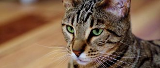
Almost every veterinary specialist knows that an animal’s eyes are a “mirror” by which a specialist can visually determine if the animal is sick or not. Today in medicine, many human diseases are diagnosed by the eyes.
Eye diseases in our cats are quite common today. If you do not take the necessary treatment measures in a timely manner, sometimes your pet’s disease can result in loss of vision.
Before talking about eye diseases, cat owners should have an idea of what the eye is.
A cat's eye is an organ specially adapted for the animal to perceive light waves. With the help of vision, your cat navigates the world around it, perceives the intensity of light, color, shape of objects, distance to them, as well as the movement of objects in space. The eyeball of a cat has a shape close to a ball and consists of an optical part and a photoreceptor part. Light rays, before reaching photoreceptor cells, pass through a complex optical system of transparent media. The first thing the rays encounter on their way is the cornea, part of the white outer shell - the sclera. Externally, the cornea resembles a slightly convex watch glass; Unlike the sclera, the cornea is devoid of blood vessels and is almost transparent. A ray of light passing through the cornea enters the anterior chamber of the eye (the space between the cornea and the lens). This space is filled with liquid (chamber moisture). Adjacent to the inner part of the sclera is the second layer of the eye - the vascular layer, which is rich in arterial and venous vessels. In the front part of the eye, the choroid turns into the iris, which contains the pigment that gives the eye its color (yellow, green, sometimes blue).
The iris, just like the aperture in a camera, regulates the amount of light entering the eye. The iris is equipped with two layers of ciliary muscles: annular and radial. In the middle of the iris there is a hole - the pupil, through which a ray of light enters the back of the eye. If a cat is in the dark and is suddenly illuminated, then as a result of contraction of the annular muscle, the pupil narrows and the flow of rays entering the eye decreases.
After passing through the pupil, a ray of light enters the lens - a transparent body similar to a small, biconvex magnifying glass. Along the edge of the entire circumference of the lens, the ligament of zinn is attached to it. The lens itself is enclosed in a capsule and attached to the ciliary muscle through the ligament of cinnamon.
From the lens, the light beam passes through the vitreous body of the eye, which mainly fills the eyeball. The vitreous body is transparent and consists of the finest fibers, which are a skeleton, between which there is liquid.
The cornea, aqueous humor of the anterior chamber, lens and vitreous body constitute the optical, or refractive, system of the eye. Before affecting the retina, the light beam changes its direction many times, due to the fact that each of these media has its own refractive index.
Adjacent to the vitreous body is the third layer of the eye, the retina. Its outer layer consists of pigment cells that prevent the scattering of light rays, while the inner layer consists of cells called rods and cones. They are the receptor, or perceptive, department of the visual analyzer.
When examined with an ophthalmoscope, the back wall of the eyeball, the so-called fundus, reveals a pale colored area from which blood vessels radiate. Experts call this area a blind spot because there are no light-sensitive cells in it. From the entire retina, nerve fibers converge to the blind spot to form the optic nerve. The total number of light-sensitive cells in a cat's eye is very large. At the same time, rods, unlike cones, have a higher sensitivity to light.
Under the influence of light, the metabolism of the retinal pigment cells changes and electrical phenomena occur. The reddish pigment rhodopsin, or visual purple, contained in the rods, turns into the yellow pigment retinen in the light. In the dark the reverse transformation occurs. Both of these pigments consist of protein and vitamin A. The quantitative ratios of rhodopsin, retinene and vitamin A are established depending on the degree of illumination of the retina.
Today, all existing eye diseases in cats are divided by experts into two large groups:
- Diseases and damage to the protective devices of the visual organs.
- Diseases and damage directly to the eyeball itself.
Veterinary specialists include diseases and damage to the protective devices of the visual organs:
- Bruises and other mechanical damage in which the integrity of the skin is not damaged.
- Mechanical injuries and wounds accompanied by skin damage and bleeding.
- Inflammation of the eyelids (blepharitis).
- Turning of the eyelids.
- Eversion of the eyelids.
- Inability to close the eyelids (lagophthalmos).
- Upper eyelid incontinence (ptosis).
- Tumors and various neoplasms.
Diseases and injuries of the eyeball include:
- Prolapse of the third eyelid.
- Conjunctivitis.
- Blepharitis (inflammation of the eyelids).
- Keratitis (inflammation of the cornea).
- Corneal ulcer.
- Cataract (clouding of the lens).
- Glaucoma (increased intraocular pressure).
- Iritis or iridocyclitis (disease of the ciliary body and iris).
- Dacryocystitis (impaired patency of the lacrimal canal).
- Exophthalmos (prolapse of the eyeball).
- Enophthalmos (sinking of the eyeball inwards).
- Panophthalmitis (inflammation of all membranes of the eye).
All of the above eye diseases can be of infectious or non-infectious etiology.
Eye diseases in cats are characterized by the presence of similar symptoms of the disease:
- lacrimation.
- redness and swelling of the eyelids and conjunctiva.
- various discharges from the eyes.
- soreness.
- photophobia.
- Sometimes in the morning the cat is unable to open the affected eye.
- itching appears in the affected eye.
Diagnosis of eye diseases.
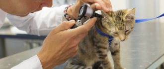
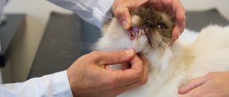
Diagnosis of a particular eye disease in cats should be carried out by specialists from veterinary clinics. At the veterinary clinic, specialists will conduct a clinical examination of your cat to rule out the presence of infectious diseases that have led to damage to the visual organs. The affected eye is examined more deeply (the size and shape of the pupils, the size of the palpebral fissure, the size of the eyeballs, the presence of injuries, etc.). If necessary, smears will be taken for culture on nutrient media. Blood will be taken to rule out infectious diseases.
Diseases of the eyes and protective devices of the visual organs
Bruises and mechanical damage without compromising the integrity of the skin.
Cause. They occur in a cat as a result of a blow with a blunt object or as a result of mechanical damage (bumping into blunt objects, falling).
Clinical signs. During a clinical examination, the veterinarian notes the presence of bruises and hematomas in the eye area. In the area of injury, upon palpation, an increase in local temperature is noted, and a change in the size of the eyeball occurs.
Treatment. A cat with an eye injury should be taken to a veterinary clinic. To relieve pain in a sick animal, a 2% solution of novocaine, antimicrobial drops or eye ointment is instilled into the conjunctival sac. If there is a hematoma, it is necessary to apply cold to the hematoma (only on the eyelid area, not on the eyeball). If there are serious complications, it is sometimes necessary to resort to surgery.
Rules for carrying out medical procedures at home
At home, the owner can independently perform ophthalmological procedures.
First of all, this is eye washing, which is carried out in the following way:
- An antiseptic solution, a decoction of a medicinal plant, or special drops are applied to a cotton pad or piece of gauze. With a light and smooth movement from the outer corner of the eye to the inner corner, secretions are removed.
- A napkin generously moistened with an antiseptic is applied to the stuck eyelids of an adult animal or kitten and held for 2-3 minutes. After this, the eyes open randomly, and the softened pus from the fur is removed with the same napkin.
- The eyeball is washed using a syringe without a needle.
- Also, the owner can independently instill a medicinal solution into the lower conjunctival pocket. To do this, he must pull back the cat's lower eyelid, carefully introduce the drug with a pipette, and then close the animal's eyelids. In a similar way, you can put medicinal ointment behind the eyelid.
Wounds and open injuries to the eye.
Cause. Most eye injuries and wounds in cats occur as a result of fights with their fellow cats, falling on sharp objects, or strong blows leading to damage to the skin.
Clinical signs. During a clinical examination, the veterinarian notes a violation of the skin in the area of the injured eye, the presence of scratches, wounds, and bleeding from the injured areas.
Treatment. The damaged area is washed with hydrogen peroxide, bleeding is stopped; in the presence of large wounds, pain relief and suturing are used, and antimicrobial therapy is administered locally. For complex and large eye injuries, eye microsurgery and sometimes removal of the damaged eye are performed.
Blepharitis
Blepharitis is inflammation of the eyelids.
Cause. Traumatic damage to the eyelids, the presence of a bacterial or fungal infection, allergies in animals, diabetes (diabetes mellitus in animals, diabetes insipidus in animals), liver disease.
Clinical signs. During a clinical examination, a veterinarian notes redness of the eyelids, swelling, photophobia, profuse lacrimation, itching and burning in the area of inflammation of the eyelids, loss of hair and eyelashes around the eyelids.
Treatment of blepharitis should be aimed at eliminating the cause that led to blepharitis. The sore eye is washed with a decoction of calendula or chamomile, a solution of boric acid, potassium permanganate or furacillin. Eye drops are instilled into the conjunctival sac. If infection is present, antibiotics are used.
Entropion of the eyelids
Cause. Entropion of the eyelids in cats occurs as a result of certain eye diseases (conjunctivitis, blepharitis, etc.) and exposure to certain chemicals. A genetic predisposition in some cat breeds (Sphynxes, Persians) leads to bloat.
Clinical signs. During a clinical examination, the veterinarian notes that the eyelid turns inward. As a result of mechanical irritation, redness of the eyeball and symptoms of blepharospasm are noted. When examining the cornea, we record the presence of ulcers in the place of constant contact with the eyelid, and lacrimation.
Treatment. Carrying out a surgical operation.
Eversion of the eyelids
Cause. Eversion occurs as a result of certain eye diseases, when they become chronic. Eversion of the eyelids is common in some cat breeds. It is quite rare in clinical practice.
Clinical signs. During a clinical examination of a sick cat, a veterinarian notes blepharospasm (spastic closure of the eye), conjunctivitis, lacrimation, and at the site of ectropion the veterinarian diagnoses the area of the mucous membrane.
Treatment. Surgical intervention.
Fusion of eyelids
Cause. Eyelid fusion in a cat can be either congenital or acquired. Owners notice congenital fusion of the eyelids after the birth of a kitten. Acquired fusion in a cat occurs after injuries to the head and eyes, with chronic blepharitis, chemical and thermal burns of the eyelids.
Clinical picture. During a clinical examination, the veterinarian cannot separate the eyelids, there is a continuous strip of skin between the eyelids, and there is a scar between the fused eyelids.
Treatment. Surgical intervention aimed at separating the eyelids.
Lagophthalmos
Lagophthalmos in a cat is manifested by the inability to completely cover the eyes with the eyelids.
Cause. Eversion and inversion of the eyelids, paralysis of the facial eyelid, hereditary shortness of the eyelids.
Clinical signs. During a clinical examination of a sick cat, a veterinarian notes a constantly slightly open palpebral fissure, lacrimation, and lag of the lower eyelid from the eyeball.
Treatment. Treatment of this pathology in cats is surgical, and it is necessary to first use antimicrobial eye drops and corneal protector medications.
Treatments
Both special veterinary drugs and medications developed for humans are used to treat cats. Therapy with traditional methods also shows good results.
Medications
Among the medications for the treatment and prevention of ophthalmic drugs in cats, the most effective ones have proven themselves:
- "Bars" - washing for injuries, treatment and prevention of keratitis, conjunctivitis, blepharitis.
- “Iris” - acute and chronic inflammation of the cornea and anterior segment of the eye.
- "Tsiprovet" - therapy and prevention of infectious inflammation of all ocular structures.
- Decta-2 - treatment of allergic and infectious conjunctivitis.
- Lakrikan - hygienic treatment and treatment of inflammatory processes.
- Neoconjunctivitis - treatment of advanced forms of conjunctivitis.
- Sofradex is an antibacterial drug for the treatment of inflammation of the eyes and ears.
ethnoscience
Traditional medicine suggests treating eye pathologies in cats using the following herbs:
- chamomile;
- calendula;
- green tea;
- St. John's wort;
- sage.
All of them have some anti-inflammatory effect and are used for therapeutic and prophylactic treatment of the eyelids and eyeball.
Uevit
Uevitis is an inflammation of the choroid of the eye.
Cause. Uevitis in cats occurs as a result of eye injuries, metabolic disorders, eye tumors, autoimmune diseases, toxoplasmosis, rickettsiosis, diseases caused by viruses (feline leukemia, feline immunodeficiency, infectious peritonitis, etc.).
Clinic. Uevitis in a cat is accompanied by photophobia, lacrimation, eye pain, blepharospasm, strabismus, the cat's pupil is greatly reduced or takes on an uneven, blurry shape. The eyes have a red and cloudy appearance on clinical examination. There is a change in the color of the iris.
Treatment of ueviitis in a cat should be aimed at the cause that led to the disease. Eye drops are instilled into the eyes and painkillers are prescribed. Antibiotics and other antimicrobial drugs are used. If there is a threat of developing glaucoma, it is necessary to resort to surgical removal of the diseased eye.
Diagnostics
It is carried out by a veterinarian in a specialized clinic, as it requires special tools. It is carried out in several stages. First, the organ is examined and the following parameters are assessed:
- preservation of vision;
- appearance of the organ: the size and shape of the pupils, their symmetry; the same parameters are used to evaluate the eyelids and their condition;
- condition of the eye: its size, shape, position relative to the orbit, absence or presence of injuries.
After an external examination of the organ, the doctor begins research using additional equipment: examines the fundus of the animal, measures intraocular pressure, prescribes general clinical and biochemical tests of blood and urine, takes a smear to identify pathogenic microflora and its sensitivity to antibiotics, etc.
Third eyelid prolapse
Prolapse of the third eyelid in a cat is often a secondary symptom of a number of infectious and parasitic eye diseases; it occurs when the optic nerve becomes inflamed.
Clinic. Cat owners and veterinary specialists use eyelids, which sometimes reach up to 1/3 of the entire visual area.
Treatment. Third eyelid prolapse in cats can be treated symptomatically or surgically. Treatment should be aimed at eliminating the primary disease that led to the loss of the third eyelid. Surgical treatment involves surgical excision of the prolapsed third eyelid.
Short description
Unfortunately, a disease in which part of a cat's eyelid is covered with a white, opaque film is common. The film bears the name of the third century and is of great importance. It removes foreign particles and dust, and also distributes tear fluid. The third eyelid (conjunctival fold or nictitating membrane) is located at the inner corner of the eye. It is almost impossible to notice it in a healthy animal. If the cat is sick, the membrane becomes inflamed and turns white.
Keratitis
Keratitis is inflammation of the cornea.
Cause. Keratitis in cats is caused by infectious diseases (infectious rhinotracheitis in cats, feline chlamydia, mycoplasmosis in cats, calicivirus infection of cats), helminthic infestations, damage to the cornea by a foreign body, and eye injuries. Often the cause of keratitis can be inflammatory processes in nearby tissues.
Clinic. Keratitis in a cat can be superficial, deep and ulcerative. With keratitis, a cat may experience vision loss. During a clinical examination of a sick animal, a veterinarian notes redness of the eye, photophobia, clouding of the cornea, pain, and visible blood vessels. Cloudiness of the cornea occurs due to cellular infiltration (by leukocytes) and changes associated with cell swelling, their degenerative decay, as well as changes in the intercellular connective tissue. The intensity of turbidity can be different and depends on the density and nature of the infiltrate or edema in the interstitial tissue. The gray-smoky color of the cloudiness is formed by a small accumulation of leukocytes. With an increase in the number of leukocytes, the color of the cornea becomes white; a yellowish tint characterizes a purulent infiltrate. The opacification may be diffuse or limited to isolated areas in the form of dots or spots in the anterior layers (superficial keratitis) in the parenchyma (parenchymal keratitis). If the surface layers are damaged, the cornea loses its specular properties and becomes matte. With keratitis, vascularization of the cornea also occurs (ingrowth of blood vessels into the cornea). At the same time, during a clinical examination, the veterinarian notes a pericorneal injection, which is characterized by overflow of densely located vessels in the limbus area. The reaction from the conjunctiva, mainly its scleral part, is manifested by edema and hyperemia. With keratitis, experts note a reaction from the iris. The membrane becomes hyperemic, the pupil narrows, and serous, fibrinous or purulent exudate sometimes appears in the anterior chamber. With acute keratitis, a cat experiences photophobia, spasm of the eyelids, lacrimation and pain. In eosinophilic keratitis, veterinarians upon clinical examination detect the presence of white plaques on the cornea that extend from behind to the anterior wall. Slight lacrimation is noted. When performing a cytological examination, we find a large number of eosinophils.
Treatment. Symptomatic treatment is carried out. Eliminate the underlying disease that caused the keratitis. The affected eye is washed with antimicrobial solutions, and eye ointments and drops are used. If a purulent process occurs, antibiotics are used; for deep and ulcerative keratitis, eye microsurgery is used. For eosinophilic keratitis, they resort to the use of immunomodulators and hormones.
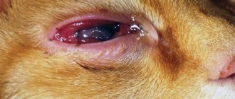
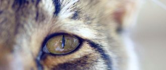
Conjunctivitis
This disease is known to everyone - it is one of the most common. Conjunctivitis is an inflammation of the conjunctiva, the mucous membrane of the eyelids. It is often a symptom of other global pet health problems.
Symptoms
In the photo of sick animals you can clearly see which symptoms of conjunctivitis are most active. These include watery eyes, swelling and redness of the eyelids, and noticeable clouding of the eye.
Causes
Many factors can cause inflammation of the conjunctiva. This is why conjunctivitis is so common. The main reasons for its appearance include:
- fungi, bacteria and viruses. They can affect only the membrane of the eyelid, or they can affect the entire body of the animal;
- damage and injury that violate the integrity of the protective membranes, thereby allowing infection to pass through;
- allergens (in this case, conjunctivitis is just one of the symptoms. Itching, runny nose, and sneezing may also occur);
- colds, weakened immunity;
- parasites;
- ultraviolet radiation.
Types of conjunctivitis
Based on the manifestation and source of inflammation, conjunctivitis is divided into four types that can transform into each other.
- Catarrhal . It is easily treated if measures are taken in time. Characterized by swelling, lacrimation and mucous discharge. Possible eversion of the eyelid. If you do not fight the disease, it goes into the next form.
- Purulent . Identified by the discharge of pus. Depending on the severity, it can be just yellow crusts in the morning, or obvious yellow-green pus. The eyelids may stick together. Serious complications and loss of vision are possible if you do not contact a veterinarian promptly.
- Phlegmonous . With this type of conjunctivitis, pus accumulates under the skin, so independent treatment is impossible.
- Follicular . This type is chronic. With it, not only the conjunctiva becomes inflamed, but also the inside of the third eyelid. Treatment is often impossible without surgery.
The first form can be cured on your own. Tetracycline ointment 1%, which can be easily purchased at any pharmacy, will help with this. Use a cotton swab to apply it to the lower eyelid of the animal. Repeat the procedure several times a day.
If treatment does not bring results, or a purulent form of conjunctivitis is possible, you should immediately consult a veterinarian.
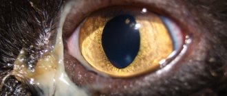
Corneal ulcer
A corneal ulcer in a cat is accompanied by tissue destruction and poor healing. Superficial disintegration of the epithelium is called erosion.
Cause. The causes of the development of corneal ulcers can be both exogenous and endogenous. Exogenous causes include various traumatic injuries caused by the entry of hard and uneven foreign bodies into the conjunctival sac or as a result of direct injury to the cornea. Ulcers in cats occur when the eyelids or eyelashes roll up, when the cornea is constantly subjected to friction by the rolled-up edge of the eyelids or eyelashes. Chemical and thermal burns are always accompanied by an ulcerative process in a cat. Purulent conjunctivitis can cause the formation of corneal ulcers. Corneal ulcers can be observed when there is a violation of its innervation (neuroparalytic keratitis). Endogenous causes of ulcers in cats include a number of infectious diseases of cats, metabolic diseases, hypovitaminosis diseases, etc.
Clinical picture. Large ulcers are detected by a veterinary specialist at the clinic during examination - a defect, clouding of the cornea and discharged exudate are revealed. Small and superficial ulcers are determined by a veterinarian using side lighting and clouding of the cornea. Ulcerative processes in cats are in all cases accompanied by spasm of the eyelids, their swelling, mucopurulent, and more often purulent discharge, and the cat’s body temperature rises.
After the ulcers heal, scars form.
Treatment. Treatment of corneal ulcers is symptomatic or surgical. In the case when the eyeball is dissolved by a purulent process, it is removed. In veterinary clinics, sick animals are given antimicrobial therapy, including antibiotics, eye drops and ointments, and pain relief (novocaine blockades). Eye microsurgery is also performed to remove damaged areas of the cornea.
Iritis (iridocyclitis)
Iritis (iridocyclitis) is inflammation of the iris and ciliary body.
Cause. Traumatic injury, inflammation spreading from the cornea, complications after eye surgery, infectious diseases.
Clinic. During a clinical examination, a clinic veterinarian notes a constriction of the pupil, dimming of the iris, photophobia, pain, and cloudy fluid in the anterior chamber of a sick cat. When conjunctivitis is associated with the disease, we note serous-purulent discharge.
Treatment. In order to restore pupil contraction, a 1% atropine solution, novocaine blockade, and antibiotics are prescribed. During treatment, eye drops and ointments with anti-inflammatory and antimicrobial effects have a good effect.
Prevention
The owner of the animal must be attentive to his well-being and behavior, and notice any changes in this.
If a cat rubs its eyes and shows anxiety, it is worth examining it, the cause may be a foreign body. If this is the case, you need to remove it and rinse the eye with plenty of clean water, furatsilin, a weak solution of potassium permanganate, and then apply any eye drops. After this, you need to take your pet to a veterinarian for a more thorough examination.
For preventive purposes, it is worth wiping your pet’s eyes daily with boiled water or chamomile decoction. Animals do not really like hygiene procedures, and therefore it is necessary to accustom them to this from childhood.
If crusts, discharge, or any changes in the pet’s behavior are detected, it is necessary to show it to a veterinarian as soon as possible.
1111
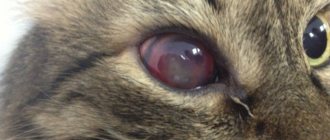
Glaucoma
Glaucoma is an increase in intraocular pressure, which is accompanied by an enlargement of the eyeball in a cat.
Glaucoma in cats is usually not a primary disease, but often accompanies other eye diseases. If treatment is not started promptly, glaucoma causes vision loss in cats. Cats are usually asymptomatic. Glaucoma can be open-angle or closed-angle.
Clinic. Angle-closure glaucoma is characterized by redness of the eye, swelling of the eyelids, severe pain, clouding of the cornea, weakness, nausea, and vomiting.
Treatment. To normalize intraocular pressure, pilocarpine is used, to improve the outflow of excess fluid in the cat’s body, diuretics, retrobulbar administration of aminazine, and the prescription of painkillers. Sometimes it is necessary to resort to surgical intervention (extirpation of the eyeball).
Panophthalmitis
Panophthalmitis is a serious condition that causes severe inflammation of the eyes. In this case, damage to all tissues of the animal’s visual organs is observed. The disease is a complication of injuries and injuries to the eyeball.
The development of panophthalmitis is caused by a purulent infection that enters the eye through damage. The causative agents of this pathological process can be various bacteria:
- white and golden staphylococci;
- pneumococci;
- streptococci;
- in rare cases, Escherichia coli and Pseudomonas aeruginosa.
Symptoms of panophthalmitis include:
- manifestation of sharp pain in the eye;
- fear of bright light;
- increased fluid secretion;
- frequent uncontrolled closing of the eyelids;
- severe swelling of the eyelids of the conjunctiva of the eyeball;
- pinching of the mucous membrane of the eye;
- When examined, the cornea may be very cloudy and swollen. Sometimes purulent discharge may be present;
- decline in visual function.
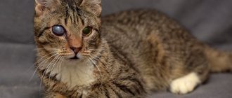
Cataract
Cataract is clouding of the lens. The disease often leads the cat to blindness. Cataracts in cats are associated with metabolic disorders in the lens and are accompanied by increased breakdown of protein components.
Cause. Trauma, infectious diseases, and chronic inflammatory eye diseases lead to cataracts in cats.
Clinic. At the last stage of the disease, the cat has difficulty seeing in the affected eye. Upon examination, the lens has a white or bluish color. The dilated pupil does not respond to light.
Treatment. The use of eye drops for cataracts can only slow down the degenerative processes developing in the lens.
Dacryocystitis
Dacryocystitis is an inflammation of the lacrimal sac, accompanied by a violation of the patency of the nasolacrimal canal due to its narrowing.
Cause. The disease in cats occurs as a result of chronic conjunctivitis.
Clinic. During a clinical examination, a veterinarian notes mucous or purulent discharge from the eyes of a sick cat. In the outer upper corner of the eye orbit, a swelling is visible, which is painful on palpation. The upper eyelid is swollen, the lacrimal gland is enlarged on palpation. Sometimes there is an accumulation of pus in the tissues around the eyes.
Treatment. Treatment of dacryocystitis in cats can be symptomatic or surgical. Symptomatic treatment consists of relieving inflammation and pain by using novocaine blockades and absorbable ointments (Vishnevsky ointment, ichthyol). Surgical treatment consists of cleaning the lacrimal canal or extirpation of the lacrimal gland.
Obstruction of the lacrimal duct
Tears must flow through a special channel that is located inside the nose, but if the outflow path is disrupted, the tears come out through the eyes. The main symptom is different types of lacrimation and the appearance of brown fur around the eyes (tear tracks). A similar problem can be congenital (found in some breeds - Sphynx, Persian or Scottish cats) or acquired (obstruction can be caused by various infections).
If patency occurs due to blockage or fusion of the nasolacrimal duct, surgical intervention may be prescribed. Although the use of special rinses and preparations is usually sufficient.