Otitis externa is an inflammation of the outer ear, which includes the pinna and external auditory canal (EA).
Author:
- Filatova Evgenia Vladimirovna
otolaryngologist, rhinosurgeon
3.67 (Votes: 9)
- Causes of a boil in the external auditory canal
- Causes of development of bullous otitis media
- Treatment
Otitis externa is an inflammation of the outer ear, which includes the pinna and external auditory canal (EA). Like most inflammatory diseases of the ENT organs, otitis externa can be acute or chronic.
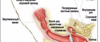
Features of the structure of the outer ear
The auricle and ear canal are covered with skin that extends to the eardrum. There are two sections in the ESP: cartilaginous and bone; at the border of these sections, a physiological narrowing is determined - the isthmus, which is why the three-dimensional model of the external auditory canal resembles an hourglass. Under the skin of the auricle and the cartilaginous part of the external auditory canal there is elastic cartilage, due to which this part of the auditory canal is mobile during chewing and articulation. In the bony section, under the skin, there is periosteum and bone. The skin of the external auditory canal differs from any other skin in that its thickness contains cerumenous glands that produce earwax (cerumen). The function of cerumen is to protect the skin from constant moisture and microorganisms, and the presence of a cerumen film is an important factor in the normal functioning of the outer ear. The dimensions of the external auditory canal may vary markedly depending on individual characteristics. Often in the area of the isthmus there are osteophytes - bony protrusions that reduce the lumen of the ESP, and sometimes completely block it. Anterior, posterior and inferior to the auricle in the subcutaneous tissue there are lymph nodes that can become inflamed with otitis externa.
Acute external otitis is an inflammation of the external ear that occurs within 1 month.
Forms of external otitis:
- diffuse external otitis;
- local external otitis or NSP boil;
- acute bullous (hemorrhagic) external otitis;
- myringitis;
- dermatitis of the auricle;
- erysipelas of the auricle;
- malignant (necrotizing) external otitis;
- chondroperichondritis of the auricle.
The most common is acute diffuse external otitis, in which bacterial inflammation of the skin of the entire ear canal occurs, often spreading to the eardrum or the skin of the auricle.
Causes of the disease
Otitis media in cats can be caused by factors related to the functioning of the body, as well as environmental influences. These include:
- fleas and ticks. They are carriers of infection. Inflammation and irritation are also caused by waste products of parasites.
- foreign body. Once in the ear canal, small objects cause local inflammation and swelling.
- bacterial infection. Its development is facilitated by hypothermia, lack of vitamins, viral diseases, and weakened immunity.
- allergens. Individual intolerance to certain foods, medications, and detergents causes a release of histamine in the cat, which can result in allergic otitis media.
- parasitic fungi. They cause inflammation, which most often affects the outer and middle parts of the ear.
- neoplasms. Verrucous nevus and malignant tumors under certain conditions provoke an inflammatory process in the tissues of the auditory analyzer.
- injuries. In the absence of proper treatment, damaged skin turns into a fertile environment for infection. This is how bacterial otitis occurs. Inflammation of the internal part can occur due to injury to the temporal bone.
- autoimmune diseases. As a result of the rejection of one's own tissues, inflammation begins in the ear.
- hormonal disbalance. A change in the concentration of hormones (primarily produced by the adrenal glands, gonads, and thyroid gland) can provoke an inflammatory process.
- growth of fur in the ears. The high density of hairs stimulates the ceruminous glands, which produce earwax. This causes irritation and, as a result, scratching of the integument.
global $ads_google; //data-ad-slot=”2475549904″ $ads_google = empty($ads_google) ? false : true; ?> if ($ads_google == false) {?>
$ads_google = true; ?> } ?>
Acute diffuse otitis media
Provoking factors: excessive toilet of the ears, water getting into the ear, microtrauma of the skin of the NSP, factors of decreased immunity (hypothermia, excessive tanning, ARVI, chronic diseases). Pathogens: Staphylococcus aureus, Pseudomonas aeruginosa.
Symptoms
This is severe pain in the ear, ear congestion, liquid discharge from the ear canal in a small amount, there is also an increase in the behind-the-ear or parotid lymph nodes, inflammation of the skin of the auricle, there may be an increase in temperature, and symptoms of intoxication. On examination, hyperemia and swelling of the skin of the NSP are noted, the skin is uneven, with overlays of transparent yellow or purulent discharge, and desquamated epidermis is visible. The lumen of the ESP is often sharply narrowed and the eardrum may not be visible. If it is visible, it is usually also uneven and moderately hyperemic. Enlargement and tenderness of regional lymph nodes, thickening, tenderness and hyperemia of the auricle are often detected. Hearing, while maintaining the lumen of the lumen, is slightly reduced or normal.
Diagnostics
To make a diagnosis, in addition to examination, it is necessary to exclude middle ear disease, for which tuning fork tests (a hearing test using a set of tuning forks), tympano- and impedance measurements (measurement of the mobility of the eardrum), and a study of hearing thresholds (audiometry) are performed. These studies are carried out to exclude diseases of the middle ear. Laboratory diagnostics includes a general blood test, blood sugar, microflora culture from the diseased ear to prescribe antibacterial therapy.
Treatment
The main direction of treatment is antimicrobial and anti-inflammatory therapy. Antibiotics and anti-inflammatory drugs (non-steroidal anti-inflammatory drugs, corticosteroids) are prescribed locally or systemically. For severe pain, there is a good effect from behind-the-ear blockades with corticosteroids and local anesthetics. A gentle ear toilet and rinsing is carried out to free the ear from discharge and epidermal masses. Additionally, you can use physical treatment methods: quartz tube or laser therapy.
Signs of ear problems
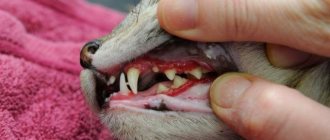
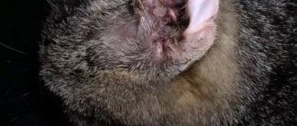
Normally, cats' ears are shiny and clean, but not wet. The skin inside the ear is a uniform pale pink color. There is no visible discharge in the ear; at most, there may be a small accumulation of light yellow wax without a strong odor. If a kitten develops an ear disease, it will manifest itself with some of the following symptoms:
1. The cat shakes its head and purses its ears. 2. The cat actively scratches its ears and even scratches them until they bleed, resulting in crusts and wounds appearing. 3. There is no (or weakened) reaction to sounds. 4. If you touch the ears at the base, the cat presses them and tries to escape, bites and scratches. 5. Swelling, unnatural growths and bumps are found inside.
Read more: The cat has a swollen ear: what to do
6. The skin in the ear is red, peeling, there are signs of inflammation and crusting. 7. Discharge in the ears ranges from whitish to dark brown. 8. A putrid or sweetish (unnatural) odor comes from the ears. 9. Shortness of breath and nasal discharge are observed. 10. There may be problems with motor coordination and balance.
Limited (local) otitis externa or boil of the external auditory canal
A furuncle is an inflammation of the hair follicle that spreads to the subcutaneous tissue. Bristly hairs grow at the entrance to the NSP and around it, and inflammation develops in their follicles.
Causes of a boil in the external auditory canal
The most common pathogens are streptococci. Provoking factors and symptoms are similar to those of diffuse otitis media.
Diagnostics
Upon examination, a narrowing of the ESP is visible due to infiltration in the area of one of its walls. The boil goes through three stages in its development:
- Infiltration stage: at this stage, a painful infiltrate of the NSP skin is formed, local hyperemia and thickening of the skin about 0.5-1.5 cm in size appear.
- The abscess formation stage is the appearance of a purulent cavity and a necrotic core in the area of infiltration. At this stage, the presence of an infiltrate is also determined, in the center of which a yellow-green purulent core is visible, usually with a hair in the center; when pressing on the infiltrate, fluctuation (the presence of fluid in it) can be determined.
- The resolution stage is the release of pus from the cavity with gradual recovery.
Treatment
Treatment varies and depends on the stage. At the infiltration stage, systemic and local antibacterial and anti-inflammatory therapy is prescribed. In the stage of abscess formation, it is worth performing surgical treatment of the boil - opening and draining, cleaning the cavity from pus, dressings are performed within several days. Antibacterial and anti-inflammatory drugs are also prescribed. During the resolution stage, it is sufficient to use only local antibacterial drugs and ear toilet.
Veterinarian help
After the veterinarian has determined the causes of the cat's anxiety and made a diagnosis, he prescribes treatment. First of all, with the help of medications, the veterinarian relieves ear pain and itching in the cat. If there is discharge, dirt, or excess wax in the ears, then clean them. It is performed in the following sequence:
- The auricle is turned outward to provide access to the ear canal.
- The fur on the inside of the ear is carefully trimmed.
- A cotton swab is dipped in oil and the plaque is removed within sight.
- The second ear is treated in the same sequence.
For external otitis in a cat, the veterinarian treats the damaged surface and instills liquid medicine, after which he gently massages the auricle to evenly distribute the solution. In case of deep purulent otitis media, the cat's eardrum is pierced to remove pus from the distant parts of the ear. After deep cleaning, the ears are treated with systemic antibiotics. Owners of furry patients are advised to take note of the veterinarian's advice from the following video:
global $ads_google; //data-ad-slot=”2475549904″ $ads_google = empty($ads_google) ? false : true; ?> if ($ads_google == false) {?>
$ads_google = true; ?> } ?>
Acute bullous or hemorrhagic otitis externa
This is a form of inflammation in which blisters (bullas) containing bloody fluid appear on the skin and on the outer surface of the eardrum. If such a bubble is opened, then bloody discharge flows out of the ear.
Causes of development of bullous otitis media
Most often, bullous otitis occurs against the background of acute respiratory viral infection or influenza, but a bacterial infection can also be the cause.
Treatment
An ear canal toilet is required. Antibacterial and anti-inflammatory drugs are prescribed locally, and drugs that strengthen the vascular wall and reduce vascular permeability should also be prescribed systemically. Opening bullae is not recommended.
Diagnostics
A comprehensive approach is required to make a correct diagnosis. The effectiveness of treatment depends on the results of the research, because bacterial otitis and otitis caused by ticks cannot be treated in the same way.
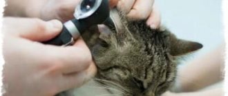
The doctor must collect all the data about the conditions of keeping and feeding the pet, chronic diseases, and behavior. A complete examination of the animal is carried out, the outer ear and ear canal are examined using an otoscope, and the doctor takes a swab from the ear for laboratory examinations.
Additionally, an x-ray may be prescribed, as well as an MRI; tomography will help determine inflammation of the middle ear at the very initial stages, when x-rays are uninformative.
Dermatitis of the ear
It can be separated into a separate form, since inflammation of the skin of the auricle often occurs, without involving the NSP. The causes of the disease are a bacterial infection plus a factor of sensitization of the body to bacterial toxins. With this type of inflammation, redness and thickening of the skin occurs, with the formation of small serous blisters, similar to those in eczema. When the bubbles open, yellow crusts form in their place. Diagnosis is made based on examination, and consultation with a dermatologist may be required. Treatment is also antibacterial and anti-inflammatory, and antihistamines may be prescribed.
Erysipelas of the auricle
Acute erysipelas is an infectious disease and is caused by streptococcus pyogenes; later, other flora may join. With such inflammation there are symptoms of skin damage and a general inflammatory syndrome - weakness, fever, chills. Factors provoking the disease: decreased immunity, abrasions and burns of the skin, swimming in polluted waters, contact with a carrier of pyogenic streptococcus or a sick person. Depending on the depth of damage to the skin, three forms are distinguished:
- Erythematous, when pain occurs, redness of the skin (it has a “varnished” appearance) slightly rises above the level of healthy skin and has a clear border with it.
- The bullous form, more severe, is characterized by the same skin lesions as in the erythematous form, but with the formation of large blisters above the surface of the skin with transparent or cloudy contents.
- Necrotic form. With this form, deep damage to the skin occurs, right down to the reticular and papillary layers, and deep ulcers with purulent plaques form. Due to the addition of various microflora, it is more severe than other forms. Typically, patients with this form require surgical debridement of areas of necrosis.
Erysipelas
This is a serious disease that often causes complications, and its treatment should take place in an infectious diseases hospital. If erysipelas is suspected, consultation with an infectious disease specialist is required. Isolated erysipelas of the auricle is rare and is usually combined with lesions of the skin of the face or neck.
Treatment
Massive antibacterial and anti-inflammatory therapy, detoxification therapy, and antistreptococcal gamma globulin are prescribed.
Prevention
To prevent ear diseases in cats, hygiene procedures must be carried out regularly.
Cleansing is done using lotion. A few drops are placed in the ear and lightly massaged. After some time, remove dirt with a clean soft cloth.
The sleeping place for pets should not be in a draft or near a frequently opened window.
Particular attention should be paid to the cat's ears while bathing. Water should not get into the ear. If this happens, then the water must be wiped off with a dry cloth. Drying your ears with a hairdryer is not recommended.
Malignant otitis externa (necrotizing otitis externa)
This form of external otitis is distinguished as a separate one, but in fact it is a negative course of ordinary diffuse external otitis. During the course of the disease, inflammation spreads to the structures of the temporal bone, damage to the cranial nerves and the development of intracranial complications may occur. The name of the disease is not associated with the development of a malignant tumor, it simply reflects the course of the disease. Fortunately, this disease is rare; it affects patients with severe immune disorders, such as AIDS, conditions after radiation and chemotherapy, decompensated diabetes mellitus, and patients receiving immunosuppressive and cytostatic therapy. The leading pathogen is Pseudomonas aeruginosa, but any infection is polymicrobial. During the process of inflammation, necrosis of the skin of the ear canal occurs and inflammation passes to the periosteum and bone, followed by its destruction. Complaints in this disease include severe pain, discharge from the ear of a purulent-inflammatory nature, with a putrid odor, and hearing loss quickly occurs. Later, symptoms of damage to the cranial nerves appear: difficulty swallowing, choking when eating, numbness of the face, paralysis of the facial muscles. When the process spreads to the middle and inner ear, systemic vertigo may develop. When inflammation spreads into the cranial cavity, cerebral and meningeal symptoms develop. On examination, one notices pronounced inflammation of the skin of the NSP, skin with foci of necrosis, a large amount of purulent-necrotic discharge, the eardrum and the structures of the middle ear are often indefinable. Symptoms of damage to the cranial nerves are determined - facial asymmetry, ptosis of the lower eyelid on the affected side, asymmetry of the soft palate, impaired mobility of the epiglottis. In diagnosis, culture for bacteriological examination and computed tomography of the temporal bones are important. Treatment is always carried out in a hospital setting. Massive antibacterial and detoxification therapy is carried out. Infusions of human gamma globulin, antipseudomonas serum or hyperimmune human plasma are prescribed. Surgical treatment is also performed - necrotomy (removal of necrotic areas of skin and bone), and subsequently, upon recovery, plastic surgery of the defect is performed using one’s own tissues.
Treatment of otitis media in cats
Ear inflammation is a very serious disease, so self-treatment with drops or ointments at home without consulting a veterinarian is not recommended. If you suspect otitis in a cat, you should contact the clinic. After establishing the diagnosis, the doctor will prescribe detailed treatment based on the type of disease.
Purulent otitis media
The prescription of drugs for this type of inflammation depends on the nature of the pathogen. If bacteria are the culprit of the pathology, then otitis media is treated with antibiotics. When using such medications, you should be aware of your cat's sensitivity to them, as some medications cause side effects in animals. For cats, antibacterial agents of the class of cephalosporins, penicillin, and macrolides are used. The dosage is calculated individually for each four-legged patient, based on age, body weight, and severity of the disease.
If the eardrum is damaged, do not use ototoxic antibacterial drugs (Norfloxacin, Rifamycin, Ciprofloxacin), as they negatively affect hearing.
Fungal otitis media
For otomycosis, the cat is treated with fungicidal drops or ointments based on nystatin. Treatment of parasitic otitis necessarily includes hygiene procedures aimed at cleansing the surface of the outer ear. The ointments are applied to the clean surface for sanitation. In addition to local medications, the cat is prescribed general antifungal medications.
Treatment of otitis in cats should be carried out in compliance with general rules of care. The animal should be protected from drafts and hypothermia. It is necessary to minimize stressful situations. The pet should be at rest and warm.
Allergic otitis media
If the inflammatory process is associated only with increased histamine synthesis in the cat’s body, then antihistamines are used to relieve unpleasant symptoms. The animal is treated using Tavegil, Fexofenadine, Suprastin, Loratadine, Cetirizine. In case of severe inflammation, the use of anti-inflammatory corticosteroids (Prednisone, Prednisolone) is justified. In combination with them or separately, the doctor may prescribe Dexamethasone. Ear mites often cause an allergic reaction. In this case, antiparasitic treatment is necessary in parallel.
Parasitic otitis media
This common form of otitis in cats requires the use of special drugs, the action of which is aimed at destroying ear mites. Treatment is carried out using anti-inflammatory and acaricidal drugs. They can be in the form of ointments, aerosols, drops. Dekta, Amitrazine, Otopheronol Gold are suitable for this purpose. Therapy also includes regular cleansing of secretions from the auditory organ. Owners need to treat their pets for fleas and ticks on a schedule.
global $ads_google; //data-ad-slot=”2475549904″ $ads_google = empty($ads_google) ? false : true; ?> if ($ads_google == false) {?>
$ads_google = true; ?> } ?>
Chondroperichondritis of the auricle
This disease is an inflammation of the perichondrium and cartilage of the auricle caused by bacterial flora. Chondroperichondritis can be caused by trauma to the auricle with damage to the skin, suppuration of an auricular hematoma, deep burns of the auricle, a boil or suppurating atheroma of the ear, and erysipelas. There are infiltrative and purulent forms. A few days after the described traumatic factor, infiltration and hyperemia of the skin of the auricle develops, accompanied by pain, with or without fever. An important diagnostic criterion is that the earlobe remains unaffected, since there is no cartilage in its thickness. Treatment: antibacterial and anti-inflammatory therapy. If there are purulent cavities, an opening with removal of pus and gentle necrotomy of the destroyed cartilage is performed. The disease is difficult to treat and can last for weeks or months. Often the consequence of chondroperichondritis is cicatricial deformation of the auricle; sometimes there may be complete destruction of the cartilage.