For the longevity and cheerfulness of a pet, its owner is obliged to closely monitor the health of his own pet. One possible problem could be a brown spot on your cat's eye. The appearance of such a pathology may indicate disturbances in the functioning of the cat’s organ of vision. So, if symptoms of the disease occur, it is recommended to contact a veterinarian in a specialized clinic to examine the animal, make an accurate diagnosis and prescribe the correct treatment.
Reasons for appearance
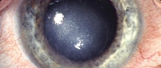
A cloudy spot on the iris of a cat occurs when the cornea of the eye is damaged. If a brown spot, black dots or a cataract appears in a cat’s eyes, this is evidence that the animal is losing visual acuity. The main reasons influencing the appearance of spots are considered to be the following pathological conditions:
- Corneal injury. It can occur with pathologies, inflammation, infections and injuries in the iris.
- Cloudiness of intraocular fluid. It appears when there is an accumulation of many leukocytes, proteins, and lipids.
- Diseases of the vitreous body.
- Cloudy lens. Develops during the formation of cataract disease.
Types of stains
Both a thorn and formations of different colors and pigment on the eyelid may appear on the cornea of the eye. They may not appear immediately, but after 2 years. Initially, the spot is small and unnoticeable, but over time it increases in size, bringing more and more discomfort to the cat. There are several varieties of such spots:
Black dots on the organ indicate cancer in the animal.
- Light. They occur when there are problems with the spine, if the animal has kidney, liver, or mental and nervous disorders.
- Brown spots. Appear on the iris or eyelids as a result of infectious diseases.
- Black dots. Formed during oncology.
- With a red tint. They appear when there are problems with the liver and enzymes in the blood.
- Growths of the “presentation” tobacco type. Develop with constipation and gastrointestinal problems.
Eyesore
Syphilis
Herpes
13256 August 17
IMPORTANT!
The information in this section cannot be used for self-diagnosis and self-treatment.
In case of pain or other exacerbation of the disease, diagnostic tests should be prescribed only by the attending physician. To make a diagnosis and properly prescribe treatment, you should contact your doctor. An eyesore: causes of occurrence, what diseases it occurs with, diagnosis and treatment methods.
Definition
An eyesore, or leukoma, is externally manifested by clouding of the cornea of the eye that occurs after injury or an inflammatory process. The cornea is the anterior, most convex part of the eye capsule, consisting of a thin avascular membrane through which refracted light enters the posterior parts of the eye. There are three stages of corneal opacification depending on the size and depth of the lesion: a cloud, a spot and a cataract, but all of them affect visual acuity, and in some cases a person can become completely blind in the affected eye.
The success of treatment largely depends on timely seeking medical help.
Types of eyesores
There are congenital and acquired eyesores:
- congenital cataract is formed during the period of intrauterine development of the embryo and is often combined with other congenital eye diseases;
- the acquired form is the most common, occurring equally often in both men and women.
Depending on the depth of the lesion, the following types of cataracts are distinguished:
- corneal cataract without affecting other parts of the eye; there are scars of different shapes and lengths in the cornea;
- a thorn welded to the iris; a cloudiness appears on the cornea, sprouting with small vessels, spreading to the area of the iris;
- a cataract in combination with true clouding of the lens, that is, cataract;
- cataract with secondary cataract;
- intense clouding of the cornea of the eye, which is characterized by adhesions of the cornea with the iris and/or lens, clouding of the lens;
- a thorn complicated by retinal detachment and the development of eye atrophy.
Possible causes of eyesores
Doctors consider keratitis, an inflammation of the cornea of the eye, to be one of the possible causes. The most common cause of keratitis is a viral infection (herpes viruses, chickenpox). Often, bacterial infections, as well as non-compliance with the rules of wearing contact lenses, lead to an inflammatory process in the cornea.
Keratitis is characterized by redness of the eye, pain, lacrimation, photophobia, clouding of the cornea already in the initial stages of the disease, and decreased visual acuity.
If treatment is not started on time, the inflammation will spread deeper and an eyesore will begin to form.
Corneal injuries are another common cause of eyesores. Injuries can be mechanical, thermal or chemical (alkali or acid burns). Alkaline burns are the most dangerous.
The first symptoms of injury are pain, lacrimation, sensation of a foreign body, drooping of the eyelid.
Any injury to the cornea requires immediate medical attention.
Infectious diseases of the conjunctiva can lead to damage to the cornea and the appearance of a cataract. The conjunctiva is the connective membrane of the eye that covers the inner surface of the eyelids and extends from the outside to the cornea. One of the most dangerous diseases of the conjunctiva is trachoma. It occurs when a chlamydial infection enters the eye, is chronic and causes blindness in almost two million people in the world. Children are most susceptible to this eye disease.
Tuberculosis is an infectious disease that, despite the successes of domestic medicine, is still often diagnosed in patients of all ages. Tuberculosis can affect any organs and systems of the body, including the infection that can spread to the eyes, lead to deep damage to the cornea and cause the formation of a cataract.
Cases of cataract formation after eye surgery have been described. This is due to a violation of the integrity of the cornea, which increases the risk of infection.
Diseases leading to the formation of eyesores
- Congenital pathologies, for example, limbal dermoid, sclerocornea.
- Keratitis.
- Injuries (chemical, thermal burns, penetrating wounds).
- Trachoma.
- Corneal ulcers.
- Tuberculosis, syphilis.
Which doctors should you contact if an eyesore appears?
An ophthalmologist deals with the treatment of eyesores. He will order tests and conduct additional examinations. If you suspect an infectious etiology of the cataract, you may need to consult an infectious disease specialist or phthisiatrician.
Diagnostics and examinations for the appearance of an eyesore
To clarify the diagnosis, the doctor may prescribe the following examinations:
- a clinical blood test with a detailed leukocyte formula will help identify inflammatory changes in various infectious and inflammatory diseases;
Symptoms
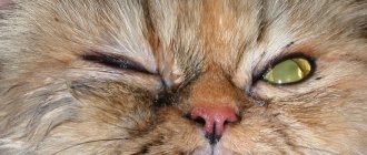
If brown spots and formations appear on the cat's eyes, symptoms may appear, which are definitely recommended to be closely monitored. Visual acuity may decrease, the animal may not get into the bowl, it may skid when turning, and orientation in space may also be disturbed. The second aspect is considered to be increased irritability and nervousness of the pet. The cat begins to behave aggressively towards its owners and other pets in the house. Often a person notices the development of strabismus in a cat, the appearance of watery discharge from the eyeball. There is severe swelling of the cornea and redness on the iris and eyelids.
If such symptoms appear, it is recommended to immediately take your pet to an appointment with a veterinarian. When examining a cat, a specialist will prescribe appropriate treatment and tell you about methods for preventing the development of such diseases. Self-medication is prohibited.
Corneal melanosis.
Corneal melanosis, which is also called pigmentary keratitis (pigmentous keratitis), is one of the common ophthalmological pathologies of brachycephalics. The essence of the pathology is that with chronic irritation of the cornea (keratitis), melanin cells are introduced into its epithelium and the surface layers of the corneal stroma, migrating from the limbus zone. It is worth noting that pigmentary keratitis is a very general diagnosis. The thing is that the cause of this condition is chronic inflammation of the cornea (keratitis). And the main goals of treatment will be to eliminate the causes that led to keratitis.
Diagnostic and treatment methods
When visiting a specialist, blood must be taken from the animal for testing.
When examining a furry patient, a veterinarian, namely an ophthalmologist, will make an accurate diagnosis and prescribe appropriate treatment. To confirm the diagnosis, the following procedures are prescribed:
- taking tests and examining them, especially blood and serology;
- measurement of intraocular pressure and comparison with healthy indicators;
- performing a cytology procedure;
- taking a smear from the eyeball and examining them;
- culture examination of the microflora of the eye and intraocular fluid.
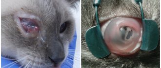
A darkened iris in a kitten can only be cured with medication, after examining the test results by a professional doctor. Once the diagnosis becomes known, such diseases can be treated by carrying out procedures such as washing the eyeball with Metrogyl, or you can administer the drug Gamavit parenterally. It is also possible to lubricate the inflamed area with tetracycline ointment.
If, after testing, glaucoma is detected, then surgery may be necessary. Only the treating doctor should treat spots on an animal’s cornea, otherwise it can cause harm to the pet. It is not recommended to self-medicate, since the appearance of spots is considered a symptom of serious pathological changes in the cat’s body. Timely medical care will help protect your pet from discomfort, illness and dangerous consequences.
Types of spots on the eyeball
In medicine, a blind area in the visual field that is not connected to the peripheral borders is called a scotoma. A spot is an area that is unable to transmit light, although light sensitivity around it is usually normal. Lack of visual perception can manifest itself in different ways:
- Focus becomes difficult when looking forward at near or distant objects;
- the darkening is only in one place and does not change its position when the eyes move;
- the spots seem to flicker. They are also called “flies”;
- color perception is impaired. Previously saturated shades look faded and faded. It is also difficult for a person to determine exactly what color is in front of him.
Let's consider two types of cattle:
It should be said that the shape of pathological spots can determine a particular disease. For example, a hereditary and rather rare visual impairment - retinal pigmentary degeneration - is characterized by a scotoma in the form of rings. And with glaucoma, which occurs with increased eye pressure and leads to cessation of the functioning of the optic nerve, arc-shaped spots are most often observed.
Eye neoplasms – what are they?
The delicate and sensitive tissues of our eyes, unfortunately, are vulnerable to many carcinogenic factors: radiation, chemicals, some viruses, burns and injuries. There may also be hereditary reasons for their degeneration. Tumors of the organs of vision and surrounding tissues are so diverse that they are studied by a special branch of clinical medicine - ophthalmo-oncology. Due to the many variations of eye tumors, diagnosing the problem is not always easy; this requires a comprehensive examination by a highly qualified ophthalmologist. But, whatever the diagnosis, there is no need to be afraid - modern treatment methods have come so far that there is a suitable method for every case.
Classification of tumors of the eye and surrounding tissues
The same classification applies to tumors of the visual organs as to tumors in general. They are divided into:
- Benign - slowly growing, does not metastasize, and does not have a toxic effect. They can degenerate and become malignant. Among eye tumors, this type includes: papilloma, senile wart on the eyelid, benign nevus and a number of others
- With locally destructive growth - those that do not metastasize, but have invasive growth (intermediate category). Basal cell carcinoma and progressive nevus are characterized by such growth
- Malignant - fast-growing, destroying other tissues and releasing toxins. Their cells are transported with the blood to other parts of the body and can give rise to secondary lesions (metastases). Examples: cancer of the conjunctiva, meibomian glands, melanoma and sarcomas of the eyelids.
Eye tumors are also classified according to their location in the affected organ.
:
- Tumors of the orbit (eye socket)
- Tumors of the adnexal apparatus of the eye (eyelids, conjunctiva)
- Intraocular tumors (choroid and retina).
Treatment of ocular tumors
Like many serious pathologies, tumors require an integrated approach to treatment. The choice of the necessary methods depends on the diagnosis, the stage of development of the tumor, and the individual characteristics of the Patient. It is important to understand that removing a tumor may not always be sufficient for subsequent health safety. In some cases, regular prevention of its re-development will be necessary.
Methods for removing tumors
:
- Laser evaporation
- Radiosurgical removal using robotic systems
- Cryotherapy (freezing cancer cells with liquid nitrogen)
- Thermotherapy (several sessions of heating tissues up to 45 degrees)
- Brachytherapy (contact radiation technique)
- Photodynamic therapy (destruction of cancer cells by the reaction of their product to light)
- Chemotherapy
- Surgical removal
The need for surgery usually arises only in the later stages of the disease. Until then, treatment may be limited to absolutely safe and minimally invasive techniques (a technique aimed at minimizing the area of intervention in the body and the degree of tissue injury).
The appearance of a neoplasm is a reason for active action, but not for panic. Your attending physician will select a combination of techniques that will achieve the most favorable result for your health. Check your vision regularly, contact a specialist if alarming symptoms appear - and be healthy!
Symptoms and signs
Almost all types and types of keratitis are characterized by the so-called. corneal syndrome, including increased lacrimation, increased sensitivity to light and uncontrolled closure of the eyelids (blepharospasm). As a rule, redness is pronounced, the vascular network becomes noticeable, and in severe cases, neovascularization (vascular neoplasms) occurs. The pain can be quite intense. The corneal layer swells, the surface loses its smoothness and often ulcerates (sometimes with a tendency to peel off), and infiltrates appear. Cloudiness of the cornea becomes one of the main symptoms and complaints with keratitis, since the opacity or translucency of the cornea has an extremely negative effect on the acuity and clarity of vision.
Keratitis is often accompanied by concomitant symptoms, the severity of which varies from mild to moderate discomfort (dry nasal and oral mucosa, headache, difficulty swallowing) to severe complications from the periodontium, gastrointestinal tract, etc. Various types of keratitis are characterized by their own specific features.