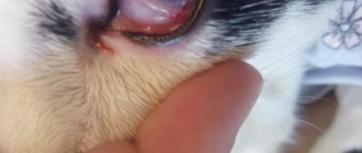
The term hyphema describes the presence of blood in the anterior chamber of the eye - what to do if you find blood in your cat's eye. Bleeding usually comes from the iris blood vessels, but can also come from the ciliary body (tissue behind the iris), from the choroidal vasculature (the layer of tissue under the retina), or from the blood vessels of the retina.
Many cats suffering from hyphema experience decreased visual function or eye damage. However, if only one eye is involved, the cat's behavior is usually normal. Eyes affected by hyphema may be painful, appear half-closed, and be characterized by increased tearing and persistent blinking of the eyelids. Hyphema caused by trauma is often accompanied by bleeding or bruising of the conjunctiva and tissues surrounding the eye.
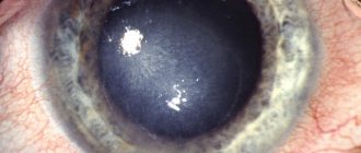
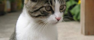
Blood in a cat's eye
Bleeding, if extensive or recurrent, may completely fill the anterior chamber, resulting in complete hyphae and blindness. Eyes with severe hyphema are at greater risk of developing glaucoma (high intraocular pressure) and should be closely monitored for this potential complication.
Initially, the blood inside the eye appears bright red. Blood may remain uncoagulated and settle at the bottom of the eye. The blood may also clot and become dark brown or bluish-gray in color over time.
The presence of a hyphema may be a symptom of severe ocular pathology or a manifestation of an internal problem located in other areas of the body. Although hyphema is often caused by trauma to the eye, spontaneous hyphema can occur in the presence of many different ocular and systemic diseases. Therefore, immediate identification of the cause is of paramount importance.
Regarding visual function, the prognosis depends on the degree of the disease (mild or severe), as the pupil may become dilated during treatment and there may be bleeding or damage to the back of the eye. The presence of severe hyphema, lack of pupil dilation, hemorrhages in the back of the eye, or retinal detachment often leads to blindness.
In what cases is it necessary to contact a veterinarian?
If there is no appetite and lethargy, you should show the animal to the veterinarian. If the cat, in addition to the desire to hide in a dark place, has the following deviations, it is necessary to show the animal to the veterinarian:
- Lack of appetite;
- Increased thirst;
- Refusal of water;
- Heavy discharge from the mouth, nose, or eyes;
- Purulent discharge;
- Changes in the consistency and smell of excrement;
- Change in the color and odor of urine;
- Dull coat, baldness;
- Skin rashes, suppuration;
- Frequent scratching, licking;
- Cough, sneezing, vomiting;
- Lethargy, drowsiness;
- Loss of coordination;
A cat’s desire to hide in a dark place, especially when it is not eating or drinking, is caused by many reasons. An attentive and caring owner will respond in a timely manner to changes in the pet’s behavior and will do everything possible to restore a comfortable environment.
Causes
In general, reasons can fall into different categories:
- Hyphema caused by closed or penetrating trauma.
- Chronic or severe uveitis. This is an inflammation of the iris, ciliary body and choroid.
- Coagulation disorders are caused by changes in platelet number or functionality and other clotting factors.
- Systemic hypertension, that is, increased blood pressure.
- Systemic diseases affecting coagulation or ocular blood vessels, such as some viral infections (feline infectious peritonitis), some leukemias, and acute increases in serum proteins.
- Retinal detachment or lacerations of retinal vessels.
- Neoplasia of the eye or other areas of the body.
- Specific causes that may be associated with the development of hyphema include:
- Closed trauma or trauma passing through closed eyelids, such as road traffic accidents, gunshots to the head.
- Penetrating injuries or injuries that pierce the eye.
- Ocular prosthesis, or protrusion of the eyeball out of the orbit, usually as a result of closed head trauma.
- Uveitis with hemorrhage that occurs from the blood vessels of the iris, from the ciliary body and from the choroidal tissues.
- Systemic hypertension (high blood pressure) is associated with diseases such as chronic kidney disease and hyperthyroidism (an overactive thyroid gland).
- Accidental ingestion of toxins from rodenticides such as warfarin or brodifacoum, or ingestion of dead rodents with such toxins.
- Chronic liver disease with decreased production of clotting factors and vitamin K.
- Coumadin toxicity is caused by an overdose of this drug (prescribed to treat certain cardiovascular diseases).
- Low platelet count (thrombocytopenia) or decreased platelet function.
- Hyperviscosity syndrome, which develops when the level of protein macromolecules in the bloodstream is extremely high.
- Primary neoplasms of the iris and ciliary body or other neoplasms that arise in the eye.
- Metastatic tumors that have spread to the eyes from other areas of the body.
- Systemic hypertension (high blood pressure), leading to hemorrhage in the choroid, retina and vitreous body and retinal detachment.
- Retinal detachment with hemorrhage originating from the choroid or torn blood vessels of the retina due to ocular trauma, systemic hypertension, uveitis, postoperative uveitis (for example, after cataract surgery) and chronic glaucoma, resulting in an enlarged globe with crystalline shift.
Main symptoms and treatment methods
For timely and adequate treatment, it is necessary to know the characteristic signs of eye diseases.
One of the common diseases in pets is conjunctivitis. Inflammation of the mucous membrane occurs for the following reasons:
- injuries;
- infectious diseases;
- allergic reactions;
- infection with worms and ectoparasites;
- lack of vitamins in the body.
There are several forms of conjunctivitis: catarrhal, follicular, ulcerative and purulent. The main symptoms indicating the development of conjunctivitis are:
- redness of the mucous membrane;
- swelling of the eyelids;
- lacrimation;
- the appearance of pus;
- eyelid gluing;
- photophobia;
- cloudiness.
Purulent conjunctivitis is indicated by bilateral inflammation. In addition, the owner notes the following symptoms of the disease:
- general depression of the pet. He becomes lethargic, does not want to play and frolic;
- high body temperature. Normally, the body temperature of an adult animal is 38-39°C; in children, the figure may be half a degree higher (38.5-39.5);
- poor appetite or its complete absence also indicates a painful condition;
- the cat tries to avoid inspection and palpation, this process causes him pain in the area of the eyeball;
- in advanced cases, inflammation spreads to the cornea.
Treatment steps are as follows:
- A visit to the veterinary clinic to remove a foreign object from the eye.
- Washing can be done at home every 3-4 hours. For the procedure, use a warm solution of manganese, furatsilin, chamomile or calendula decoction. Herbs help reduce inflammation, eliminate swelling and itching. In addition, natural decoctions have anti-inflammatory properties. Manganese copes well with infection, but at the same time dries out the mucous membrane. Therefore, a pale pink solution is used. It is recommended not only to thoroughly dissolve the manganese crystals, but also to filter the resulting composition through several layers of gauze. This guarantees the absence of crystals in the water, the accidental entry of which can cause a burn to the mucous membrane. Furacilin is diluted in hot water (1 g per 5 l). Used for washing while warm.
- Applying ointment. At a pharmacy or pet store you need to buy any eye ointment, for example, Tetracycline, Syntomycin or Erythromycin. The composition is placed behind the eyelid. The procedure is carried out wearing gloves. The ointment should be applied carefully with a special glass rod. Applying with fingers or cotton swabs is unacceptable. After each application of the ointment, the stick is disinfected in boiling water. This is necessary in order not to spread the infection in the future. Some people use an ointment tube to apply the ointment. This must be done extremely carefully, otherwise bacteria will settle on the spout of the tube and, during the next treatment, will again get into the eyes.
- Instillation is carried out only in washed eyes. It is necessary to instill 2-3 drops every 4 hours.
- If the disease is caused by an infection, a course of antibiotic therapy is necessary.
- A solution of novocaine and hydrocortisone (ratio 1 ml/0.2 ml) helps relieve swelling. The composition is instilled into the conjunctival sac.
- Chronic pathology is recommended to be treated with silver-based drugs.
- In a veterinary hospital, a novocaine blockade is carried out. It reduces pain and improves tissue nutrition, which leads to a speedy recovery.
- For follicular conjunctivitis, surgical intervention is performed. After the operation, ointments and antibiotics are prescribed.
We suggest you read: Muscovy ducklings and their cultivation
Keratitis and its types
Inflammation and clouding of the cornea in cats indicate the development of keratitis. The disease is acquired and requires proper treatment. The reasons for the development of keratitis are:
- mechanical injuries of the cornea. The ingress of solid dust particles, grains of sand, blades of grass, and fibers leads to mechanical damage;
- conjunctivitis. The combination of diseases leads to the formation of keratoconjunctivitis;
- thermal or chemical burns;
- infections and viruses. Typically, keratoconjunctivitis develops against the background of a general infection of the body (herpes, adenovirus). In this case, complex treatment is required, otherwise it will not be possible to get rid of keratitis;
- allergic reactions to food, dust, pollen, household chemicals;
- inflammatory processes in the lacrimal glands, leading to drying out of the cornea;
- lack of vitamins in food;
- genetic predisposition to the disease (in British, Sphynx, Persian and Siamese cats).
The appearance of the following symptoms should alert the cat owner:
- clouding of the cornea, its dullness and roughness;
- accumulation of blood vessels on the cornea;
- the discharge causes the fur around the eye to become wet;
- accumulation of pus;
- fear of bright light;
- the appearance of scars indicates the irreversibility of the process. The animal may go blind.
Treatment is prescribed after a thorough examination and confirmation of the diagnosis. The initial stage of the disease can be treated with drugs. In advanced cases, laser correction of the cornea is used.
The following groups of drugs are used as complex therapy:
- antibacterial agents (Tetracycline, Erythromycin, Gentamicin eye ointment, Tobrex);
- antiviral and immunomodulatory drugs (Cycloferon, Gamavit).
Keratitis requires long-term treatment and special care for your pet. If the veterinarian's instructions are not followed, the animal may go blind.
The nictitating membrane in cats is a kind of protection provided by nature. The third eyelid is a whitish film located in the inner corner of the eye. The prolapse of this membrane can be noticed by the following signs:
- uncontrollable trembling, closing eyelids;
- increased lacrimation;
- redness around the eye;
- copious discharge of mucus or pus.
Prolapse of the nictitating membrane is associated with the following diseases:
- conjunctivitis;
- eye damage;
- gastrointestinal diseases;
- fungal pathologies;
- allergic reactions;
- helminthic infestations.
If you suspect third eyelid prolapse, you should wash your pet's eyes with a warm decoction of chamomile or calendula. If after a few hours the nictitating membrane has not returned to its place, a visit to the veterinarian is necessary. Surgery may be required.
There are several types of blepharitis in cats:
- simple;
- with the formation of ulcers;
- scaly;
- meibomian;
- demodectic;
- allergic.
If left untreated, simple blepharitis develops into ulcerative blepharitis. The plaque that appears along the growth of the eyelashes disappears, and ulcers appear in its place. The progression of the disease is accompanied by loss of eyelashes, further damage to the conjunctiva and eyes, and panophthalmitis develops.
The initial form of blepharitis is similar to the manifestations of conjunctivitis. The cat is worried about the following symptoms:
- lacrimation;
- itching;
- swelling and redness of the eyelids;
- reduction in eye size.
The causes of pathology include:
- viral diseases;
- tick-borne infection;
- lichen;
- injuries;
- allergic manifestations;
- the appearance of dandruff;
- endocrine and autoimmune disorders.
After the tests, long-term treatment will be required:
- the eyelids are treated with brilliant green, alcohol-ether;
- to soften the scales, use saline solution and petroleum jelly, then remove them with a cotton-gauze swab (necessarily sterile);
- Sofradex, Betnesol, Iris are prescribed for instillation;
- the conjunctival sac is filled with one of the suspensions - Gentamicin, Syntomycin, Methyluracil.
To prevent eye scratching, a protective collar is needed. You can wash your eyes with herbal decoctions (chamomile, calendula, hyssop, sage, St. John's wort).
Symptoms
Redness in the eye located between the cornea and the pupil. Collecting blood may hide part of the iris or pupil. It may settle to the floor of the anterior chamber due to gravity or form a blood clot in the chamber.
Other signs of injury (bruises, wounds), inflammation or irritation (redness, discharge from the eyes) of the eye.
Pain may occur, accompanied by the presence of half-closed or closed eyes.
Decreased vision or monocular blindness (if only one eye is affected) or binocular blindness (if the condition affects both eyes).
Signs of conjunctivitis in children
Symptoms vary depending on the type of disease. However, there are general signs characteristic of any inflammation of the conjunctiva:
- swelling of the eyelids;
- swelling of the conjunctiva;
- fear of light;
- increased lacrimation;
- Pain in the eyes;
- sensation of a foreign body in the eyes;
- blepharospasm.
In infants, the disease can be suspected by frequent crying, restlessness, and frequent attempts to rub their eyes.
Signs of diseases, depending on the type:
- Bacterial has a typical symptom in the form of viscous or mucopurulent discharge from the eyes. The child's eyelids stick together and crusts dry out on the eyelashes. The color of the pus can be light yellow or even yellow-green.
- Gonorrheal is characterized by bluish-purple skin color, dense swelling of the eyelids, swelling of the conjunctiva, and copious discharge of pus.
- Chlamydial - swelling of the mucous membrane of the eyelids, drooping eyelids, liquid pus in the conjunctival cavity.
- Diphtheria - thickening of the eyelids, painful swelling, discharge of cloudy secretions, gray films on the surface of the conjunctiva, after removal of which the mucous membrane bleeds.
- Viral - copious watery discharge plus symptoms of ARVI.
- Herpetic - blistering rashes on the eyes.
- Allergic – excessive lacrimation, redness and swelling of the eyes.
Diagnostics
Complete medical history and in-depth physical examination.
It is recommended that you inform your veterinarian of any potential exposure to toxins or poisons, head or eye injuries, patterns of (sudden or gradual) bleeding, medications currently being administered to your cat, pre-existing medical conditions, or recent physical abnormalities observed in the cat.
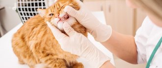
Complete ophthalmological examination.
Typically, this includes: examining the inside of the eye using appropriate magnifying glasses, fluorescein staining of the cornea, and tonometry to determine the presence of glaucoma. It is important to determine the extent of the hyphema, check whether it affects only the anterior chamber or even the back of the eye, and whether it affects one or both eyes. The veterinarian, if deemed necessary, may refer the owner to a veterinary ophthalmologist for further evaluation of the hyphema using appropriate instruments.
Laboratory research
A complete blood count, which includes a platelet count (to look for any infection or inflammation and to ensure there are enough platelets).
Serum biochemical panel to evaluate organ function and to measure the level of proteins present in the serum.
Thyroid function test. May be prescribed for older cats with hyperthyroidism.
Specialized blood tests. They are necessary to assess blood clotting.
Taking blood pressure to determine if your cat has high blood pressure.
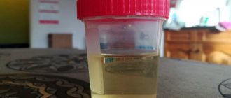
General urine test for suspected renal pathology.
X-rays of the chest and abdominal cavity. They may be recommended if blood test results show other organic abnormalities.
Your veterinarian or veterinary ophthalmologist may recommend additional diagnostic tests to look for other body conditions or to assess the extent of eye damage, including:
Ocular ultrasound
This is an imaging technique that shows the structures present at the back of the eye and behind the eye. This test is especially useful when the hyphema is so severe that it prevents examination of the back of the eye with conventional instruments. Ultrasonography can help detect the presence of abnormal masses in the eye, lens luxation, retinal detachment, or vitreous hemorrhages.
Radiographs of the skull and orbit.
They may be necessary to identify the presence of fractures in cats that have suffered head trauma. Plain radiographs (white) are also useful in identifying any metallic foreign bodies.
Ultrasound of the abdominal cavity.
This may be recommended if laboratory tests reveal abnormalities in certain abdominal organs or the potential presence of a tumor in the abdominal cavity.
Bone marrow aspirate (collection of cells from the bone marrow). This may be performed to evaluate the bone marrow's ability to produce platelets or to detect bone marrow cancer.
Treatment
The goals of therapy are to treat eye inflammation due to bleeding and any underlying causes of bleeding. Treatment of hyphema itself provides the following:
Topical corticosteroids, in the form of eye drops or ointment. They are used to reduce inflammation present in the anterior chamber of the eye.
Topical atropine, in the form of eye drops or ointment. It is used to dilate the pupil. The expansion of the latter helps relieve pain and minimize adhesion between the iris and the crystal.
Treatment of glaucoma, if the latter has caused hyphema or developed as a result of edema, is prescribed if intraocular pressure is high.
What to do at home
Keep your cat at home as quiet as possible to stop bleeding, allow the hyphema to settle in the eye, and reduce the risk of further bleeding. It may be necessary to limit your cat's physical activity for 7-10 days.
Since in some cases vision deteriorates, it is recommended not to allow the cat to go outside without supervision until complete recovery.
Do not give your cat over-the-counter human medications such as Visine or other ophthalmic medications designed to reduce eye redness or irritation, as these products are not effective for hyphema.
Contact your veterinarian as soon as possible, because some causes of hyphema are dangerous not only for the eyesight, but also for the life of the animal.
Disease prevention
Basic preventive measures:
- disinfect the room in which the child is located;
- observe the rules of personal hygiene;
- strengthen immunity;
- Avoid contact with anyone who has conjunctivitis.
Do not delay going to the doctor if you notice signs of this disease in your child. The earlier treatment is started, the lower the risk of complications. Make an appointment with a SM-Clinic specialist.
Sources:
- N.N. Arestova, L.A. Katargina, E.V. Yani. Conjunctivitis and dacryocystitis in children: clinical characteristics, modern treatment options // Russian Pediatric Ophthalmology, 2016, No. 11(4), pp. 200-206.
- L.D. Ksenzova. Treatment of allergic conjunctivitis in children // Issues of modern pediatrics, 2008, vol. 7, no. 5, pp. 135-140.
Ageev Vladimir Sergeevich Clinic
Author of the article
Ageev Vladimir Sergeevich
Candidate of Medical Sciences
Specialty: ophthalmologist
Experience: 16 years
The information in this article is provided for reference purposes and does not replace advice from a qualified professional. Don't self-medicate! At the first signs of illness, you should consult a doctor.