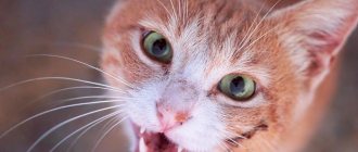
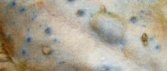
Inflammatory diseases
Lymphoplasmacytic rhinitis (LP rhinitis) is the most common inflammatory disease of the nasal cavity in cats. This type most often occurs in middle-aged and older cats, but it can also occur in young animals. It is not known why some cats suffer from this type of inflammatory disease and others do not. When examined with modern imaging techniques (such as CT or MRI), diseases of the nasal cavity of this type usually do not show destruction of the turbinates (tiny bony curls inside the nose). Sometimes CT, MRI and rhinoscopy show thickening (hyperplasia) of tissue. Rhinoscopy may show other changes, such as redness (flushing). To make a definitive diagnosis of this disease, tissue from the nasal cavity must be sent for histological examination (examination under a microscope by a histologist).
Steroid medications, such as prednisolone or prednisone, can be used to reduce inflammation in the nasal cavity. First, we prescribe these drugs in high doses for a short time to reduce inflammation. If the cat shows signs of improvement, we slowly reduce the dose as much as possible to control the symptoms. The exact dose reduction schedule depends on your cat's condition, but in general, if the condition improves, we usually reduce the dose by 25% every 2-3 weeks. In some cases, it is possible to stop taking steroids completely. Some cats require a long course of low dose steroids. If this is your case, we will try to keep your dose to the minimum required to control your symptoms.
Steroids have several side effects. At high doses, thirst and urination increase. It is very important that the animal always has enough water. A high dose will likely increase your cat's appetite, but you should not feed her more than usual. In rare cases, steroids cause gastrointestinal ulcers, which may be accompanied by vomiting, diarrhea, decreased appetite, blood in the stool, or black, tarry stool. If you notice any of these symptoms, be sure to tell your veterinarian. Steroid medications should not be stopped abruptly as this can lead to a life-threatening reaction (Addisonian crisis), and therefore changes in the dosage of these medications should only be done under the supervision of a veterinarian.
Allergic rhinitis . Cats can suffer from allergic rhinitis, but it is less common in them than in humans. With CT/MRI and rhinoscopy, the picture may be similar to lymphocytic-plasmacytic rhinitis, and a final diagnosis requires a biopsy with histological examination (examination of a tissue fragment under a microscope, carried out by a histologist). In allergic rhinitis, different types of inflammatory cells are visible under the microscope than in lymphocytic-plasmacytic rhinitis. If your pet has allergic rhinitis, we recommend trying environmental control measures similar to those for people with allergies (using air purifiers, avoiding allergy triggers). It is possible to prescribe specific medications depending on the patient.
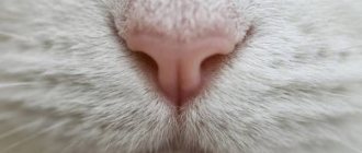
Rhinitis is inflammation of the nasal mucosa.
The main risk factors for the development of AR are: 1. Family history - hereditary factors, allergic diseases in the family. 2. Allergic sensitization. Year-round AR is characterized by exposure to allergens characteristic of enclosed spaces (household allergens, house dust mite allergens, fungal allergens, pet allergens) 3. Contributing factors (smoking, quality of inhaled air in the home, air pollution, climatic factors). 4. “Lifestyle” factors. 5. Relatively high socio-economic status.
Distinguishes the following main stages of AR: 1. Vasotonic stage
.
It is characterized by a constant change in vascular tone and is clinically manifested by variable nasal congestion. 2. Stage of vasodilation.
Nasal congestion is constant due to dilation of the mucous vessels.
3. Stage of chronic edema.
The nasal mucosa changes from pale marble to bluish, and nasal congestion is almost constant.
4. Stage of hyperplasia.
The nasal mucosa grows, polyps form, the paranasal sinuses are often involved in the process, secondary otitis develops, and a secondary infection is almost always associated.
These stages of development of a single pathological process are realized due to markers of inflammation in the territory of the nasal mucosa.
Biological markers of inflammation in allergic rhinitis
1. Cytokines: - Monokines (GM-CSF, TNF, IL1, IL5, IL6, IL8, IL12, IL15) - Lymphokines (IL2, IL4, IL13, gamma-IFN) 2. Leukotrienes: - Cysteine (LTC4, LTD4, LTE4 ) - Non-cysteine (LTB4) 3. Granular proteins: - Eosinophil (cationic protein, peroxidase, protein X, neurotoxin) - Mast cell tryptase 4. Adhesion molecules: - Endothelial adhesion molecule - ELAM-1 - Vascular adhesion molecule - VCAM-1 - Intercellular adhesion molecule - ICAM-1
Clinical forms of allergic rhinitis:
The definition of
“mild”
means that the patient has only minor clinical signs of the disease that do not interfere with daytime activities and/or sleep.
The patient is aware of the presence of manifestations of the disease and may be ready to be treated, but here everything depends on his personal desire. The definition of “moderate”
means that the symptoms disturb the patient’s sleep, interfere with work, study, and sports.
The quality of life deteriorates significantly. The term "severe"
means that the symptoms are so severe that the patient is unable to work, study, play sports or leisure activities during the day, or sleep at night unless treated.
The term “episodic (or intermittent)”
means that the manifestations of AR bother the patient less than 4 days a week or less than 4 weeks a year.
The term “frequent (persistent) presence of symptoms”
means that the patient reports symptoms of the disease more than 4 days a week or more than 4 weeks a year.
Perennial allergic rhinitis (PAR) (persistent)
caused by the following allergens:
- House Dust Mites
House dust mites are microscopic arachnids (Arachnids) found in indoor dust, especially in bedrooms, upholstered furniture, carpets, curtains, etc. The sizes of the most common species, Dermatophagoides pteronyssinus and Dermatophagoides farinae, do not exceed 0.4 mm. At all stages of their development, these mites are keratinophages, i.e. they feed on desquamated epidermis (dead flakes of the upper layers of human or animal skin, which sometimes make up up to half the volume of house dust). Under conditions of optimal humidity (relative humidity 70-80%) and temperature (20-300C), the life cycle of ticks is approximately one month and includes the following stages: egg, larva, protonymph, tritonymph and adult. During her life, a female tick lays 70-100 eggs. o Under favorable living conditions, the mite population can grow very quickly, and tens of thousands of individuals can be found in 1 g of dust.
- Microscopic fungi are distributed almost everywhere. Fungal spores were found even at an altitude of more than 2000 m. The concentration of fungal spores in the air, even during the period of intense flowering of plants, is a thousand times (1000!) higher than the concentration of pollen grains. Molds consist of a system of small threads - mycelium - and reproduce by spores. The spores are small enough to enter the lower respiratory tract and even reach the alveoli. Yeasts are single-celled fungi that do not form filaments. In reality, a person comes into contact with 100 species of mushrooms. The species composition of mushrooms differs indoors and outdoors. In the air of cities in warm weather there is an average of 3100 spores/m3 of air. In the open air, Cladosporium and Alternaria predominate, in smaller quantities - Penicillium and others. These fungi are characterized by great seasonal variability with a maximum in summer and a minimum in winter. But, since the concentration of their spores is increased throughout the warm period, the flowering and spore formation calendar does not help diagnose allergies. In house dust and indoor air, the fungi Aspergillus and Penicillium are more often released, in smaller quantities - Cladosporium, Alternaria, Mucor, Candida, etc. Aspergillus and Penicillium reproduce all year round. Indoors, higher levels are observed in autumn and winter. They are called storage fungi and cause rotting of stored grains, fruits and vegetables. Penicillium can often be seen on items stored in basements as patches of green mold. There are no clear standards for the content of fungi in the air, only a conditional standard - no more than 500 spores/m3, but during research it turned out that in more than 80% of rooms it was exceeded, and in some the concentration of spores in the air reached tens and hundreds of thousands per m3. For Cladosporium and Alternaria, the content norm is 102 and 3 x 10³/m3. The highest concentrations of fungi were found in old houses, on the first floors of buildings and in rooms with various leaks. Mold loves damp and warm places, bathroom walls, shower stalls, trash cans, refrigerators. The source of mold can be moldy products, old paper wallpaper, linoleum. Fungi can colonize household appliances such as humidifiers or air conditioners. Flower pots are often the source of Cladosporium and Alternaria, which live on rotting parts of plants, but the connection between indoor flowers and the number of fungi is not as great as thought. Yeasts can also live in the soil.
- In the dwellings of central Russia, there are mainly three species of cockroaches, the allergens of which are the cause of the development of allergic diseases in humans: Blatta orientalis, Blattella germanica and Periplaneta Americana. The high antigenic activity of insect body particles is mainly associated with anthropodin (a water-soluble protein that makes up 15% to 50% of the cuticle). Sensitization to cockroaches can develop through bites, direct contact with insect bodies and cockroach metabolites. House dust from infected apartments contains cockroach allergens: saliva, feces, insect body tissue. The most pronounced allergenic properties are found in fragments of the head, cuticles and excrement.
- The main allergen in cats is Fel d1 (1-10 µm, average 3.5 µm). The main source of Fel d1 is animal saliva; it is also found in the sebaceous glands of the skin. In cats, but not in cats, Fel d1 is also excreted in urine. In general, cats produce significantly more allergens than cats. After removing the cat from the premises and/or carrying out elimination measures against the cat allergen, a decrease in the level of allergens and the symptoms they cause is usually observed no earlier than 6 months. Because Fel d1 is very resistant to the environment. Do you like to visit prostitutes? And even at an age? There are mature prostitutes from Tyumen here: https://sex-tumen.prostitutki72.com/zrelye Intimacy for the evening is guaranteed. Dog allergens are found in animal dander, saliva, urine and serum. The main dog allergen is Can f1. Different breeds of dogs emit allergens that vary in spectrum and quantity.
| Profession | Allergen |
| Baker | Flour |
| Laboratory worker | Laboratory animals |
| Painter painting with spray gun | Isocyanates |
| Workers exposed to plastics and resins | Anhydrides |
| Carpenter | Western Red Cedar Wood |
| Medical workers | Latex |
Food products as etiological factors of food allergies, according to the degree of allergenic activity (without taking into account individual characteristics)
| Activity level | Products |
| High | Cow's milk, fish, crustaceans, eggs, chicken meat, strawberries, raspberries, wild strawberries, blackcurrants, blackberries, grapes, pineapples, melons, persimmons, pomegranates, citrus fruits, chocolate, coffee, cocoa, nuts, honey, mushrooms, mustard, tomatoes , carrots, beets, celery, wheat, rye |
| Average | Pork, turkey, rabbit, potatoes, peas, peaches, apricots, red currants, bananas, green peppers, corn, buckwheat, cranberries, rice |
| Weak | Horse meat, lamb (low-fat varieties), zucchini, squash, turnips, pumpkin (light colors), green and yellow apples, white currants, gooseberries, plums, watermelon, almonds, green cucumbers |
In addition to antigens, clinical manifestations of AR
can be provoked by such trigger factors as infection, nonspecific irritants, tobacco smoke, pollutants, cold air, drafts, which indicates the formation of nonspecific hyperreactivity of the nasal mucosa.
Diagnostic measures for CAP:
1. Taking an anamnesis. 2. General blood test. 3. Examination by an allergist-immunologist, ENT doctor. 4. Skin testing with a set of mixed allergens followed by additional examination with the corresponding group of allergens. Determination of total serum IgE and specific IgE to specific allergens. 5. Conducting provocative nasal tests 6. Cytological examination of a smear-imprint from the nasal mucosa, washings for bacteriological examination. 7. Determination of the function of the ciliated epithelium of the nasal mucosa. 8. X-ray examination of the nose and/or paranasal sinuses.
Preventive and therapeutic measures for CAP:
1. Elimination of causally significant allergens.
(detailed information is presented 2. SIT 3. Antihistamine therapy, taking into account the need for long-term use of antihistamines (it is advisable to prescribe 3rd generation drugs, since they have minimal side effects and can be used for a long time) 4. Prescription of cromones (for example, cromohexal in the form of a nasal spray, practically has no side effects) . Immunology and allergology. Algorithms for diagnosis and treatment. - M.: Geotar-Med, 2003. 2. Pukhlik B. M. Elementary allergology. - Vinnitsa, 2002. 3. Gushchin I.S., Ilyina N.I., Polner S.A. Allergic rhinitis. - M., 2002. 4. Stanley M. Nagua, M. Eric Gershwin. Secrets of allergology and immunology. M.: Binom, 2004. 5. Khaitov R.M. Clinical allergology. - M.: "MEDpress-inform", 2002. 6. Roy Patterson, Leslie K. Gremmer. Allergic diseases. - M., Geotar Medicine., 2000.
Infectious diseases
Fungal infections. Cats sometimes suffer from cryptococcal nasal infections. They are caused by fungi present in the environment and occur in both indoor and outdoor cats. Fungal infections can be accompanied by severe inflammation and destruction of nasal structures.
The diagnostic method is to determine the titer of antibodies in the blood, which increases during infection; cytological examination of the nasal mucosa; PCR diagnostic method. The preferred treatment is the use of internal antifungal drugs (their type depends on whether the brain is affected).
Side effects of internal antifungals include vomiting, diarrhea, decreased appetite, and liver problems. Often a course of treatment lasting 3–12 months or even longer is required.
Bacterial infections – Primary bacterial infections, which are the main cause of nasal problems, are very rare in cats. However, often bacteria, taking advantage of the fact that the tissues of the nasal cavity are damaged by the disease, cause secondary infections that accompany damage to the nasal cavity for any other reason. Because of these secondary bacterial infections, cats often improve when antibiotics are given, but when the antibiotics are stopped or simply over time, the symptoms worsen again because the underlying cause is not addressed. Because cats normally harbor bacteria in their nasal cavity, culture of deep tissue samples is necessary to diagnose a bacterial infection.
Mycoplasma infections (mycoplasmas) are a special type of bacteria. These microorganisms can infect any part of the respiratory system, including the nasal cavity. Diagnosis of mycoplasma infection requires cultivation under special conditions. Unfortunately, mycoplasmas grow very poorly in culture, so a negative microbiological culture result does not exclude mycoplasma infection. Only certain types of antibiotics can kill mycoplasmas. These include: azithromycin (Zithromax), doxycycline and enrofloxacin (Baytril).
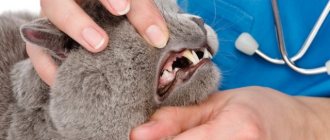
Tooth root abscess . Infection of the tooth root can lead to the formation of an abscess. Sometimes in such cases, the abscess ruptures into the nasal cavity, and not into the oral cavity or onto the surface of the skin. In some cases, we detect tooth root abscesses during clinical examination. It can be diagnosed by dental x-ray, CT scan or MRI. These abscesses often require dental surgery and antibiotics to treat.
Viral infections. These infections, including calcivirus and herpesvirus infections, may be accompanied by discharge from the eyes and nose, as well as ulceration of the oral mucosa. Cats often become infected with these viruses when they are still kittens, and symptoms return when stressed. Diagnosis can be made based on viral isolation. An L-lysine supplement can be used for treatment.
Cancer (neoplastic)
Various types of cancerous tumors occur in the nasal cavity. The most common type in cats is adenocarcinoma, but other types, including soft tissue sarcoma and lymphoma, also occur. Cancerous tumors can cause problems in one or both nostrils. Possible deformation of the muzzle. Usually, with neoplasms in an animal, changes can be seen on MRI or CT. Ultimately, to determine the type of tumor, it is necessary to do a biopsy, taking a piece of tissue for histological examination (evaluating a section of tissue under a microscope), or a puncture for cytological examination (evaluating cells in a smear). Treatment for cancer depends on its type. Endoscopic removal of the tumor, rhinotomy, radiation and chemotherapy are likely.
Foreign bodies
When foreign materials enter the nose, inflammation and irritation may develop around them. Sometimes foreign material can be seen during rhinoscopy. In some cases, foreign objects can be removed from the nose using grasping instruments or rinsing. To rinse the nose of an animal under anesthesia, a liquid (saline solution) is injected. Sometimes visual examination methods: CT or MRI are required to detect foreign material.
Diagnostic methods
As discussed above, various methods are used to diagnose the cause of nasal abnormalities. A complete examination for nasal disorders includes:
- MRI or CT - these highly effective visual diagnostic methods can better distinguish the details of the nasal cavity than x-rays.
- Rhinoscopy is an examination of the nasal mucosa (the outermost layer) using a rhinoscope.
- A biopsy of tissue from the nasal cavity for histological examination (where a histologist examines the tissue under a microscope) and microbiological culture.
These procedures are performed under anesthesia, so additional baseline blood tests are often required to ensure the absence of systemic disease. If systemic diseases are present, it may be necessary to change the drugs used for anesthesia.
- Complete clinical analysis to evaluate red blood cells, white blood cells and platelets.
- Biochemical analysis - assessment of liver and kidney function indicators, protein concentrations, electrolytes.
- Urinalysis - assessment of liver and kidney function.
- Blood pressure measurement. We try not to biopsy tissue from the nasal cavity if the patient has high blood pressure, since this makes it more difficult to stop the bleeding.
- Coagulogram is a blood test for clotting. It is important to make sure that the blood is capable of normal clotting before performing a biopsy.
- Chest X-ray. We recommend a chest x-ray to identify possible spread to the lungs and to ensure that there are no other lung problems that could increase anesthetic risk.
- Echocardiography is mandatory for all cats to exclude structural and hemodynamic pathologies.
3. Symptoms and diagnosis
As shown above, typical manifestations of erysipelas of the nose include erythema and inflammatory swelling of the soft tissues, clearly demarcated by a roller from healthy skin. The swelling can be so severe that the patient’s face becomes deformed and changes beyond recognition. Blisters filled with mucopurulent exudate (bullous form of erysipelas) are often observed.
As a rule, the general symptoms of infectious intoxication are pronounced: malaise, fever, hyperthermia (up to 40° and above), headache, weakness, soreness and swelling of the nearest lymph nodes. After a few days, body temperature may drop just as sharply.
It should be noted that in the absence of timely adequate therapy, erysipelas tends to expand to neighboring areas (skin of the face, neck, chest, ear, etc.); with the involvement of the mucous membranes and submucosal layers of the pharyngeal structures, the development of such serious complications as abscess formation, phlegmon, sepsis, and intracranial inflammation is possible.
The clinical picture of erysipelas is quite specific and usually does not create any difficulties in diagnosis. Examination of the ENT organs reveals hyperemia of the mucous membranes of the nasal cavity. Laboratory diagnostic methods are used to identify the pathogen(s) and assess drug sensitivity. In more complex cases, they resort to instrumental imaging diagnostics (endoscopy, ultrasound, etc.).
About our clinic Chistye Prudy metro station Medintercom page!