Tumor-like growths in the oral cavity are the scourge of veterinary dentistry. It would seem that a harmless “pea” can turn into an incurable disease for a pet and shorten its life. Unfortunately, as practice shows, owners suspect something is wrong when the process affects not only the oral cavity but also other internal organs. The specificity of oral cancer is its dependence on the anatomical structure of this part of the animal’s body. Brachycephalic dogs (dogs with short muzzles) oral tumors than dogs with medium or long muzzles. Factors that provoke the growth of tumors include feeding canned food, constantly wearing an anti-flea collar (due to contact of the mucous membrane with pesticides included in the collar), as well as smoking by the animal owner!
Tumor lesions include odontogenic and non-odontogenic tumors. The most common odontogenic tumors are peripheral odontogenic fibroma
and
acanthomatous adamantinoma
.
The most common nonodontogenic neoplasms are malignant melanoma
and
squamous cell carcinoma
.
Unfortunately, malignant tumors in the oral cavity are recorded much more often than benign ones.
What signs should make an owner think about a possible tumor-like process in his pet’s body?
At the beginning of the development of the tumor process, noticeable symptoms are usually not observed. Sometimes general symptoms of the development of a tumor process may attract attention: weakness, loss of appetite, exhaustion, depression.
As a dog develops cancer of the jaw , gums, tongue or other oral organs, symptoms that are noticeable to the owner appear:
- Excessive salivation
- Unpleasant odor from the mouth
- Loose or tooth loss
- Complicated eating
- Bleeding in the mouth
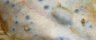
- Growths in the mouth
- Exfoliation of tooth enamel
- If the upper jaw is affected - nasal discharge
- Sometimes there is swelling of the mandibular lymph nodes
- A sharp change in body weight downward
Sometimes the main reason for contacting a veterinarian is a pronounced deformation of the animal’s muzzle. It is very important not to miss them, because already at this stage the effectiveness of further treatment noticeably decreases over time.
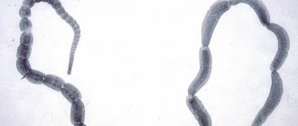
In inductive odontogenic tumors, the cells form hard dental tissues that can be easily identified on x-rays.
Adamantinoma
It is a neoplasm of epithelial tissue, such as enamel, that has not differentiated to the extent that enamel can form. It develops either at the gingival margin (peripheral ameloblastoma, manifested by epulis) or from within the bone (central ameloblastoma). Some of the central ameloblastomas appear as cystic lesions within the bone. Although biologically this tumor is benign and does not metastasize, locally it is extremely infiltrative and aggressive, causing extensive bone resorption, tooth displacement and even resorption of tooth roots.
Treatment
The specificity of the disease is expressed in the fact that chemotherapy is not used for treatment - in this case it does not produce results. Gamma therapy is prescribed to stop the spread of the tumor and reduce its size. 3 weeks after irradiation, surgical removal of the jaw (resection or disarticulation) is performed. At a later stage, more extensive surgical intervention may be necessary: exenteration of the orbit, sanitation of the paranasal sinuses, lymphadenectomy.
Rehabilitation, restoration of speech, chewing and swallowing functions takes a long time. 2 years after complete recovery, correction is made using bone grafting methods, as well as using special splints.
Odontoma
It is a benign neoplasm of mixed origin, in which both epithelial and mesenchymal cells are fully differentiated. Typically, enamel and dentin in this case are distributed in a pathological manner. Odontoma is usually detected in young animals, and it can develop in any part of the dental arch. Complex odontoma is a disorganized amorphous volumetric formation of hard dental tissues that has no resemblance to normal dental tissue. Mixed complex odontoma consists of several small tooth-like structures. Both types of tumor are encapsulated and often associated with an unerupted tooth. They are benign in nature, but can cause tooth decay and sometimes spread very actively.
Lacerations
Lacerations of the mouth are most common in pets that often walk outside, in animals that like to sort things out with other cats. Owners of such pets often turn to the veterinarian at the clinic with a request to heal wounds after a fight. Treatment of wounds consists primarily of stopping the bleeding, for which the cat is immobilized and, if necessary, sedatives are used. Small cuts are not sutured, but lacerations and deep wounds are stitched, especially if the bleeding does not stop. In cases where the wound is caused by an incorrect position of the tooth, it is removed. During treatment, the cat’s mouth must be washed twice a day with a weak antiseptic solution and given specially prepared food, mostly soft.
Malignant melanoma
It is a rapidly growing tumor, usually accompanied by ulceration and/or necrosis. Malignant melanoma can be pigmented or non-pigmented (amelanotic melanoma). Amelanotic melanoma is often difficult to diagnose and has a very aggressive course.
It is considered the most common oral malignancy in dogs and accounts for 30–40% of all oral malignancies in this species. In most cases, it occurs in older dogs due to some degree of oral pigmentation. Malignant melanoma is rare in cats, but its biological behavior in this species is the same as in dogs.
The most common localizations are the gums and mucous membranes of the lips, cheeks, but other localizations are also possible (on the palate, dorsum of the tongue). When gums are affected, teeth are often damaged and bone invasion is common.
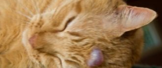
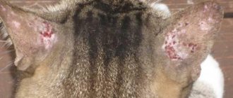
Squamous cell carcinoma in a cat
When surgically treating carcinoma in situ, it is usually enough to make a small indentation from the borders of the tumor to completely remove it. If the lesions are multiple in nature or located in places difficult to access for surgical treatment, then imiquimod (Vartotsid, Keravot) can be used - this is an immunomodulator with antiviral activity. Imiquimod is used as a 5% cream to treat actinic keratoses, basal cell carcinoma and genital warts in humans.
In one study, imiquimod was used to treat Bowen's disease or carcinoma in situ in 12 cats. Treatment was carried out every other day or three times a week (the same effect was observed). All animals showed a significant response to treatment, but 9 animals developed new lesions over time and also responded well to treatment with imiquimod. Of the side effects, three cats had peeling and erythema of the skin, and one had neutropenia and increased alkaline phosphatase activity, which disappeared after discontinuation of the drug (Gill VL. et. al., 2008).
Imiquimod was used to treat sun-induced squamous cell carcinoma of the auricle in one cat, similar to the treatment of solar keratoses in humans (Peters-Kennedy K. et. al., 2008; Samaro A. et. al., 2013). However, there are many treatment options for this form of SCC in cats that have some effectiveness: surgical resection, photodynamic therapy, cryodestruction, diathermy, plesiotherapy and radiation. The choice of treatment depends on the location of the tumor, the stage of the disease and the capabilities of the doctor, clinic and owners.
If the tumor is located in the area of the auricle, then surgery is usually the method of choice. To achieve adequate resection margins, it is recommended to make an indent of 1 cm from the tumor borders. For better wound healing, it is necessary to stitch the skin edges over the ear cartilage.
When the tumor is localized in the eyelid area, it is necessary to make an indentation of 4-5 mm from the visible edges of the tumor (Ayres SA. et. al., 2012). Removing a tumor in this location often requires plastic reconstruction of the eyelid defect, which is sometimes not the easiest task. In one study, when following the recommended resection margins in cats with SCC in the eyelid area, the median relapse-free period was 319 days (range 95 to 1510 days) (Schmidt K. et. al., 2005). In another study, 5 out of 6 animals had a disease-free period of more than 12 months (Hunt Gd. 2006).
The choice of treatment for tumors in the area of the nasal planum depends on the stage of disease of the primary tumor. Invasive tumors at stage T3 or T4 may respond well to treatment with radiation therapy. The most effective way to completely cure cancer in this area is to remove the tumor tissue with a minimum distance of 5 mm from the boundaries of the tumor, which means complete removal of the nasal planum. This operation leads to a cosmetic defect, but does not significantly impair the animal’s respiratory function or quality of life. More superficial tumors in stage Tis or T1-3 can be successfully treated with photodynamic therapy, cryosurgery, strontium-90 plesiotherapy and radiation (Bexfield NH., et. al. 2008; Buchholz J. et al., 2007; Lana SE. et al., 1997, Clarke RE. 1991; Jarrett RH. et. al., 2013; Goodfellow M. et al., 2006; Hammond GM. et al., 2007; Theon AP. et al., 1995; Melzer K et al., 2006).
Photodynamic therapy (PDT) uses a photosensitizer drug that is absorbed by tumor cells. The active substance is activated by a wave of light of a certain length, which leads to cell death with minimal impact on surrounding tissue. In one study, topical application of photosensitizing cream or 5-aminolevulinic acid resulted in 85% remission in 55 cats, relapse was observed in 51% of cases, and the disease-free period was 157 days (Bexfield NH., et. al. 2008). In another study of 61 cats with cutaneous SCC, PDT with an intravenous photosensitizer resulted in remission in 49% of animals (61% had 1-year disease control), and in another study of 18 cats there was 100% remission and 1-year disease control in 75% of animals. (Buchholz J. et al., 2007). The problem with photodynamic therapy is the limited penetration of the rays and this treatment is more suitable for superficial tumors (in all studies, the best result was obtained for superficial tumors).
Various results have been obtained with the use of cryosurgery, which is due to the difficulty of assessing the exact limits of the effect. The best results were obtained for superficial tumors (stages T1-T2). The disease-free period was 254 days in a study of 11 cats with SCC in the area of the nasal planum and auricles (Lana SE. et al., 1997). In another study of 102 cats, the median remission was 26.7 months, and tumors located in the area of the nasal planum and eyelids responded best to treatment (Clarke RE. 1991).
In one study from New Zealand, 34 cats with pathologies affecting less than 50% of the nasal planum were treated with curettage and diathermy for three cycles. 16 animals had actinic changes, 9 had carcinoma in situ and the rest had full-blown squamous cell carcinoma. In 94% of animals, the relapse-free period was more than a year (Jarrett RH. et. al., 2013).
Strontium-90 plesitherapy is a successful and cosmetic therapy for stage Tis, T1 and T2 squamous cell carcinoma, as beta rays penetrate only a few millimeters into the tissue. In one study, 13 of 15 cats had a median lifespan of 692 days, and two animals that did not respond to treatment responded after the second course (Goodfellow M. et al., 2006). In another study, 88% of animals responded to treatment with a median survival time of 1071 days (Hammond GM. et al., 2007).
Radiation therapy (orthovoltage, megavoltage, electron, and proton therapy) has also been studied in cats with nasal planum SCC with similar success. Cats with stage T1 RCC had an 85% chance of surviving more than 1 year, compared with animals with stage T3 disease, of which 45% of animals survived more than 1 year (Theon AP. et al., 1995). In another study, 16 of 17 cats with stage T1-2 disease had a median disease-free period of 414 days. In this study, electrons were used (10 fractions of 4.8 Gy over 5 days, total dose 48 Gy) (Melzer K. et al., 2006).
There are publications on the use of chemotherapy in cats with RCC, either alone or in combination with radiation therapy. In one study, animals with stage T2-T4 disease were treated with intratumoral carboplatin and orthovoltage therapy. All animals responded to treatment, and the relapse-free period was 52-562 days (4 of 6 animals were in remission at the time of writing (de Vos JP. et al., 2004). In another study, 7 out of 9 animals treated with electrochemotherapy and intratumoral bleomycin had remission for up to 3 years (Spugnini EP. et al., 2009).
Immunohistochemical studies have revealed the expression of receptors for COX-2 in cutaneous squamous cell carcinoma cells, but its role is not fully understood (Bardagi M. et al., 2012).
Sarcoma
It is a malignant tumor and, depending on the effect on the bone, is divided into osteoclastic and osteoblastic. In osteoclastic sarcoma, predominantly destruction and destruction of bone are observed, and in osteoblastic sarcoma, in addition to these phenomena, there are signs of chaotic bone growth and multiple needle-like shadows located perpendicular to the length of the bone (periosteal “visors”). In some cases, mixed forms of osteoclastic and osteoblastic sarcoma are possible.
Oral osteosarcoma develops primarily in dogs of medium and large breeds and, as a rule, in middle or older age. Osteosarcoma is more common in the lower jaw and less common in the upper jaw. Sarcoma should be differentiated from osteomyelitis, tuberculosis and cystic degeneration of the jaw bone.
When visiting a doctor is unavoidable
What to do if it is not possible to cure a cat at home, if the pet’s condition worsens day by day (appetite disappears completely, in addition to ulcers in the mouth, pus forms)? Home treatment should be stopped and contact a specialist immediately. There is a possibility that the doctor will recommend the use of antibiotics (Amoxicillin, Oxytetracycline and other drugs that should only be prescribed by a specialist), immunomodulators (Gamavit, Hemobalance), and vitamins.
It is also important to identify the cause of the disease. So, for example, treatment of ulcerative stomatitis caused by, say, rhinotracheitis, will include taking antihistamines
And if inflammation of the oral mucosa is a consequence of liver problems, the animal will probably need to take hepatoprotectors and painkillers. In other words, it is better to entrust the selection of medications for the treatment of stomatitis to a veterinarian.
Therapeutic techniques
So, how is epulis treated in cats? As a rule, there is only one way out - surgery . The affected areas of tissue are excised, and “faulty” teeth are removed. Naturally, everything is done exclusively under general anesthesia. If there is a possibility of developing a secondary infection (and in an exhausted and weakened cat it is very high), additional antibiotic therapy is resorted to. In addition, anti-inflammatory corticosteroids are sometimes prescribed, but this should only be done after weighing the pros and cons.
What it is?
This unusual term refers to a tumor of fibrous tissue of a benign nature . Despite its benign origin, this type of neoplasm often behaves quite aggressively. In particular, one type of epulis causes complete or partial destruction of not only the fibrous tissue by which the teeth are attached to their alveoli, but even the bone. Moreover, the degree of destruction is such that in severe cases it is necessary to completely (!) remove the upper or lower jaw. You understand that an animal with such an injury definitely becomes disabled, since it no longer has a chance for a normal life.
Important! There is still no reliable information about the exact causes of the disease, and therefore even prevention of this disease is impossible. The genetic and viral theories have the most supporters.
Interestingly, it is not uncommon for practicing veterinarians to encounter gum pathologies during medical examinations of animals. But there are quite a lot of aspects to their treatment, it all depends on the specific case and the condition of the sick pet. So, in some cases it is necessary to trim the gums, in others it becomes necessary to “extract” a tooth, sometimes all teeth have to be removed ... Moreover, you need to decide how much theoretically healthy tissue needs to be left along the edge of the gums. Finally, after all these issues have been resolved, the question of the degree of operability of the tumor and the need to use radiation/chemical therapy arises. Thus, epulis is a rather “complex” disease that requires a balanced and differentiated approach to treatment.