Veronica Igorevna Sharipova
veterinarian Petstory
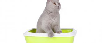
Feline pancreatitis as an independent disease is a fairly rare pathology. Oddly enough, there are many myths and prejudices around this disease. For example, it is believed that if a cat has acute or chronic vomiting, then he has pancreatitis and needs a low-fat diet. But this is not at all true. We will look at the features of pancreatic inflammation in cats in this article.
The pancreas is a unique organ in the body, which is a gland of both exogenous and endogenous secretion. The exocrine function ensures optimal digestion of food and absorption of nutrients in the intestine and consists of the production of special digestive enzymes (trypsin, lipase, amylase). The endocrine function of the pancreas is to maintain glucose levels in the body and is ensured by the production of the hormone insulin. Thus, inflammation of the pancreas - pancreatitis - is a dangerous disease that affects the functioning of the entire body.
According to a new large study conducted on 6504 cats, it was found that only in 0.6% of cases the death of cats was caused by pancreatitis, and in 0.4% of cases the disorders were characteristic of chronic pancreatitis, and in 0.2% of cases - for acute. Thus, the rare intravital diagnosis of this disease is associated with difficulties in making a diagnosis and a fairly rapid fatal outcome for the pet.
Anatoly Chernikov
Chief veterinarian of the veterinary clinic 4 Lapas (Khimki), graduate of the Moscow State Academy of Veterinary Medicine named after.
K. I. Scriabin (2009). Specialist in the field of dentistry, gastroenterology, endoscopy. Regularly takes part in domestic and international Veterinary Conferences: Moscow International Veterinary Congress, National Veterinary Conference (NVC), Congress of the European Association of Veterinary and Comparative Dietetics (ESVCN), Southern European Veterinary Conference (SEVC), Conference of the International Community Felinological Medicine (ISFM), Conference of the Russian Gastroenterological Academy (RGA), International Veterinary Conference Purina Partners. The term “triaditis” refers to the simultaneous occurrence of three pathological processes in cats: pancreatitis, cholangitis and inflammatory bowel disease (IBD). Despite its wide popularity and frequent occurrence in various scientific literature sources, data on the true prevalence of triaditis and its etiopathogenesis are still lacking. To date, the prevalence of this problem according to various sources is estimated in the range of 17-39% among cats with inflammatory lesions of the organs of interest (cholangitis, pancreatitis or IBD). (1, 2, 3) Triaditis has also been reported to be found in 50–56% (4, 5) of cats diagnosed with pancreatitis and 32–50% of cats diagnosed with cholangitis/inflammatory liver disease. There is currently no reliable information about the gender, age or breed predisposition of triaditis. (6, 7)
Symptoms of the disease
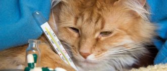
Pancreatitis in the initial stages is practically asymptomatic, so it is difficult to detect it at home.
Among the main symptoms:
- sluggish and apathetic state,
- diarrhea,
- temperature increase,
- sudden weight loss,
- dehydration,
- lack of appetite,
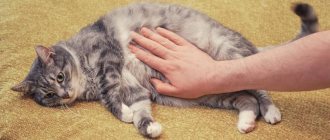
- belly hard to the touch
- icteric mucous membranes,
- general weakness.
Note! Typically, an attack of vomiting or diarrhea is observed 10-15 minutes after the animal eats food.
Are you observing one or more signs in your pet? Do not hesitate, contact a veterinary clinic for professional help. The sooner pancreatitis is diagnosed, the faster and more effective its treatment!
Possible complications and concomitant diseases
In this article, we will review the most current information on the diagnosis and treatment of triaditis, focusing on individual comorbid pathologies: pancreatitis, cholangitis and IBD.
Pancreatitis
Pancreatitis in cats is considered an idiopathic disease, but can be triggered by specific etiological factors such as viral infection, toxoplasmosis, fluke infection, trauma and organophosphorus poisoning. (7, 
Histologically, pancreatitis can be divided into acute and chronic. Acute pancreatitis (AP) is characterized by neutrophilic inflammation with associated interstitial edema and mesenteric fat necrosis, while chronic pancreatitis (CP) is characterized by lymphocytic inflammation, fibrosis, and acinar atrophy. Because cats with CP are more likely to have comorbidities than cats with AP, CP may be more common in triaditis. Inflammation of the pancreas can extend to the pancreatic duct and even the sphincter of Oddi, causing cholangitis and possibly extrahepatic biliary obstruction. Cholangitis occurring as a primary disease can cause inflammation extending to the sphincter of Oddi and pancreatic duct, and therefore, in turn, is a risk factor for the development of pancreatitis. (9)
Cholangitis
The diagnosis of cholangitis involves inflammation of the bile duct; if the inflammatory process spreads to the liver parenchyma, a diagnosis of cholangiohepatitis is made. Cholangitis is a fairly common disease in cats. One British study examined 1,500 feline liver biopsy samples. (10) The most common histopathological diagnoses were neutrophilic cholangitis (NC) and reactive hepatitis, while lymphocytic cholangitis (HL) was only fourth.
The most common type of cholangitis in cats is NH, which is characterized by infiltration of the bile ducts by large numbers of neutrophils. (11) It is believed to occur as a result of a bacterial infection originating from the intestines. NC can be divided into acute and chronic forms. Acute neutrophilic cholangitis (ANC) is characterized only by neutrophilic inflammation, while chronic neutrophilic cholangitis (CNC) is accompanied by tissue infiltration not only by neutrophils, but also by lymphocytes and plasma cells. In the chronic process, fibrosis and proliferation of the bile ducts are more pronounced, which is the main predisposing factor to the formation of extrahepatic biliary tract obstruction (HEBO). (11) VOT, in turn, is a risk factor for the development of triaditis. (12)
HL is characterized by wall infiltration of lymphocytes and plasma cells, with varying degrees of fibrosis and biliary hyperplasia. The nature of the inflammatory infiltrate suggests an immune-mediated pathogenesis. (13) The disease affects cats of different age groups. HL was most frequently diagnosed in Persian cats, indicating a possible genetic predisposition. (14)
Chronic cholangitis associated with liver fluke infestation (family Opisthorchiidae), the third distinct type of feline cholangitis, is rare. Its role in the development of feline triaditis has been little studied.
Inflammatory bowel disease (IBD)
The exact cause of IBD remains unknown. However, the gut microbiome, food antigens, genetic predisposition, and dysregulation of the intestinal immune system are thought to play important roles in pathogenesis. (15, 16) Feline IBD primarily affects middle-aged animals, although it can also be detected in cats under 2 years of age. It is difficult to identify specific breeds susceptible to the disease, although among Siamese cats, IBD is the most common in the literature. (17)
The severity of inflammation of the gastrointestinal tract is usually assessed on a simple four-point scale: normal (0), mild (1), moderate (2) or severe (3). (18)
Chronic pancreatitis
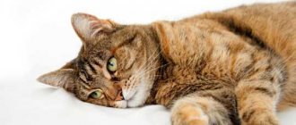
This form of the disease is more dangerous and insidious. Pancreatitis slowly develops and “destroys” the cat’s body. Without any noticeable symptoms, the animal gets worse and worse. A chronic process can develop for years, and the owner of the animal will not know about it - this is its main danger.
The disease is indicated only by changes in the pet's behavior (the animal becomes lethargic and drowsy), incessant rumbling in the stomach, dulling of the fur and yellowish stools.
Prevalence of triaditis
Several studies have examined the prevalence of triaditis. In one of them, 78 cats were studied. It was found that 39% of cats with cholangitis also had IBD and pancreatitis (ie, triaditis), and the prevalence of IBD and pancreatitis was higher in cats with cholangitis (83% and 50%, respectively) compared with cats with non-inflammatory liver disease . (1)
In another study of 44 cats diagnosed at necropsy with moderate to severe cholangitis, the majority had CNC (n = 33) or ONC (n = 7); 60% also had pancreatitis, 50% also had IBD, and 32% had both (ie, triaditis). (2)
In a study of 115 cats, 45% of apparently healthy cats had pancreatic lesions at postmortem examination, (19) highlighting the difficulty of diagnosing the disease.
A more recent prospective study aimed to examine the incidence of IBD, cholangitis, pancreatitis, or a combination of these conditions in symptomatic and asymptomatic cats by comparing clinicopathologic features with histopathologic and laboratory findings. (3) In 57% of cats, inflammation affected more than one organ; 34% had concurrent IBD and cholangitis; 17% were diagnosed with triaditis; and 6% had IBD and pancreatitis. It is important to note that triaditis was only identified in symptomatic cats (20%). A positive correlation was also found between the severity (score) of IBD lesions and the number of comorbidities. (3)
Diagnosis of the disease at the private medical clinic “Medunion”
Diagnosing this disease is not difficult, since the first signs speak for themselves. However, in order to prescribe adequate treatment, it is necessary to determine the form of the disease. To do this, the doctor performs laparoscopy - a method that allows you to examine the abdominal cavity from the inside using a special instrument.
If acute pancreatitis is suspected, laboratory tests are performed:
- General blood analysis
- Blood chemistry
- Analysis of urine
- Stool analysis
- Ultrasound, MRI or radiography of the abdominal organs
- Computed tomography according to indications
In the chronic form, the same studies are carried out, but it is better to take tests during the period of exacerbation of the disease.
Etiology of triaditis
The etiology of feline triaditis is poorly understood. The disease can occur due to an infectious or autoimmune process, as well as a physical (mechanical) problem such as a blocked duct or injury. It is also unclear whether each pathology develops separately or whether they have a common etiology. The anatomy of the cat's gastrointestinal tract may also play an important role: the small intestine is comparatively shorter than that of the dog; a higher concentration of bacteria is observed in the duodenum; The pancreatic duct joins the common bile duct before entering the duodenum at the papilla. (20) Such conditions increase the risk of bacterial penetration from the duodenum into the liver and pancreas, resulting in parenchymal inflammation. Vomiting can cause chyme to reflux into the pancreatic duct and/or bile duct, spreading gut microbiota. Intestinal inflammation can promote the translocation of intestinal bacteria into the liver and pancreas. (21, 22)
Impaired flow of bile or pancreatic juices may also play a role in triaditis. The sphincter of Oddi is a muscular valve that controls the flow of bile and pancreatic juices. Sphincter blockage or dysfunction may predispose cats to pancreatitis and cholangitis. In medical practice, dysfunction of the sphincter of Oddi, in particular its spasm, in connection with IBD is diagnosed quite often. (23) One study reported six cats with sphincter of Oddi obstruction, and three of these cats had concurrent IBD. (24)
The pathophysiology of triaditis may also have an autoimmune component. (21) Changes in intestinal mucosal permeability that occur in IBD may also allow bacterial cell antigens to cross natural barriers, promoting the production of autoreactive antibodies. (25)
Clinical signs of triaditis
Clinical signs associated with feline pancreatitis are vague and nonspecific (Table 1). Anorexia was reported in 63-97% of cases and lethargy in 28-100%. Weight loss, dehydration, pallor and jaundice are often detected only when examined by a doctor. (2, 6, 9) Unlike dogs, fever is less common with pancreatitis in cats. In a study of cats with pancreatitis, only 25% of animals had hyperthermia, while in 50% the temperature was, on the contrary, below normal. (8)
In many cases of severe triaditis, vasculitis develops and signs of blood clotting disorders are observed.
Most cases of mild CP remain subclinical. (14) In one study, 45% of apparently healthy cats showed signs of pancreatic disease at postmortem examination. (26) Pancreatitis is common in diabetic cats and may be a contributing factor to the worsening of the disease and/or leading to the development of diabetic ketoacidosis. Cats with NH are often young (3-5 years old) and have an acute illness. Clinical signs are often nonspecific, including anorexia, lethargy, vomiting, and weight loss, with jaundice, dehydration, fever, hepatomegaly, and abdominal pain occurring in less than half of all cases. (27) Cats with HL may exhibit clinical signs of anorexia, lethargy, vomiting, and weight loss. As with NH, severe cases may develop signs of hepatic encephalopathy or coagulopathy. (28)
Clinical signs of IBD are similar to those of other chronic gastrointestinal diseases (eg, lymphoma), and include diarrhea, vomiting, hyporexia, and/or weight loss. (29) However, clinical manifestations may be less significant. So in many cats with IBD, only hyporexia appears.
There is also a connection between kidney inflammation and triaditis. When cholangitis and/or pancreatitis occurs, inflammatory mediators can exert their influence on the kidneys. (1, 2) In one study, 63 cats, 27% of cats with AP and 15% of cats with CP, had kidney disease. (thirty)
Table 1. Symptoms of triaditis
| Pancreatitis | Cholangitis | IBD | |
| Hyporexia/anorexia | There is | There is | There is |
| Polyphagia | There is | ||
| Vomit | There is | There is | There is |
| Diarrhea | There is | There is | There is |
| Weight loss | There is | There is | There is |
| Lethargy | There is | There is | |
| Dehydration | There is | There is | |
| Ictericity | There is | There is | |
| Hyperthermia | There is | There is | |
| Hypothermia | There is | ||
| Abdominal pain | There is | There is | There is |
Signs highlighted in yellow are most characteristic of the pathological process.
What are the symptoms of pancreatitis?
Depressed general condition of the animal, reduced or completely absent appetite. The appearance of constant or temporary attacks of anxiety, or pain on palpation of the abdominal wall, most often in the navel area and on the left side. Pain and anxiety may increase after feeding dogs and cats fried and fatty foods, low-quality dry and canned food. There is frequent vomiting, a swollen abdomen, and a tense abdominal wall. The animal develops and intensifies diarrhea. Increased body temperature is observed, especially with acute pancreatitis, which is a complication of infection. Weight loss progresses to the point of extreme exhaustion of the body.
The animal takes a characteristic “praying” pose
Possible disturbances in endocrine function and the development of symptoms of diabetes mellitus, dryness of the oral mucosa, frequent urination, skin itching and scratching, hyperglycemia in the blood, and glucosuria in the urine. Feces become liquid or very dense with excess starch. Amylase activity in the blood is sharply increased. ESR is increased. As a rule, leukocytosis with a shift to the left is observed in the acute form and exacerbation of chronic pancreatitis.
Diagnosis of triaditis
Considering the large number of factors involved in the etiopathogenesis of the disease, the diagnosis of triaditis must be approached comprehensively. The diagnosis is made based on the clinical signs listed above (Table 1), the results of blood tests and visual diagnostics (Table 2). The gold standard for assessing the nature of the pathological process and making a final diagnosis is a biopsy, however, given the invasiveness of the method and the often serious condition of the patients, its intravital implementation in most cases seems very difficult. The table below shows the main diagnostic findings characteristic of the inflammatory process in each of the examined organs of interest.
Table 2. Used diagnostic tests and pathognomonic signs
| Type of study | Pancreatitis | Cholangiohepatitis | IBD |
| OKA blood | Neutrophilia, Neutropenia, Thrombocytopenia | Anemia, Neutrophilia | Neutrophilia |
| Blood biochemistry | Hypocalcemia, Hypoalbuminemia, Increased fPLI/fPL, | Increased ALT, AST, GGT, alkaline phosphatase, bilirubin, globulin | Deficiency of cobalamin, albumin, folates |
| Radiography | Deterioration of tissue visualization, dilation of the duodenum, intestinal obstruction, abdominal, pleural effusions. | Hepatomegaly, cholelithiasis | Usually uninformative |
| Ultrasound | Changes in the size of the pancreas, hypo-/hyper-echogenicity of tissues, dilatation of the pancreatic duct, abdominal effusion | Increased echogenicity of the liver, hepatomegaly, dilation of the bile flow, cholelithiasis, biliary sludge, changes in the gallbladder wall | Thickening of the intestinal walls, hypertrophy of the muscle layer, lymphadenitis of the mesenteric lymph nodes |
| Diagnostic procedures performed under ultrasound guidance | TIB/TIAB: necrosis, inflammation, neoplasia | TIB/TIAB: liver (lipidosis, inflammation, infection (bacteria, toxoplasma), gall bladder (cytological and myrobiological study) | TIB/TIAB: lymphadenopathy, lymphoma. |
| Endoscopy | Uninformative | Uninformative | Change in mucosal color or texture, biopsy |
| Laparoscopy | Changes in size, shape, color, texture of an organ; biopsy | Changes in size, shape, color, texture of an organ; biopsy, cholecystocentesis | Laparoscopy with biopsy |
| Diagnostic laparotomy | Thorough examination of the pancreas, biopsy | Thorough examination of the liver, gallbladder, biliary tract, tissue biopsy, bile sampling | A thorough examination of the intestines followed by a biopsy, taking lymph nodes for histology. |
Feeding cats with pancreatitis
In the general therapy of pancreatitis, a special place is given to animal nutrition. Against the background of severe vomiting in a cat, a starvation diet is necessary. This allows you to reduce the production of enzymatic substances in the inflamed gland itself.
A strict fasting diet should not last more than 48 hours, otherwise there may be a risk of developing lipidosis of the liver structures. A sick animal that is in a deplorable condition and is unable to eat food on its own is fed using a special tube.
It is necessary to feed a cat with pancreatitis in small portions and often. The basis of the diet is light, non-fat food, which is absorbed easily and without special expenses for the body. Changes in the body are especially acute after the acute stage of the inflammatory process.
Against the background of pathology, part of the tissue structures of the pancreas is replaced by coarse scar tissue, and the organ itself cannot function completely normally and produce the necessary hormones and enzymatic substances.
The chronic type of the disease includes dietary nutrition to control the onset of attacks. The diet should consist of elements low in lipids. If after treatment and recovery the cat has poor digestion of food, it is necessary to administer enzymes such as Creon 1000 or Pancitrate.
Other types of enzymatic preparations are not recommended for use in cats, since in addition to enzymes they contain bile acids. The best solution for the owner would be to select high-quality cat food for pancreatitis.
Super-premium ready-made food contains all the necessary nutrients in the quantities allowed for inflammatory processes in the pancreas.
For convenience, wet prepared food can be diluted with warm, clean water. Animals whose owners prefer to feed their own prepared food should receive porridge (preferably rice) cooked in beef broth.
Treatment of triaditis
Since triaditis is a simultaneous occurrence of pancreatitis, cholangitis and IBD, treatment should also comply with the standards for the treatment of these pathologies, taking into account the severity of the general pathological process. (31)
Cats with mild clinical signs, without hemodynamic compromise, can be treated as an outpatient, but cats with severe clinical signs require hospitalization and aggressive treatment, including fluid therapy, analgesics, antiemetics, and force feeding, to prevent the development of hepatic lipidosis. Intravenous fluid therapy helps restore hydration and normal organ perfusion, and eliminates fluid losses due to vomiting, diarrhea, gastrointestinal motility disorders and ascites. When conducting infusion therapy, it is important to monitor acid-base and electrolyte balances. So calcium and potassium levels are subject to careful monitoring. Hypokalemia is most common in cats with triaditis and can be treated parenterally by adding potassium chloride to the electrolyte solution or orally by adding potassium gluconate (initial dose is 2.2 mmol/L per 4.5 kg every 12 hours).
The use of synthetic colloids has been questioned in recent years. (32) The use of plasma may be beneficial for cats with severe liver disease and/or AP who have developed coagulopathy or disseminated intravascular coagulation.
Table 3. Drugs used for feline triaditis
| Antiemetics | — Maropitant 1 mg/kg SC or IV once a day — Ondansetron 0.5-1 mg po or i/v 2 times a day — Metoclopramide 0.2–0.5 mg/kg IV, SC, PO 2 times a day or 1–2 mg/kg/day IPA (*may have a negative effect on the sphincter of Oddi) |
| Analgesics | — Buprenorphine 0.02–0.03 mg/kg IV, SC, PO, every 6-12 hours — Methadone 0.1–0.3 mg/kg IV, every 4-6 hours — Gabapentin 5–10 mg/kg IV, SC, PO, every 8–12 hours — Maropitant 1 mg/kg SC or IV once a day — Fentanyl 5 mcg/kg IV bolus or 2-4 2-4 mcg/kg/hour IPA |
| Antibiotics | — Amoxicillin with clavulonic acid 10-20 mg/kg s.c., p.o., i.v., every 8-12 hours — Tylosin 7–15 mg/kg po 1-2 times a day — Metronidazole 7.5–15 mg/kg po 1-2 times a day |
| Immunosuppressants | — Prednisolone 1–2 mg/kg po 1 time per day — Chlorambucil 2 mg/kg po every 2-7 days — Cyclosporine 5 mg/kg po 1 time per day |
| Vitamins, dietary supplements, etc. | — Cobalamin 250 mcg per cat subcutaneously or intramuscularly once every 7 days — Folic acid 400 mcg–1 mg per cat po 1 time per day — Vitamin K 0.5–1.5 mg/kg s.c. or i.m. 2 times a day for 2-3 days (coagulogram control) — SAMe 100 mg/cat orally once a day — UDCA 10-15 mg/kg orally once a day for 2-3 months |
| Appetite stimulants | — Mirtazapine 1.9 mg/cat po every 48 hours — Capromorelin 2 mg/kg orally once a day |
In cats with triaditis, it is important to assess the level of pain (for example, using the Glasgow Feline Pain Scale) and promptly manage it. If opioid medications are not available, gabapentin or lidocaine as a continuous infusion can be used to control pain.
Antiemetic therapy plays an important role in the treatment of triaditis. Although vomiting may not be apparent, patients may experience nausea, which may worsen anorexia. Maropitant citrate is a very effective antiemetic and is also useful in reducing visceral pain, further justifying its use in pancreatitis in cats. (33) Ondansetron acts at the serotonin 5-HT3 receptor and is a very effective antiemetic in cats. Metoclopramide may also be effective, but its use in cats is currently questionable. (34)
Nutritional support is critical in treating triaditis. Insufficient enteral nutrition can cause atrophy of the intestinal mucosa and an increase in infectious complications due to bacterial translocation from the intestine. Also, with insufficient nutrition, cats can develop liver lipidosis. (8, 27) Enteral nutrition stabilizes gastrointestinal barrier functions, improves enterocyte health and immune function of the intestinal mucosa, improves gastrointestinal motility, and ultimately reduces mortality.
Appetite stimulants can help maintain natural nutrition and thereby reduce the need for an esophagostomy (or gastrostomy). Mirtazapine is widely used as an appetite stimulant in cats. Because mirtazapine is metabolized by the liver, it is recommended that it be used with extreme caution in cats with hepatic diseases. However, in cases of severe anorexia and vomiting, placement of a feeding tube is the first choice. Forced oral syringe feeding is not recommended as it can cause stress and lead to food aversions.
Pancreatitis
Cats with pancreatitis should be monitored for the development of exocrine pancreatic insufficiency and hypocobalaminemia. If necessary, cobalamin should be administered parenterally or orally. A recent study examining the use of corticosteroids in dogs for the treatment of pancreatitis reported an earlier reduction in C-reactive protein concentrations and an earlier improvement in clinical signs after initial treatment with prednisolone (1 mg/kg/day) compared with the non-prednisolone group. (35) However, there is currently insufficient evidence to support the use of corticosteroids in the treatment of pancreatitis in cats.
Cholangitis
Treatment of cholangitis is ideally based on bile culture results and histopathological diagnosis; however, symptomatic therapy is also important. Because NH is often associated with bacterial infection, the use of antibiotics is warranted. If possible, bacterial culture and antibiotic susceptibility testing of bile aspirate or liver biopsy/aspirate are performed. As a rule, a 4-week course of antibiotics is indicated, and in acute cases the positive effect appears faster than in chronic cases.
While awaiting bacterial culture results, empirical treatment with amoxicillin with clavulanic acid is acceptable. (36.37)
Drugs metabolized by the liver (for example, metronidazole) should be used with caution.
Symptomatic adjunctive therapy may include ursodeoxycholic acid (UDCA), S-adenosylmethionine (SAME), silybin, silymarin and their derivatives (38) or other antioxidants such as N-acetylcysteine or vitamin E (tocopherol). Cats with liver disease or severe IBD have a high prevalence of coagulopathies. As a rule, the use of vitamin K in such cases has a positive effect. (39)
HL is believed to be an immune-mediated disease and many cats will require immunosuppressive therapy with drugs such as prednisolone, cyclosporine or chlorambucil. A retrospective study comparing UDCA (alone) and steroids (also alone) in the treatment of LC showed that the former was inferior. (40)
IBD
Treatment of IBD (Table 3) is best performed after a biopsy, when the type of disease (lymphocytic, eosinophilic, neutrophilic, granulomatous) and the degree of structural changes (predominant villous smoothing, fusion) have been determined, and after lymphoma has been excluded. Mild lymphocytic-plasmacytic enteritis (which does not cause significant structural changes) often responds to dietary therapy (selection of a hypoallergenic or hydrolyzed diet). Patients who do not respond to diet monotherapy and who have moderate or severe lymphocytic-plasmacytic enteritis usually respond to diet and antibiotic therapy (tylosin), or diet therapy, antibiotic therapy (tylosin) and immunosuppressive therapy (prednisolone).
Lymphocytic-plasmacytic enteritis is the most common form of IBD in cats. Treatment for this disease usually involves changing the diet with a potential new protein or using a diet containing hydrolyzed protein. A new protein is a protein source that the cat has not previously eaten, such as rabbit or salmon, and therefore is not sensitized to it. It is expected that such a product will not cause a hyperimmune response in the cat, aggravating the course of IBD, although this possibility still remains. Also on sale are specialized industrial diets with hydrolyzed proteins and refined carbohydrates. The protein in hydrolyzed diets is broken down by enzymes into small peptides that are less allergenic than whole proteins. (41) The use of such feeds as a treatment for animal IBD is currently the gold standard, facilitating early reduction of doses of immunosuppressive therapy, alleviating the course of the disease and improving the prognosis. Thus, the Pro Plan® Veterinary Diets HA ST/OX Hypoallergenic diet based on hydrolyzed soy protein has proven itself well as a therapeutic food for cats with IBD, both in mono mode for mild cases of the disease, and in complex therapy.
Immunosuppression plays an important role in the treatment of IBD and is considered for treatment when dietary interventions fail to respond. First of all, we are, of course, talking about the use of prednisolone, however, in case of recurrence of clinical signs, when the dose of prednisolone is reduced, therapy with other immunosuppressive drugs, such as chlorambucil or cyclosporine, can be started. (29) Chlorambucil or cyclosporine should also be used as first-choice treatment over prednisolone in cats with concomitant diabetes.
The use of probiotics in chronic gastrointestinal diseases has been proven to be beneficial, so the clinically proven probiotic Pro Plan® Veterinary Diets® Fortiflora® can be used as an adjuvant therapy for IBD. (42) If cobalamin deficiency is detected or suspected, the vitamin must be replenished by parenteral administration.
Antimicrobial therapy using tylosin or metronidazole may be justified in the presence of intestinal infiltration by macrophages or neutrophils, with risks of translocation of intestinal microflora and the development of a generalized septic process. Metronidazole should be given at a lower dose to patients with severe liver disease because it is metabolized by the liver. (43)
Fecal microbiota transplantation may be considered in cases of intractable diarrhea.
In the absence of severe autoimmune inflammation of the intestines, the use of easily digestible gastroenterological diets, such as Pro Plan® Veterinary Diets EN ST/OX Gastrointestinal, is allowed. The use of wet gastrointestinal diets will also be more appropriate when feeding animals through a feeding tube. Of course, when choosing a diet, it is important to rely on the results of histological examination and clinical effectiveness.
How is pancreatitis diagnosed in animals?
The diagnosis is made comprehensively, taking into account the medical history, clinical signs, and laboratory results. First of all, this is a general and biochemical blood test, a urine test. As additional research methods, the doctor may prescribe radiography and ultrasound diagnostics. A specific test for pancreatitis is the pancreatic lipase test . The veterinarian himself determines what tests should be done and then prescribes specific individual treatment for pancreatitis.
In the differential diagnosis, the doctor should exclude acute cholecystitis, cholelithiasis, gastric and intestinal ulcers, diseases that occur with the appearance of gastrointestinal colic, and some others.
Correct diagnosis is half of successful treatment. Pancreatitis in animals requires more complex examination than in humans.
Forecast
The prognosis for cats with triaditis depends on the severity of their disease. Cats with mild disease can be treated as an outpatient and have a good prognosis. However, cats with acute severe disease, especially those with systemic complications (systemic hypotension, hepatic encephalopathy, coagulopathy, vasculitis, disseminated intravascular coagulation, etc.), may have a poor prognosis and require more aggressive therapy.
Several risk factors have been reported in patients with triaditis. Hypoalbuminemia and hypoglycemia are markers of a severe and life-threatening condition. (15) Many affected cats develop a chronic form of the disease with recurrent episodes of triaditis. Chronic pancreatitis may predispose to exocrine pancreatic insufficiency and/or diabetes; chronic/severe cholangitis may predispose to cholangiohepatitis, cholelithiasis, chronic biliary cirrhosis and possibly cholangiocarcinoma; Chronic lymphoplasmacytic IBD may predispose to intestinal lymphoma.
Prevention
- Follow the vaccination schedule, regularly deworm and do not neglect preventive examinations.
- Strictly adhere to the diet, monitor the quality of food and ready-made commercial feed, and stop attempts at home to “treat” your pet with food from the table.
- Monitor your pet's weight and prevent obesity.
- Strictly adhere to the recommendations of veterinary specialists during treatment of chronic pancreatitis in the acute phase and during remission. Do not replace medications or change dosages of medications.
- Get rid of all poisonous houseplants.
- Place household chemicals out of the reach of cats.
Strict adherence to these simple rules will help your pet stay healthy longer.
Bibliography:
- Weiss DJ, Gagne JM and Armstrong PJ. Relationship between inflammatory hepatic disease and inflammatory bowel disease, pancreatitis, and nephritis in cats. J Am Vet Med Assoc 1996; 209: 1114–1116.
- Callahan Clark JE, Haddad JL, Brown DC, et al. Feline cholangitis: a necropsy study of 44 cats (1986–2008). J Feline Med Surg 2011; 13:570–576.
- Fragkou FC, Adamama-Moraitou KK, Poutahidis T, et al. Prevalence and clinicalopathological features of triaditis in a prospective case series of symptomatic and asymptomatic cats. J Vet Intern Med 2016; 30: 1031–1045.
- Swift, NC, Marks, SL, MacLachlan, NJ, et al. (2000) Evaluation of serum feline trypsin-like immunoreactivity for the diagnosis of pancreatitis in cats. Journal of the American Veterinary Medical Association 217, 37-42
- Forman, MA, Marks, SL & De Cock, HE, et al. (2004) Evaluation of serum feline pancreatic lipase immunoreactivity and helical computed tomography versus conventional testing for the diagnosis of feline pancreatitis. Journal of Veterinary Internal Medicine 18, 807-815
- Hill RC and Van Winkle TJ. Acute necrotizing pancreatitis and acute suppurative pancreatitis in the cat. A retrospective study of 40 cases (1976–1989). J Vet Intern Med 1993; 7:25–33.
- Nivy R, Kaplanov A, Kuzi S, et al. A retrospective study of 157 hospitalized cats with pancreatitis in a tertiary care center: clinical, imaging and laboratory findings, potential prognostic markers and outcome. J Vet Intern Med 2018; 32: 1874–1885.
- Köster LS, Shell L, Ketzis J, et al. Diagnosis of pancreatic disease in feline platynosomosis. J Feline Med Surg 2017; 19: 1192–1198.
- Julien Bazelle and Penny Watson. Pancreatitis in cats: Is it acute, is it chronic, is it significant? Journal of Feline Medicine and Surgery 2014 16: 395
- Bayton WA, Westgarth C, Scase T, et al. Histopathological frequency of feline hepatobiliary disease in the UK. J Small Anim Pract 2018; 59:404–410.
- Van den Ingh TS, Cullen JM, Twedt DC, et al. Morphological classification of biliary disorders of the canine and feline liver. In: WSAVA standards for clinical and histological diagnosis of canine and feline liver diseases. St Louis, MO: WB Saunders, 2006, pp. 61–76.
- Mayhew PD, Holt DE, McLear RC, et al. Pathogenesis and out- come of extrahepatic biliary obstruction in cats. J Small Anim Pract 2002; 43: 247–252.
- Day MJ. Immunohistochemical characterization of the lesions of feline progressive lymphocytic cholangitis/cholangiohepatitis. J Comp Pathol 1998; 119: 135–147.
- Lucke VM and Davies JD. Progressive lymphocytic cholangitis in the cat. J Small Anim Pract 1984; 25: 249–260.
- Inness VL, McCartney AL, Khoo C, et al. Molecular characterization of the gut microflora of healthy and inflammatory bowel disease cats using fluorescence in situ by hybridization with special reference to Desulfovibrio spp. J Anim Physiol Anim Nutr 2007; 91:48–53.
- Janeczko S, Atwater D, Bogel E, et al. The relationship of mucosal bacteria to duodenal histopathology, cytokine mRNA, and clinical disease activity in cats with inflammatory bowel discomfort. Vet Microbiol 2008; 128:178–193.
- Jergens AE, Moore FM, Haynes JS, et al. Idiopathic inflammatory bowel disease in dogs and cats: 84 cases (1987–1990). J Am Vet Med Assoc 1992; 201:1603–1608.
- Day MJ, Bilzer T, Mansell J, et al. Histopathological standards for the diagnosis of gastrointestinal inflammation in endoscopic biopsy samples from the dog and cat: a report from the World Small Animal Veterinary Association Gastrointestinal Standardization Group. J Comp Pathol 2008; 138 Suppl 1: S1–S43.
- De Cock HEV, Forman MA, Farver TB, et al. Prevalence and histopathologic characteristics of pancreatitis in cats. Vet Pathol 2007; 44: 39–49.
- Sutton SC. Companion animal physiology and dosage form performance. Adv Drug Deliv Rev 2004; 56:1383–1398.
- Simpson KW. Is there a direct link between IBD, cholangitis, and pancreatitis in cats? Proceedings of the 22nd ECVIM-CA Congress; Maastrich, September 6–8, 2012.
- Ishida T. Feline triaditis: inflammatory diseases of the liver, pancreas and small intestine. Proceedings of the 36th World Small Animal Veterinary Association World Congress; Korea, October 14–17, 2011.
- Evans PR, Dowsett JF, Bak YT, et al. Abnormal sphincter of Oddi response to cholecystokinin in postcholecystectomy syndrome patients with irritable bowelsyndrome. The irritable sphincter. Dig Dis Sci 1995; 40: 1149–1156.
- Furneaux R.W. A series of six cases of sphincter of Oddi pathology in the cat (2008–2009). J Feline Med Surg 2010; 12: 794–801.
- Jergens AE. Feline enteric triaditis – is it real? Proceedings of the 79th Western Veterinary Conference; Las Vegas, February 18–22, 2007.
- Habtezion A. Inflammation in acute and chronic pancreatitis. Curr Opin Gastroenterol 2015; 31: 395–399.
- Owens JM, Drazner FH and Gilbertson SR. Pancreatic disease in the cat. J Am Anim Hosp Assoc 1975; 11:83–89.
- Lisciandro SC, Hohenhaus A and Brooks M. Coagulation abnormalities in 22 cats with naturally occurring liver disease. J Vet Intern Med 1998; 12:71–75.
- Jergens AE. Feline idiopathic inflammatory bowel disease: what we know and what remains to be unraveled. J Feline Med Surg 2012; 14:445–458.
- Ferreri JA, Hardam E, Kimmel SE, et al. Clinical differentiation of acute necrotizing from chronic non-suppurative pancreatitis in cats: 63 cases (1996–2001). J Am Vet Med Assoc 2003; 223:469–474.
- Simpson KW. Pancreatitis and triaditis in cats: causes and treatment. J Small Anim Pract 2015; 56:40–49.
- Yozova ID, Howard J, Sigrist NE, et al. Current trends in volume replacement therapy and the use of synthetic colloids in small animals – an internet-based survey. Front Vet Sci 2017; 4: 140. DOI: 10.3389/fvets.2017.00140.
- Nioym S, Boscan SNP, Twedt DC, et al. Effect of maropitant, a neurokinin 1 receptor antagonist, on the minimum alveolar concentration of sevoflurane during stimulation of the ovarian ligament in cats. Vet Anaesth Analg 2013; 40: 425–431.
- Jovanovic-Micic D, Samardzic R and Beleslin DB. The role of alpha-adrenergic mechanisms within the area postrema in dopamine-induced emesis. Eur J Pharmacol 1995; 272:21–30.
- Okanishi H, Nagata T, Nakane S et al. Comparison of initial treatment with and without corticosteroids for suspected acute pancreatitis in dogs. J Small Anim Pract 2019; 60: 298–304.
- Wagner KA, Hartmann FA and Trepanier LA. Bacterial culture results from liver, gallbladder, or bile in 248 dogs and cats evaluated for hepatobiliary disease: 1998–2003. J Vet Intern Med 2007; 21: 417–424.
- Walker A, Jang SS and Hirsch DC. Bacteria associated with pyothorax of dogs and cats: 98 cases (1989–1998). J Am Vet Med Assoc 2009; 216:359–363.
- Center, SA, Randolph JF, Warner KL, et al. The effects of S-adenosylmethionine on clinical pathology and redox potential in the red blood cell, liver, and bile of clinically normal cats. J Vet Intern Med 2005; 19: 303–314.
- Center SA, Warner K, Corbett J, et al. Proteins invoked by vitamin K absence and clotting times in clinically ill cats. J Vet Intern Med 2000; 14: 292–297.
- Otte CM, Penning LC, Rothuizen J, et al. Retrospective comparison of prednisolone and ursodeoxycholic acid for the treatment of feline lymphocytic cholangitis. Vet J 2013; 195:205–209.
- Cave NJ. Hydrolyzed protein diets for dogs and cats. Vet Clin North Am Small Anim Pract 2006; 36:1251–1268.
- Packey CD and Sartor RB. Commensal bacteria, traditional and opportunistic pathogens, dysbiosis and bacterial killing in inflammatory bowel diseases. Curr Opin Infect Dis 2009; 22: 292–301.
- Guilford WG. Idiopathic inflammatory bowel diseases. In: Guilford WG (ed). Strombeck's small animal gastroenterology. 3rd ed. Philadelphia: W. B. Saunders, 1996, pp. 451–486.