Kittens, like other pets, are born without teeth. As kittens grow older, they develop baby teeth one after another, which are eventually replaced by permanent teeth. The process of the appearance and change of teeth usually goes unnoticed for owners, as it usually takes place without any complications.
Knowledge of the process of changing baby teeth to permanent teeth will allow kitten owners to promptly notice and eliminate possible problems in the kittens’ oral cavity.
Formation of a cat's dental bite.
A kitten's milk teeth begin to erupt from the 2nd week of life and finish erupting by 6-8 weeks of age. In total, by the age of 2 months, a kitten has 26 milk teeth, which form the cat’s bite.
The order of eruption of baby teeth in a cat.
The kitten's incisors are the first to erupt at 2-4 weeks of age. At 3-4 weeks of age, fangs appear. Premolars appear in a kitten at the age of 6-8 weeks.
A kitten's emerging baby teeth are thinner than their permanent teeth. The baby teeth will serve the kitten for several months. If the cat has enough milk, and the kittens are given balanced supplementary feeding in a timely manner, there is no lag in weight gain and development, the replacement of milk teeth with permanent teeth will begin at the age of 3–4 months.
How do baby teeth change to permanent teeth?.
The replacement of baby teeth with permanent teeth in a kitten begins at 3-5 months of age and ends at 7-8 months, when 30 permanent teeth form a permanent molar bite. Constant contact and mechanical irritation causes destruction of the roots of baby teeth, which stimulates loosening and their loss.
The process of replacing baby teeth with permanent teeth is usually painless and goes unnoticed for cat owners.
The resulting permanent dentition in a cat is represented by:
- 12 incisors (6 incisors each on the lower and upper jaws).
- 4 fangs (two on each jaw, running along the edges of the incisors).
- 10 premolars (start behind the canines, with 6 premolars in the upper jaw and 4 premolars in the lower jaw).
- 4 molars (2 on each jaw). These 4 molars in the cat are missing in the primary dentition.
The order of replacement of baby teeth with permanent teeth.
The replacement of milk teeth with permanent ones in a cat occurs in the same order as the appearance of milk teeth.
- First, at 4-5 months, the incisors are replaced.
- Then, at 4-6 months, the fangs are replaced.
- At 5-6 months, the premolars are replaced.
- At the end of the 6th month, molars grow.
Cats teeth
In most cases, kittens are born without teeth. There are exceptions to this rule, but they are extremely rare. At the age of 4-5 weeks, the kitten begins to cut its first teeth. The first set consists of 26 pieces (14 on the upper jaw and 12 on the lower jaw). As a rule, by two months the growth process is completed. By this time, healthy young cats can feed themselves and can be given solid food. They can also suckle from a cat; this usually does not cause discomfort to mothers.
Milk teeth are covered with white enamel, they themselves are sharp and quite thin. There are six incisors on the front of the jaws, followed by canines - one on each side. After them, premolars and molars grow, three on the lower jaw and two on the upper. The sequence of eruption is as follows: first the incisors appear, then the canines, and then the molars.
When the process of growth of the first, milk teeth is completed, the kittens are sent to a new place of residence (if the owner intends to do so). Before this, the first vaccinations are given and the necessary documents are completed.
It is necessary to sell or give away a kitten before the change of milk teeth to permanent ones begins.
Moving is stressful for an animal, and, in addition, during the process of replacing teeth, its body is weakened, so it is advisable to separate these two events in time. It is better for the animal if the shift begins when it has already become accustomed to its new place of residence and owners.
How is the diagnosis carried out?
The process is not considered an independent disease, but a symptom of a serious disease affecting the body.
The exception is injuries sustained. Diagnostic measures are aimed at determining the cause of such changes.
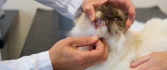
The veterinarian collects information about the cat’s diet and living conditions and listens to the owner’s complaints.
A visual examination of the pet is also carried out, and additional tests may be prescribed.
Changing teeth in kittens
At about 15-16 weeks of age, baby teeth begin to fall out and are replaced by molars. This happens in the same way as when the first set appears: first the baby teeth fall out and new incisors grow, then the lower, and then the upper canines, then the premolars and molars. The only difference is that in adult cats the full set consists of 30 teeth: 2 pieces are added on each jaw, 1 on each side.
The change process is completed at approximately seven months of age. Just like humans, cats have second molars that serve them throughout their lives. In order for them to be fully preserved in old age, the cat’s oral cavity must be properly cared for.
How to find out the age of a kitten
Every owner should know how old his pet is. It's easy to see the difference in what a 1 month or 1 week kitten looks like in appearance. It develops at a rapid pace, at each specific stage there are characteristic signs that indicate an approximate date (only a veterinarian can say for sure). There are certain ways that will help you find out how old your pet “predator” is. The time of birth can be determined using the following criteria:
- size;
- weight;
- teeth;
- eye color;
- appearance;
- behavior.
Symptoms of tooth change
The owner may not notice the loss of incisors at first: they are very small. And cats are designed in such a way that they swallow small objects that end up in their mouths rather than spit them out. In addition, if the process goes well, then the kitten does not experience much discomfort and does not complain to the person.
The only normal symptom is itching of the gums, which the kitten tries to relieve by scratching them on something. During this period, wires, furniture, clothing, shoes and other things suffer. Also, teething is accompanied by copious salivation.
The appearance of permanent teeth occurs gradually. The root presses on the milky one, dissolving its weak roots in a few days. The top of the milky one falls out, and the root one begins to grow. During this period, the gums become slightly inflamed and swollen. When the permanent tooth erupts, it becomes normal.
During the period of teeth change, the kitten may show poor appetite, as it most likely experiences discomfort when chewing. In some cases, animals develop a short-term fever: the temperature rises slightly, and the cat tries to warm up, looking for warmer places.
It happens that the permanent tooth is already visible, but the milk tooth has not yet fallen out. If the latter does not interfere with the growth of the former, and the teeth do not injure the oral mucosa, then there is no need to worry. After some time, the milk will fall out. They normally do not harm each other, since they grow from separate holes.
Could there be problems?
The process of tooth growth is completely natural, so it very rarely causes problems. Despite this, veterinarians recommend remaining vigilant and closely monitoring the symptoms that occur in your pet.
First of all, it is important to understand that due to a decrease in immunity, a kitten can easily catch a cold and get sick. If he has a fever for a long time or the symptoms listed above are complemented by others, contact your veterinarian.
Possible problems include:
- Persistence. Characteristic only for changes to radical ones. With this disorder, 2 rows of teeth are preserved in the oral cavity.
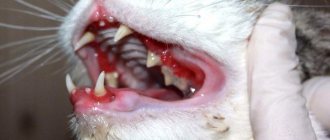
- Gingivitis, or inflammation of the gums. Accompanied by severe redness and swelling of the gums. Due to the constant dripping of saliva, the fur on the chest gets wet and takes on an unkempt appearance.
- Periodontitis, or inflammation of deep dental tissues. Distinctive signs are a pungent odor from the mouth and severe bleeding of the gums.
Treatment involves the mandatory use of mild sedatives to immobilize the animal. Persistence is eliminated strictly surgically, and in other cases, damaged teeth are tried to be preserved.
IMPORTANT!
Don't forget to maintain oral hygiene. Otherwise, your pet may suffer from caries, pulpitis and even premature loss.
Caring for a kitten during the period of teeth change
During the period of changing teeth, kittens' gums itch very much, just like people's gums (if adults remember this feeling). They try to chew on something to reduce the level of discomfort. At this time, owners need to be especially careful: the animal can harm itself, for example, by chewing electrical wires. There is also a danger of choking on a small object on which the kitten decided to scratch its gums. It is necessary to remove all such small items, wiring and other potentially dangerous things from the cat’s access area.
It is also necessary to pay attention to several issues: vaccination, nutrition and oral hygiene.
Nutrition
It is better to consult a veterinarian about the nutrition of a kitten during the period of teeth change. To build teeth, the animal’s body requires large amounts of calcium and phosphorus, so these elements can be slightly increased in the diet. However, you cannot overdo it: this is a serious burden on the kidneys. If parents or siblings had problems with the excretory system, then there is no need to introduce additional calcium and phosphorus.
If the kitten is accustomed to industrially produced food, then it should be fed dry food during this period. This will allow the pet to scratch its gums and, possibly, remove already loose teeth. There is no great danger in kittens swallowing teeth, but sometimes swallowed solids can harm the soft tissues of the digestive tract.
If the kitten has a natural diet, then it is recommended to give relatively large pieces of dietary meat. You can give fish twice a week, but you shouldn’t especially get used to such food. This is especially true for male kittens who are supposed to be castrated. In the future, fish will have to be completely excluded from the diet, and the animal should not get used to it.
You should also give your kitten plenty of dairy products rich in calcium. This can be cottage cheese, whole milk (if the animal tolerates it normally), yogurt, kefir with a low fat content.
In specialized stores you can find special bones that a kitten can chew on when its gums itch. In addition to the fact that it eliminates discomfort and helps teething, such products also contain useful additives that will improve the kitten’s health.
Vaccinations
Kittens should be vaccinated according to the schedule. The first vaccinations are given at two months; further procedures should be consulted with a veterinarian. As for vaccination during the period of teeth change, all veterinarians agree on this issue: while the milk teeth are falling out and the molars are growing, there is no point in getting vaccinated.
A visit to the veterinary clinic is stressful for the animal. And during the period of changing teeth, the kitten already feels somewhat weakened, even if everything is going fine. Therefore, vaccinations should be done before they begin to fall out, and then after the root ones grow.
Oral hygiene
To prevent tartar and other diseases of the oral cavity, teeth and gums, cats should brush their teeth regularly. Experts advise accustoming the animal to this procedure from infancy: then it will not experience discomfort, will get used to it and, perhaps, will even love cleaning.
Kittens need to brush their teeth with a special toothpaste and brush. Human toothpastes are not suitable for cats as they contain ingredients that are harmful to them. Each family member should have their own brush. There are also special gels for disinfecting the oral cavity for cats.
This should be done approximately once every three weeks. During the shift, you can purchase a special product containing anti-inflammatory and anesthetic components. Procedures with such a gel will significantly reduce the animal’s discomfort.
Itching and other associated symptoms
Most pets teething without pain. Their appearance can be recognized by the following symptoms:
- severe itching in the gums;
- profuse drooling;
- redness and swelling of the gums;
- slight and short-term increase in temperature;
- deterioration of appetite and sleep;
- loss of activity.
To suppress itchy sensations in the mouth, babies try to chew on everything that gets in their way. Furniture, shoes, and wires can be damaged. For this reason, all potentially dangerous items should be kept away from the floor. Instead, your pet should be given a special teething ring with a cooling effect - and any other chewing toys from the pet store.
IMPORTANT!
The growth and change of primary teeth weaken the immune system, so these periods are contraindications for vaccination.
In what cases is it necessary to contact a veterinarian?
The change of teeth occurs gradually, and it is necessary to regularly examine the kitten’s oral cavity. If the gums are pink, there is no inflammation or redness in the places where soft tissue joins the bone, and baby teeth do not interfere with the growth of permanent teeth, then there is no reason to worry. You should contact your veterinarian if you notice the following problems:
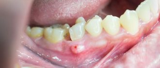
- there is redness around the baby tooth or the hole where it was located;
- pus appeared in the hole from the fallen tooth;
- a very strong unpleasant odor comes from the mouth;
- there is inflammation on the gums;
- the new tooth grows in such a way that it damages the mucous membrane, due to the fact that it is interfered with by the milk tooth or for another reason;
- The radical ones have grown, but the milk ones have not completely fallen out, and the shift time has ended.
You should also pay attention to the well-being and behavior of your pet. Warning signs:
- the kitten is apathetic, does not play;
- a plaintive meow, indicating serious discomfort and pain;
- the animal eats poorly or refuses to eat at all;
- anxiety, poor sleep.
Sometimes owners notice that the tray is empty for more than a day. This is also a cause for concern. The absence of bowel movements may be due to the fact that the kitten was trying to scratch its teeth and swallowed some object that blocked the intestinal lumen.
The owner should be aware that during the period of changing teeth, kittens' immune defenses are somewhat reduced, so changes in behavior may be due to the fact that the animal has contracted an infection. This can also happen to a kitten who lives in the house and is not outside. Owners can bring pathogenic flora into the apartment on shoes or clothes. Usually it is not dangerous for either animals or humans, however, against the background of a decrease in the immune status, the cat may get sick. Not everyone has been vaccinated by this age, and the animal does not have specific immunity to certain pathogens.
How are baby teeth different from molars?
Milk teeth in kittens differ from primary teeth in their number, color and shape. In the very first row of teeth, 4 molars are missing, distinguished by the presence of 3 roots at once.
The teeth of adult animals are dense and curved. They are colored yellowish and covered with a noticeable layer of plaque and stone. For babies, the shape is perfectly straight and pointed at the ends. Their teeth are covered with milky-white enamel, and on its inner side there are small tubercles. They look like mini copies of regular teeth and disappear with the appearance of the molars.
First aid for teething
When baby teeth appear, the baby may require not only parental care, but also medical attention. The dentist may recommend that parents use anesthetic dental gels and treat the affected areas of the gums with decoctions of sage, oak bark or soda solution. If the pain is severe, the baby may be prescribed paracetamol, ibuprofen and other systemic painkillers.
To ease the discomfort that a child experiences when teething, it is necessary to use teethers - specialized devices made of rubber or plastic that the baby can bite and gnaw on without risking damage to the soft gum tissue. In addition, it is advisable to regularly massage the child’s gums with a finger wrapped in a clean, damp bandage.
Symptoms that indicate that the process has begun:
- Some children experience an increase in temperature (up to 38). In this case, you should not hesitate; it is better to seek advice from your doctor.
- Unpleasant sensations in the gums.
- The space between the teeth increases.
- The roots begin to dissolve (The roots of baby teeth are much shorter than those of permanent teeth. When the process reaches the neck, a tooth change occurs).
- Gums are bleeding.
A modern view of the problem of helminthiasis in children and effective ways to solve it
According to the World Health Organization, of the 50 million people who die annually in the world, more than 16 million are caused by infectious and parasitic diseases. In the structure of infectious diseases, intestinal helminthiases are in third place. The World Bank estimates that the economic cost of intestinal helminthiases ranks fourth among all diseases and injuries. Given the importance of the control of parasitic diseases for many countries, the 54th World Health Assembly in 2001 approved a strategy for the control of soil helminthiasis until 2010 [5].
In the Russian Federation, more than 10 million people are examined annually for helminth infections, most of them are children. In 2002, 813 thousand infected were identified, of which 681 thousand (83.8%) were children under the age of 14 [4]. More than 15 types of helminths are found in children, of which the most common are enterobiasis, ascariasis, opisthorchiasis, diphyllobothriasis, trichocephalosis, and hymenolepiasis. In recent years, toxocariasis has been increasingly recorded, which is associated with the widespread introduction into practice of a diagnostic test system for its detection.
In the structure of helminthiasis, the leading place is occupied by enterobiasis (91%) and ascariasis (8%). Among all infected children, 92.3% of cases of enterobiasis, 71.1% of ascariasis, 61.5% of trichuriasis and 66.2% of toxocariasis occur.
The incidence of enterobiasis and ascariasis in children in rural areas is significantly higher than in cities, which is apparently due to different sanitary and hygienic conditions in child care institutions in the city and village, as well as the degree of contamination of the environment with helminth eggs (Fig. 5).
Ascariasis is one of the most common helminthiasis, in the formation of foci of which soil contamination with ascaris eggs is of primary importance. In 2002, 74,196 cases of ascariasis were identified, including 52,801 in children under 14 years of age; compared to 2001, the incidence increased by 3.5% and amounted to 217.7 per 100 thousand children.
The incidence of trichuriasis, with a clear downward trend over the last decade, in 2002 increased by 2.8% and amounted to 7.4 per 100 thousand children. Trichocephalosis is registered mainly in the Southern Federal District (Republic of Dagestan, Chechen Republic).
Enterobiasis still ranks first in terms of prevalence among other helminthiases. In 2002, 614,955 cases of the disease were registered among children, which amounted to 2535.5 per 100 thousand patients.
The maximum number of people infected with enterobiasis in 2002 was identified in the Siberian, Northwestern, Ural, Far Eastern, and Volga federal districts.
A feature of most helminthiasis is the chronic course of the disease, associated with the long-term presence of the pathogen in the body and repeated repeated infections. Helminth infections in children, as a rule, are accompanied by a variety of nonspecific clinical manifestations: weakness, fatigue, irritability, sleep disturbances, dyspeptic symptoms, slower growth and weight gain, and decreased immune status. The most important component of the pathology of helminth infections is the sensitizing effect of metabolic products and excretion of helminths, leading to the development of allergic reactions in the form of atopic dermatitis, asthmatic bronchitis, rhinitis, blepharitis, etc.
A selective analysis of the results of clinical examination of 520 children, carried out in the Sverdlovsk region of Perm in 2002, showed that ascariasis was detected in 1.35%, and enterobiasis in 5.8% of those examined. At the same time, on average, the number of health problems per child in children with enterobiasis in the group of preschoolers was 2.5, and in schoolchildren - 2.9. More often than others, diseases of the genitourinary system (in girls), allergic dermatitis, anemia, vegetative dystonia syndrome, neuropathic conditions and diseases of the gastrointestinal tract were noted. Of the diseases diagnosed in children with ascariasis, the most common were vegetative dystonia syndrome, functional diseases of the gastrointestinal tract, pneumonia, and allergic dermatitis. Among children with ascariasis, the incidence rate per child averaged 2.0. In the control group, which included children without parasitic diseases (37 people), the average number of health problems was significantly lower than among those infected with ascariasis and enterobiasis, and amounted to 0.2. Thus, the presence of ascariasis and enterobiasis leads to a deterioration in the general health of children. At the same time, children with various underlying diseases that lead to weakening of the body are more often infected with them.
Let us dwell in more detail on the importance of the most common helminthiasis in the development of pathology in children - ascariasis and enterobiasis [1, 3].
Ascariasis. The development of the causative agent of ascariasis (Fig. 2, 3) in the human body occurs with the migration of larvae emerging from the eggs along the bloodstream through the lungs; the larvae are then swallowed with sputum and develop into adults in the intestine. The lifespan of roundworm in the human body is several months. Ascariasis has a significant impact on the quality of nutrition and immunological mechanisms in children. Ascaris allergen is the most powerful of allergens of parasitic origin. It can cause reactions in the lungs, skin, conjunctiva, and gastrointestinal tract. Allergic reactions can be so severe that they often pose a threat to the child’s life.
The immunosuppressive effect of roundworms is due to the lack of effect of vaccination and revaccination against measles, diphtheria, tetanus, and polioviruses in children.
The leading mechanisms of pathogenesis of the migratory stage of ascariasis are the traumatic effect of larvae and sensitization by parasitic antigens. In this case, 2 main types of lesions occur in different organs and tissues.
- The traumatic effect of migrating larvae in organs and tissues along the migration route. At the beginning of migration, the larvae are still small (no more than 0.5 mm long) and cause limited hemorrhages in the wall of the small intestine and in the liver. By the end of migration, the larvae reach 2 mm in size and, penetrating into the alveoli and bronchioles, and then into the bronchi, cause more significant hemorrhages.
- Eosinophilic inflammation of the tissues in which the larvae develop. The tissue phase of ascariasis occurs during the migration of ascaris larvae to the liver and lungs. The metabolites released in this process cause serious immunological changes and inflammatory reactions. In the migration phase, ascariasis can cause hepatomegaly and asthmatic syndrome. In this case, the clinical picture resembles respiratory allergosis.
In the intestinal phase of ascariasis, important pathogenetic factors are the ability of roundworms, reaching a length of 20–40 cm, to spiral forward movements and the desire to penetrate small openings (Vater's nipple, drainage tubes, etc.). The presence of invasion leads to hypertrophy of the muscular layers of the intestinal wall, a decrease in the depth of the crypts, changes in the chemical composition of the intestinal contents, and disruption of the motor-secretory function of the stomach and intestines. Roundworms secrete inhibitors of trypsin and chemotrypsin, as a result of which the absorption of nutrients, proteins, and fats worsens. With ascariasis, functional deficiency of pyridoxine develops, the level of retinol and ascorbic acid decreases, and lactase tolerance decreases. Ascariasis is usually accompanied by intestinal dysbiosis.
Often symptoms of the intestinal phase of ascariasis are nausea, vomiting, diarrhea, fatigue, dizziness, poor sleep, and abdominal pain. An increased level of eosinophils in the peripheral bloodstream is characteristic of the migratory phase of ascariasis.
Complications of the intestinal phase of ascariasis: intestinal obstruction caused by a ball of adult roundworms; peritonitis due to perforation of the intestinal wall or penetration of roundworms into the abdominal cavity through a surgical suture; obstructive jaundice during migration of helminths into the common bile duct; blockage of the pancreatic ducts; asphyxia due to the migration of roundworms into the upper respiratory tract.
Enterobiasis. The development of the enterobiasis pathogen (Fig. 1) in the human body occurs within the gastrointestinal tract. The larvae emerge from the eggs (Fig. 4) and, on average, develop into adults within 2 weeks, which parasitize the lower parts of the small intestine and the upper parts of the large intestine. The lifespan of pinworms can reach 100 days, and the state of infestation in children due to repeated infections can last much longer.
| Figure 1. Life cycle of the enterobiasis pathogen (according to HC Jeffrey, RM Leach, 1975) |
Inflammatory reactions during enterobiasis develop under the influence of larvae, which produce hyaluronidase, proteolytic enzymes, lectin-like substances that promote the activation of the complement system, the release of prostaglandins by the cells of the host tissues surrounding the helminth [1, 2, 3].
With enterobiasis, the processes of absorption and digestion of food products are disrupted. In 30–40% of infected people, the acidity of gastric juice decreases, up to anacidosis and inhibition of pepsin-forming function. In most children, the intestinal microbiocenosis changes. Impaired absorption and digestion of nutrients in the intestines lead to weight loss and delay the growth and development of the child.
An additional factor in the pathogenesis of enterobiasis is the mechanical effect of pinworms in the intestines, leading to pinpoint hemorrhages, erosions, and penetration of bacterial flora, in particular pathogens of intestinal infections.
A striking symptom of enterobiasis is perianal itching, which occurs when the female moves during oviposition (Fig. 6). Severe itching occurs, as a rule, during sleep, more often at night, from 23.00 to 1.00 am. It is at this time that helminths can, remaining unnoticed, lay eggs that will mature to the invasive, contagious stage by the morning. Despite its apparent harmlessness, perianal itching is difficult for children to tolerate and can persist for quite a long time after enterobiasis is cured as a result of the formation of a persistent focus of excitation in the cerebral cortex. Complications that arise as a result of perianal itching are skin damage when scratching, perianal pruritis, eczema, weeping dermatitis. The etiological agent of the inflammatory process is most often streptococci.
| Figure 2. Roundworm egg (70 microns) |
| Figure 3. Adult roundworms |
| Figure 4. Pinworm egg (50–60 µm) |
Abdominal pain is a common symptom of enterobiasis. Pain of a transient nature is observed in the majority of infected people. Sometimes acute abdominal pain may be the reason to seek surgical help. In such cases, it is often not possible to detect a specific pathology; only the accumulation of gases is detected.
In recent years, the number of cases of the formation of perianal granulomas or abscesses in children, inside which female pinworms or helminth eggs were found, has increased. In this regard, it is advisable to screen all children with these conditions for enterobiasis.
In many cases, enterobiasis occurs over a long period of time and is repeated many times. As a result, the intestinal biocenosis is disrupted and the antagonistic properties of the intestinal microflora in relation to pathogens of acute intestinal infections are reduced. In the majority of infected children, the number of E. coli decreases and the proportion of lactonegative intestinal flora increases. The activity of enterokinase and alkaline phosphatase in feces increases. Since the intestinal microflora is one of the factors that supports the activity of intestinal enzymes, disturbances in the processes of digestion and absorption of nutrients that develop as a result of enterobiasis lead to loss of body weight and retard the growth and development of the child. Pinworms have a mechanical effect on the intestinal mucosa, which leads to pinpoint hemorrhages, erosions, and penetration of bacterial flora, in particular pathogens of intestinal infections. The antagonistic properties of the flora in relation to the causative agents of typhoid fever and other intestinal infections are reduced [1, 3].
If pinworms migrate into the abdominal cavity, urinary and genital tracts, inflammatory and allergic reactions outside the intestine may develop.
| Figure 5. Incidence of enterobiasis and ascariasis in urban and rural children in 2002. |
One of the common complications of enterobiasis is vulvovaginitis due to the penetration of pinworms into the genital tract and the addition of bacterial infections. If vulvovaginitis develops in a girl, a parasitological examination for enterobiasis should be prescribed and, if the result is positive, this invasion should be treated with simultaneous bacteriological examination and, if necessary, antibacterial therapy.
Against the background of enterobiasis, children often develop urinary tract infections, especially girls, since enterobiasis is a factor predisposing to the development of this complication.
Parasitism by pinworms in children leads to suppression of nonspecific immunity, manifested by a decrease in the level of interferon a in the blood serum. A decrease in the nonspecific resistance of the child’s body leads to an increase in the incidence of viral and bacterial infections.
| Figure 6. Tail end of a female pinworm |
The presence of enterobiasis leads to a decrease in the effectiveness of preventive vaccinations. The immune layer against diphtheria was initially lower among children infected with pinworms. Protective immunity does not develop during primary vaccination against this dangerous infection, and during revaccination in many cases there is no immune response. It is difficult to develop immunity when vaccinated against measles and tetanus, so to increase the effectiveness of vaccinations, you first need to make sure that the child’s body is free from helminthiasis pathogens.
In children with allergic diseases, enterobiasis develops much more often. Due to the relatively high probability of detecting enterobiasis in children with allergic diseases, patients in this group should be recommended to be examined for enterobiasis and deworming if infestation is detected.
Enterobiasis negatively affects the neuropsychic development of children. This invasion leads to a lag behind the corresponding age norms. Among those infected with enterobiasis, there is a high percentage of irritable children, with a disruption in the process of falling asleep, and with negative habits (biting nails, sucking fingers, etc.).
With enterobiasis in children, the level of copper, zinc and magnesium in the blood decreases significantly. Since the lack of these microelements can negatively affect the physical and mental development of children, their loss should be compensated by introducing certain foods into the child’s diet, prescribing medications (or nutritional supplements) until these indicators are normalized after enterobiasis is cured.
Main indications for examination for helminth infections:
- stomach ache;
- frequent nausea, vomiting;
- diseases of the gastrointestinal tract;
- fatigue, irritability, restless sleep, grinding teeth in sleep;
- allergic conditions;
- perianal itching (enterobiasis);
- vulvovaginitis (enterobiasis);
- urinary tract infections (enterobiasis);
- increased level of eosinophils in the blood;
- retardation in height, weight;
- untidiness.
The diagnosis of enterobiasis and ascariasis is made only upon receipt of positive results from a laboratory parasitological examination of the patient. If enterobiasis is suspected, a perianal scraping (imprint) is examined, and fecal samples are examined for ascariasis. On the laboratory referral form, you should indicate what kind of helminthiasis the doctor suspects in the child. The choice of the most effective research method by laboratory specialists will depend on this.
Treatment of ascariasis and enterobiasis
The search for remedies for the treatment of helminthiases, including enterobiasis and ascariasis, began many centuries ago. To expel pinworms, Ibn Sina recommended taking elecampane and celandine with sugar, washing them down with water. The medicine for expelling worms (“killing worms”), indicated in the Ebers Papyrus, contains, among other components, date seeds and the plant disart, sweet beer. The Salerno Health Code, which dates back to the early 16th century, recommends another remedy: mint [3].
The modern arsenal of drugs used to treat intestinal helminthiases includes a significant number of drugs of various chemical classes. They are used both in clinical practice for the treatment of identified patients or parasite carriers, and for the purpose of mass prevention.
The Russian pharmaceutical market currently offers several anthelmintic drugs that act on the causative agents of ascariasis and enterobiasis (Table 1).
The most effective drugs for the treatment of enterobiasis and ascariasis are derivatives of carbamate benzimidazole (mebendazole, medamine) and tetrahydropyrimidine (pyrantel). In addition to the ability to influence mature forms of helminths, they are distinguished by high ovicidal and larvicidal activity. Drugs of these pharmacotherapeutic groups disrupt oxidative processes, inhibit glucose transport in helminths, act on the muscles of intestinal nematodes by depolarizing their neuromuscular junctions and block the action of cholinesterase.
The effectiveness of medicines used to treat enterobiasis and ascariasis is very high, the method of administration is very simple and is designed primarily for children. It is very important that as a result of their intake, the process of releasing the pathogen into the environment is not activated. Thus, during treatment the person does not become more dangerous to others. However, pinworm eggs that have already entered the environment, in particular indoors, persist for a long time - more than 2 weeks. Therefore, it is recommended to repeat the treatment of enterobiasis after 2-3 weeks at the same dose in case hygienic measures were not effective enough. For the same reason, simultaneously with the treatment of infested people, everything possible must be done to clear the premises of pathogens.
For many years, pyrantel has been used throughout the world to treat ascariasis and enterobiasis, and has gained popularity among pediatricians and patients. According to recommendations developed in the USA (Medical Letter, 2002), pyrantel is considered a first-line drug for the treatment of enterobiasis in children and adults.
The anthelmintic effect of pyrantel pamoate is associated with a stimulating effect on the H-cholinergic receptors of the ganglion synapses of helminths, leading to spastic paralysis and their subsequent expulsion from the human body. Clinical trials of the effectiveness and tolerability of pyrantel showed its high medicinal activity against enterobiasis and ascariasis - 94-100%, as well as good tolerability [1, 3].
Pyrantel for the treatment of enterobiasis is prescribed at a rate of 10 mg/kg per day once during or after meals. For the treatment of ascariasis, pyrantel is prescribed at a dose of 5 mg/kg once. The drug is well tolerated by children; in some cases, nausea, vomiting, diarrhea, abdominal pain may develop; very rarely, a transient increase in the activity of liver transaminases, headache, dizziness, and sleep disturbances may occur. Pyrantel is contraindicated in children with liver disease.
We have extensive experience in treating enterobiasis in children with the drug Pyrantel (tablets, suspension). Pyrantel is well known on the world market of anthelmintic drugs and is widely used for the treatment of enterobiasis in Russia by many generations of doctors. In our opinion, this drug has a number of advantages compared to other anthelmintic drugs. Firstly, the drug in the form of a suspension is easy to give to children, secondly, Pyrantel has a pleasant peach taste, as a result of which the child does not experience negative emotions during treatment, and, finally, thirdly, Pyrantel has a reasonable price and is widely sold in pharmacies. The bottle is equipped with a measuring spoon with a division scale of 2.5 and 5.0 ml, which makes it easy to dose the drug depending on the body weight of the infected child (or adult). Pyrantel suspension can be used in children from 6 months of age.
Along with pyrantel, mebendazole and medamine have a good anthelmintic effect against enterobiasis and ascariasis.
Mebendazole (Vermox) for the treatment of enterobiasis is prescribed to children 2–5 years old at the rate of 5 mg/kg per day, over 5 years old - 100 mg per day. For the treatment of ascariasis in children 2–5 years of age, the drug is prescribed at a dose of 5 mg/kg in 2 doses per day for 3 days; for children over 5 years of age, mebendazole is prescribed at a dose of 10 mg/kg per day in 2 doses for 3 days.
Mebendazole is not recommended for use in the first trimester of pregnancy. It must be remembered that the drug is contraindicated in children under 2 years of age. Side effects of mebendazole include abdominal pain and loose stools.
Medamine (2-medoxycarbanylamino-benzimidazole) is close in chemical structure and spectrum of anthelmintic action to mebendazole. For the treatment of enterobiasis, it is prescribed at a dose of 10 mg/kg per day in 2-3 doses (simultaneous administration is also possible) after eating a small amount of food; it is recommended to chew the tablets and wash them down with water. For the treatment of ascariasis, medamine is prescribed in the same doses for 3 days.
Side effects of medamine include nausea and weakness. In case of allergic manifestations, the drug is discontinued. Medamin is contraindicated in the first trimester of pregnancy.
To restore the microbiocenosis of the colon in patients with intestinal nematodes, including enterobiasis and ascariasis, and to increase the effectiveness of specific therapy, it is recommended to prescribe bificol, milk bifidum-bacterin. Food products and medicinal plants that can be used for the treatment and prevention of enterobiasis have long been known. Carrots and carrot juice have a good anthelmintic effect. You can also use the anthelmintic activity of walnuts, wild strawberries, pomegranate (especially pomegranate juice), garlic and lovage.
Among the medicinal plants, St. John's wort is used in the form of decoctions and infusions, tea, as well as elecampane (Inula helenicum). The effectiveness of herbal medicine for enterobiasis is low, but the introduction of foods with anthelmintic effects into the diet is a good measure for the prevention of enterobiasis and enhances the effect of medications prescribed by a doctor.
The criteria for the effectiveness of treatment of helminthiases are a negative result of a control parasitological study of fecal samples (for ascariasis) and a perianal scraping or print (for enterobiasis), as well as the disappearance of clinical symptoms of invasion.
Prevention of helminthiases
Features of the prevention of helminthiases depend on the characteristics of their epidemiology. With enterobiasis and ascariasis, the only source of infection is humans. Infection occurs when mature infective helminth eggs are ingested. However, the epidemiology of these helminthiases is otherwise very different. Pinworm eggs mature indoors and on the human body within a few hours and last on average up to 1 month on various household items. Ascaris eggs mature when dropped into the soil within several months and remain there for up to 10 years or more. Enterobiasis is transmitted indoors from one person to another mainly through dirty hands, bed and underwear, toys, dishes and other household items contaminated with pinworm eggs.
A person becomes infected with ascariasis by ingesting soil particles containing invasive roundworm eggs (with unwashed vegetables, herbs, and fruits). The risk of infection (in the case of ascariasis) increases if a child has such a bad habit as geophagy (tasting or eating earth, sand, clay), which occurs quite often (in 3-10% of children under 7 years of age).
Prevention of enterobiasis and ascariasis is the most important task of medical and educational institutions and parents. It can be solved by simultaneously implementing a set of measures, the main components of which are the identification and treatment of infected people and sanitary and hygienic measures. Prevention of ascariasis, enterobiasis and other helminth infections in the Russian Federation is regulated by new sanitary standards and rules approved by the Ministry of Health of the Russian Federation in 2003.
For questions regarding literature, please contact the editor.
T. I. Avdyukhina, Candidate of Medical Sciences, Associate Professor T. N. Konstantinova, Candidate of Medical Sciences, Associate Professor M. N. Prokosheva RMAPO, Moscow Children's Clinical Hospital named after. P. I. Pichugina, Perm
Treatment: what the doctor prescribes
Therapy is selected individually, taking into account the reasons that led to such changes. The main goal of its implementation is to eliminate the provoking factor and normalize the animal’s condition.
Nutrition correction
The first thing to do is to adjust the animal’s diet.
It is extremely important that the menu is balanced . It should contain a sufficient amount of vitamins and microelements.
Tartar removal
If necessary, remove hard plaque under anesthesia. The pet is conscious, but cannot offer resistance.
Upon completion of the process, the cat needs to be provided with complete care. The procedure is very stressful for him.
Treatment of the underlying disease
The main emphasis is on eliminating the pathology that provoked tooth loss. For this purpose, immunostimulating, anti-inflammatory drugs and antibiotics can be prescribed. The therapy is aimed at strengthening the gums, eliminating bleeding and pain.
What to do at home
The owner is required to strictly follow the veterinarian's instructions. Self-medication is dangerous for the cat’s health and can cause irreparable harm to it. Medicines and their dosage are determined by a specialist.
In order for the animal to lead a normal lifestyle, it is necessary to change the consistency of the food, since dry hard granules can further injure the oral cavity.
The oral cavity is regularly examined and treated with a disinfectant solution of Chlorhexidine. These procedures prevent infection of the enamel and damage to other teeth.
Preventive measures
Avoiding such problems is much easier than dealing with their consequences later. To this end, it is worth following a number of recommendations.
Regular teeth cleaning
To avoid the formation of tartar, it is recommended that your pet regularly brush its teeth.
True, cats walking outside clean their mouths themselves. They have it in their nature. We are talking about pets. For this purpose, special pastes are used, which are available at the veterinary pharmacy. There is no need to wash them off with water.
The use of human hygiene products is prohibited. Cleaning is done with a special or children's brush. The main thing is that the bristles are soft.
Balanced diet
If the owner feeds the animal with natural food, then the pet’s menu should include fermented milk products, vegetables, fish and meat.
When using dry food, the choice is made on premium products. It contains the necessary concentration of nutrients.
Regular examinations by a specialist
At least once a year you need to visit a veterinary clinic and have your pet examined. In this way, it will be possible to identify the disease at the initial stage of its development.