If a cat has a swollen eye, it can be caused by a number of factors, some of which are quite dangerous and can even lead to loss of vision. When such a problem occurs, it is important for the owner not to self-treat the animal. The first thing you need to do is to examine the cat’s visual organ, paying attention to the condition of the eyeball and the protective parts of the eye. The animal should then be taken to the veterinarian, providing him with detailed information about when the symptoms were noticed and whether injury was present.
Detailed description of possible causes
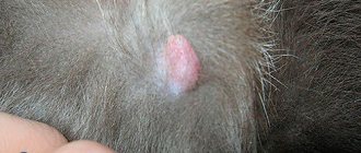
It’s more difficult when a bump on a cat’s head appears for unknown reasons, or rather, the very moment of its appearance did not come into the owner’s field of vision.
Then you have to guess and determine the cause based on the existing accompanying symptoms. If there is a possibility or need, the veterinarian will be able to figure it out faster, ruling out the following possible diseases after cytology, microscopy and histology: Trauma. The most common cause of a soft lump on the head of a cat at any age. In the first years of life, such swellings often occur due to inexhaustible curiosity, which often ends in falling from the sofa, table or windowsill during active play. There is nothing wrong with this, all animals go through this. Such seals heal within a few days even without the use of any ointments. The situation is a little more complicated with injuries received in a fight with other animals, since swelling after a bite from teeth or a scratch from claws can become a source of infection, which will provoke an inflammatory process. Such injuries must be treated with an antiseptic solution; to be safe, you can apply a bandage soaked in levomekol to the wound for several days. At the same time, do not forget to make sure that your pet does not lick the medicine. Hematoma. They were specially separated from trauma, although they are closely related. If a cat has a large bump on its head after a fall from a great height or an accident, then you need to consider the option of a hematoma. They are soft to the touch and painless. The insides are filled with dried blood and lymph fluid. Small hematomas almost always resolve on their own within 1-2 weeks. After serious collisions or if the size of the mass exceeds acceptable limits (in some cases reaching the size of a large apple), a visit to the veterinarian is considered mandatory. This is necessary so that the doctor punctures the hematoma and drains the contents from it. The cavity of the resulting wound is thoroughly disinfected, and the necessary ointments with anti-inflammatory and regenerating effects are selected for the animal. Fleas. Bites from these insects can cause small bumps that are predominantly pink or red in color to appear on your cat's head. The bites themselves often itch, but do not hurt. The pet begins to behave restlessly, you may notice atypical behavior (itches more than usual or pays a lot of attention to a particular area of the body). The situation is even worse with animals that are allergic to flea saliva. Here you have to get rid of not only parasites, but also eliminate the symptoms of an allergic reaction. The most effective way to combat fleas is considered to be regular treatment of all animals living in the same house with the help of preparations that are applied to the withers.
It does not matter whether the pet visits the street or not, since insects can get into its fur from the soles of the owner’s shoes or clothes. The best drugs today are considered to be Advocate, Stronghold and Frontline
Of the domestic ones, Barrier drops are praised. It is recommended to apply the drops monthly evenly to the animal’s withers so that he does not have the opportunity to lick it off. For medicinal purposes, a regimen of applying the drug three times every three weeks is used. Abscess. If a cat has a purulent lump on its head, there is a possibility that there is an inflammatory process caused by an infection getting under the skin. Abscesses often form after deep wounds received in a fight with other animals, when a foreign body gets under the skin. There are two options for the development of events: wait until the abscess breaks out on its own or take the pet to the veterinarian, who will open the abscess and clean it of pus. If the abscess nevertheless ruptures on its own, then it is necessary to treat the wound with an antiseptic solution and cover it with a bandage previously soaked in Levomekol ointment.
What do you need to know about the problem?
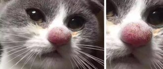
One of the most common problems veterinarians include is subcutaneous formations that can appear anywhere. In a cat, a lump on the nose may indicate the development of a cyst, an enlarged lymph node, hemorrhage, the presence of a foreign object or even a tumor. At the first signs of formation, owners need to conduct an independent inspection.
If the cat is irritated, then the animal should be examined with calm stroking movements of the hands. The purpose of this physical examination is to identify similar lesions in other locations. If there are no subcutaneous growths on the body, hemorrhage due to a blow or scratch should then be excluded. If your pet loves to get into fights with neighbor cats, then in this case there is a high probability of an ordinary wound received from the claws of rivals.
In a cat, a lump on the nose is often a benign formation. As the tumor develops, it gradually grows while maintaining its structure. A congenital bump on the nose is a nasal glioma. It does not cause pain or discomfort, but may begin to grow inside the nasal passages. In some cases, the lump makes breathing difficult, in which case it must be removed surgically.
Symptoms
During an oncological process in the body, the animal is often irritated. If the bump on the head of a cat or kitten is benign, then it is soft to the touch, rolls under the skin, and does not hurt or bother you when pressed. Internal malignant growths often appear as capsules that can be normal temperature or hot. A lump on the withers, on the side of the neck or on the mucous membrane in the throat from degenerated cells quickly increases in size. In addition, an adult cat or kitten develops the following symptoms:
- weakness;
- irritability or lethargy;
- digestive problems;
- refusal to eat;
- sudden weight loss;
- dyspepsia.
What will be offered at the veterinary center
To determine the characteristics of a tumor in the neck, the doctor:
- conduct a survey;
- examines;
- will do all the necessary tests.
The examination involves feeling the lump. Perhaps, if it is a large abscess, then it will be eliminated immediately by removing the pus and washing the wound.
If additional data is needed for a complete picture, the doctor will refer you for detailed tests and procedures:
- Ultrasound;
- Histo- and cyto-examination;
- Donation of blood and urine.
This will help determine how to further treat and care for the cat. Does he need surgery or specific therapy?
One of the most dangerous and varied in symptoms diseases is skin cancer in cats. 75% of all registered skin tumors in cats are malignant. Depending on the type of tumor cell, treatment, prognosis, and clinical signs vary greatly. In this regard, one of the most important stages of therapy is accurate diagnosis of pathology.
Places of possible education
Bumps associated with various diseases can appear on cats' paws, stomach, or throughout the body. Photos of various bumps on cats can be viewed on the Internet. This can make it easier to differentiate one lump from another.
The appearance of bumps on a cat's body can be due to various reasons.
Lipomas or fatty tissues. They have a tendency to actively grow. They have a soft consistency and cause a painful syndrome
If you do not pay attention to this occurrence in time, it can lead to the death of the animal.
Lymphadenitis or inflammation of the lymph nodes. The appearance of enlarged lymph nodes signals that the pet is infected with a number of serious diseases.
Symptoms of lymphadenitis are accompanied by signs of intoxication and a deterioration in the general condition of your pet.
Abscess. Occurs after an unsuccessful injection or injury. They are very painful and can cause an increase in the animal’s overall body temperature.
Tick bite. This type of lump can be successfully removed and often does not affect the cat’s condition.
Mastitis and mastopathy. This often occurs in nursing cats due to frostbite or the use of hormonal drugs.
They occur in the area of the nipples, have a dense structure and cause severe pain. This also includes the occurrence of breast cancer.
Presence of any skin diseases. Such as carbuncles, boils and others
A bite of an insect. May be licked or scratched by the cat herself.
Other causes of belly bumps
The appearance of bumps in cats is not always caused by benign or malignant neoplasms. Similar signs may occur with inflammation of the skin, the appearance of lipomas and hernias.
Small and large bumps throughout the animal’s body are characteristic of the following cases:
Wen (lipomas)
Wen look like bumps under the skin. With the normalization of lipid metabolism and the introduction of a balanced diet, these formations can resolve on their own. Only a veterinarian can distinguish a lipoma from cancer. This will require a biopsy, ultrasound and x-ray.
Hernias
Hernias can form under the skin of a cat. The umbilical variety is characterized by a lump in the navel area, while the inguinal variety is characterized by a lump in the groin. Other types of hernias are also localized in their “own” natural place. Their diagnosis is not very difficult. It is enough for the veterinarian to conduct a visual examination of the animal and, if necessary, perform an ultrasound.
There are many reasons for the appearance of bumps on a cat’s stomach. The owner only needs to detect them in a timely manner and contact a veterinarian. Only a doctor, using modern medical equipment, will recognize the true nature of their occurrence and prescribe effective treatment.
I like it I don't like it
Additional signs that usually accompany a bump on the nose
If the tumor is caused by a fight with other pets, it is not difficult to determine - scratches, abrasions, and even blood will be found on the cat's body. If the injury is too serious, it is best to go to the vet immediately - you may need stitches. If the abrasions are minor, you can help the animal yourself - treat it with antiseptics, apply a cold compress to the nose. In case of infectious diseases, a growth on a cat’s nose is accompanied by the following symptoms:
- a cloudy liquid with an unpleasant odor is released from the eyes;
- the pet refuses to eat;
- the animal becomes apathetic, lethargic, and shows no interest in anything;
- The coat loses its shine and bald patches appear.
Inflammatory processes are not difficult to determine - the cat is constantly sneezing, breathing is difficult, and wheezing is clearly audible. The pet often shakes its head and becomes nervous when its owners approach. Fluid is released from the nasal passages, which may be clear or yellowish, mixed with pus.
Types of bumps under the skin of a cat
Cones can have a different nature, depending on the animal’s predisposition to a particular disease.
Lipoma (wen)
At first, a small formation appears, which gradually grows and can grow to the size of a chicken egg. The cause of lipoma is improper distribution of fat after clogged pores. Lipoma is a benign neoplasm that does not metastasize and does not cause discomfort. The exception is if the lipoma occurs on the back or neck. The lipoma does not hurt even when pressed, it does not affect life processes: the appetite is preserved, the animal’s normal behavior, the temperature is normal.
In older animals, the appearance of lipomas may indicate the onset of cancer. You should definitely have your cat examined by a veterinarian.
Abscess
An abscess appears at the site of a bruise, any internal injuries, or in case of an incorrect injection.
The tumor appears approximately on the 3rd day after injury, gradually growing.
Unlike a lipoma, an abscess is a painful condition, accompanied by an increase in temperature and depression of the pet’s condition.
The abscess is firm to the touch, but not hard. You can feel accumulated pus inside. If the abscess is not completely healed, pus is released into the hole when pressed.
Lymphadenitis
Inflammation of the lymph nodes. The lump is extremely painful even with slight pressure. The animal's condition is depressed, the temperature is elevated, and the appetite is reduced.
Cyst
A cyst is a benign tumor. A cyst is a lump filled with fluid from the inside. This tumor grows slowly and does not affect the cat’s condition; the cyst is not painful.
Oncology
The most terrible type of neoplasm. This is what the owner thinks about when he feels a lump on his pet’s body. Cancer spreads very quickly and metastasizes throughout the body. Inevitably leads to the death of the animal, but in the first stages of the disease it often does not manifest itself.
Causes of subcutaneous lumps
Small round balls often appear after injection under the skin. They are called post-injection granuloma and resolve on their own a few days after the end of the injections. To avoid them, the injection site is massaged daily. Sometimes granulomas remain for life, without changing their size and position. In other cases, the reason lies in:
- blockage of the sebaceous glands;
- allergic reaction;
- infestation with parasites;
- hormonal imbalances;
- infection of open wounds;
- disturbances in the formation or functioning of connective tissues;
- injuries;
- inflammatory processes.
The risk group includes older animals, cats with chronic diseases, late castrated and sterilized pets. The risk of developing cancer increases with constant stress, low activity and poor diet. A predisposition also appears with frequent injuries in the same area of the body, infection with leukemia and regular inhalation of tobacco smoke. Cancer is often inherited and is diagnosed in animals older than 7 years.
Stages of development
The abscess proceeds like this:
- redness of the skin area, which can only be detected with a thorough examination (therefore, after a fight or suspected injury, the pet must be examined immediately);
- swelling of the affected tissue and the formation of a tissue melting zone;
- fluctuation (accumulation of pus in the capsule);
- baldness in this place;
- necrosis (or death) of tissue (with strong immunity, a fistula is formed and pus comes out through it, and if not, then the inflammation spreads further);
- blood poisoning - sepsis;
- infection;
- death...
How to treat so that the latter does not happen? But better - how to prevent complications of this common pathology? Read more about this on our blog.
Causes of the disease
Experts can give a dozen different reasons why a cat’s lower lip could be swollen. Some of them are completely harmless and go away on their own, others can be treated at home, and there are others that can lead to a fatal outcome for a member of the feline family. Below we will discuss the most common factors that can cause a pet's lower lip to swell:
Burn wounds. There are thermal, electrical and chemical burns. The first option is rare, since cats rarely eat food that is too hot for them. However, your pet is capable of licking a hot frying pan if it smells delicious. Electrical burns occur because the animal likes to chew on live wires. Chemical lesions leading to swelling of the lower lip can be caused by household chemicals or animal hygiene products. A common symptom for each of them is severe inflammation located in the lip area. Insect bites. The lip may become swollen if it is stung by a wasp or bee. The owner is able to detect, in this case, the remaining sting. Over time, severe swelling occurs at the site of the lesion. Lip injuries. Occurs if the cat has a metal food bowl with sharp, uneven edges. In parallel with this, gum damage can also be detected. An animal can also get injuries to the soft tissues of its lower lip during a fight with its fellow tribesmen on the street. Oncology localized on the upper or lower lip. Rarely encountered, but extremely dangerous. In veterinary medicine, neoplasms of this kind are called squamous cell carcinoma. According to statistics, they are more often malignant, which means they are capable of metastasizing. Additional signs characterizing this disease include a putrid odor from the cat’s mouth, ulcers on the oral mucosa, and excessive salivation. Over time, the lymph node closest to the tumor becomes inflamed. A benign tumor can be removed surgically, but a malignant one, unfortunately, leads to death in 90% of cases. Allergic reaction. If the lip is inflamed for this reason, then the first thing the owner needs to do is to detect a harmful allergen that negatively affects the pet’s body. This could be plant pollen, components of a pet’s diet, household chemicals, bites of blood-sucking parasites, pharmaceuticals. If the irritant cannot be identified on your own, you should seek help from a qualified specialist. Industrial feed. If the lip is swollen after the cat was switched to dry food, then rest assured that this is the reason. This is especially true in cases where the animal previously ate natural food. The hardness of industrial feed provokes the appearance of calluses on the delicate tissue of the lip, where inflammation occurs. Gingivitis can be another of the many factors that cause a cat to have a swollen lower lip. Its striking symptoms are the cat’s bad breath, bleeding gums, and an orange border appears around the pet’s teeth. If the owner does not react promptly, the cat is in danger of losing a tooth completely. In addition, gingivitis “walks” hand in hand with such terrible diseases as diabetes and liver failure. Stomatitis in a cat, caused by the harmful activity of a fungus that has entered the oral cavity, can be determined by the fact that the upper lip is swollen. In this case, an additional sign is the formation of white plaque on the mucous membranes of the animal. This pathology can be caused by a decrease in the cat’s immunity or long-term treatment with certain pharmaceuticals. Calcivirosis in cats also leads to swelling of the lips and mucous membranes of the mouth. The disease has an infectious etiology, additional symptoms include lameness, mouth ulcers, conjunctivitis and loss of appetite. Requires surgical and complex therapy. Acne, another name is subcutaneous pimple. It occurs due to disruptions in the hormonal system, insufficient oral hygiene of the pet, and excessive activity of the sebaceous glands. Located in the corner of the lips, it does not cause much concern to the cat. You can remove a pimple yourself; to do this, wipe the affected area with antibacterial soap, and then apply an ointment that contains benzoyl peroxide.
It is important that the cat does not lick the medicine, and that it stays on the pimple for at least 5-6 minutes. Then wash off the remaining ointment with Chlorhexidine solution
Causes
- Sometimes there is a violation of sterility during the provision of medical care in a veterinary clinic or at home.
- Fights between animals, during which damage is caused to each other (a pinpoint tear of the skin occurs and infection occurs on the fangs or claws). By the way, a cat bite is one of the dirtiest injuries; it does not heal for a long time and festers.
- Punctures or cracks on the paw pads while being outside (from broken glass, chips, thin wire) and climbing trees, buildings (from thorns), and at the same time introducing a foreign body into the wound (splinter, sand);
- Breaking off a claw due to injury (possibly purulent inflammation of the corolla, after which it will fall off on its own, or the help of a surgeon will be required).
Insect bite
Quite often, cats get injured through their own fault. For example, by hunting a bee or a wasp and achieving success. Of course, the poison contained in the sting leads to an inflammatory reaction. As a result, the cat's cheek was swollen and his eye was swollen. It looks really creepy. But it usually goes away in a few days or even hours, without causing unnecessary trouble to either the cat or the owner.
The situation is much worse with several bites or allergies. This can lead to the most dire consequences. To avoid them, you need to use an antiallergic drug – Claritin or Suprastin.
At risk
It contains:
- stray animals that most often get into fights;
- cats with weakened immune systems;
- domestic purrs, who often go out for walks (although you can sometimes get hurt at home), and especially uncastrated males, as potential rivals to each other, organizing “showdowns,” especially in March;
- Young animals are injured more often because they are more active, mobile and inquisitive.
The owner’s task is to be alert and identify this “accident” in the pet early and begin to treat it. And the sooner the better, in order to prevent sad consequences! And for this, you should know all the symptoms of an abscess.
Diagnostic methods
If you notice a lump or lump on your pet's body, you should immediately contact your veterinarian.
To make a correct diagnosis, the doctor will need some information: how long ago the lump appeared, whether it is growing rapidly, whether it affected the mood and condition of the pet, how many such lumps are on the body.
After receiving this information, the veterinarian will conduct a thorough examination of the cat's body and take the necessary tests, such as biopsy and cytology.
Then, when the test results are ready, the doctor will prescribe the necessary treatment for your cat. The content of therapy depends on the nature of the lump, so for each individual case the treatment will be different.
Remember that it is highly undesirable to treat your pet at home without consulting a veterinarian. This may not work or may be fatal.
How to care for a cat during treatment?
During the healing of an abscess, it is important to ensure that the wound is well drained and that the drainage is not clogged, for example, with dried blood. If the abscess is internal, then it is necessary to monitor the condition of the animal and its temperature. In case of any deterioration, you should immediately contact a veterinary clinic.
Recurrence of abscesses depends greatly on where the abscess occurred and what tissues were involved. For example, in the case of anal gland abscess, the gland may need to be removed. And in the case of prostate abscess, castration of the animal can prevent relapse. It is also very important to ensure that your animal does not take part in fights (for example, in a dispute between cats for a female), this can lead to repeated abscesses.
Untimely or inadequate treatment of an animal can lead to chronic suppuration of tissues or even an organ. Therefore, it is important to follow all orders and recommendations from your veterinarian.
Opening the abscess and providing adequate drainage, followed by proper care and appropriate antibiotics, plus good pain management and nutrition, should result in a full recovery for your pet.
Symptoms and course of abscess in cats
A small wound under the fur is difficult to notice. It tightens quickly, almost no traces are visible. Therefore, the first sign of malaise is often the lethargy of the animal. If the problem in the mouth is related to the teeth, then the cat stops eating or tries to chew on one side, sparing the other. Sometimes it spits out food and rubs its cheek on the litter or ground.
The cat is in pain, she is patient, she does not cry, so it is worth looking closely at her behavior. An abscess in a cat (especially in the mouth) is not only painful, but also dangerous for complications, including general blood poisoning.
Patients are also susceptible to feline AIDS (immunodeficiency); the risk of contracting it remains after recovery.
Missing the first stage will lead to an intensification of the disease process. Once a tumor appears, it can no longer be overlooked.
Without treatment, the abscess continues to worsen, its development pattern looks like this:
- introduction of infection through the site of injury (this is not yet noticeable);
- slight swelling, sometimes redness of the skin;
- the area becomes hot to the touch (local increase in temperature);
- soreness (the cat moves away and does not allow you to palpate);
- intense redness of a round shape;
- the swelling increases and is clearly visible;
- the contents of the affected cavity are visible through the skin;
- If it does not open on its own or forcibly, the purulent contents of the abscess spill into other tissues.
My cat was diagnosed with an abscess. What it is?
In simple terms, an abscess is a pocket of pus located anywhere in the body.
Abscesses are described anatomically by location, such as a tooth root abscess, submandibular abscess, soft tissue abscess of the thigh, or renal abscess. If your animal has an abscess, there will be noticeable inflammation and swelling in that area of the body, and pressing on it will be painful. The abscess may be hard or soft to the touch, like a water balloon. Of course, this applies to abscesses located on the external surfaces of the body or in the oral cavity. Abscesses that are located inside the body cavity or deep in the tissues are often not visible from the outside. The abscess may be large or small, and the skin at the site of the abscess will be red. Also, the skin at the site of the abscess may be damaged or have a fistula if the abscess begins to rupture. Breaking through, abscesses begin to emit a foul odor.
In simple terms, an abscess is a pocket of pus located anywhere in the body. Abscesses are described anatomically by location, such as a tooth root abscess, submandibular abscess, soft tissue abscess of the thigh, or renal abscess.
If your animal has an abscess, there will be noticeable inflammation and swelling in that area of the body, and pressing on it will be painful. The abscess may be hard or soft to the touch, like a water balloon. Of course, this applies to abscesses located on the external surfaces of the body or in the oral cavity. Abscesses that are located inside the body cavity or deep in the tissues are often not visible from the outside. The abscess may be large or small, and the skin at the site of the abscess will be red. Also, the skin at the site of the abscess may be damaged or have a fistula if the abscess begins to rupture. Breaking through, abscesses begin to emit a foul odor.
A cat with an abscess will often have a fever, even if the abscess has ruptured and is draining. If the abscess is located inside the body, for example, in the liver, then the animal will also have a fever, but in this case additional complications may arise in the form of purulent peritonitis, spread of infection through the bloodstream throughout the body and sepsis.
Why does the lower lip swell in cats?
Swelling of the lower lip in cats can occur for completely harmless reasons. For example, if a pet has always eaten liquid food, and dry food is included in its diet, then out of habit a callus will form, which will go away on its own after an adaptation period. In some cases, the problem may arise when there are serious disorders in the animal’s body.
Swelling of a cat's lower lip can occur as a result of the introduction of dry food into the diet.
Swelling can be caused by injury to the salivary gland or the formation of a cyst in this area. It is difficult to differentiate an ordinary callus from a tumor at home, so if you have such symptoms, it is best to consult a doctor for advice. Injury to the lip can also occur if you feed the animal fish with bones.
Bruises and insect bites
Swelling can occur as a result of the pet's active play, for example, when falling, colliding with interior objects, or trying to chew something hard. Injury is also likely if the animal is often on the street and comes into contact with other individuals who are not always friendly.
A similar symptom can also occur when bitten by insects: bees, wasps, horse flies, mosquitoes, etc. In this case, the tumor becomes pronounced and is accompanied by intense painful symptoms. In addition, a similar problem often occurs in pets with a pathological reaction to insect saliva.
A bee sting can cause a swollen lip in your pet.
Burns
Burns can cause not only swelling, but also severe inflammation. Such incidents occur infrequently, as cats avoid hot foods. However, a burn can occur if your pet accidentally comes into contact with a pot or frying pan that is on the stove. These types of damage can also be chemical and occur when a cat comes into contact with various products containing acids and other aggressive substances.
Allergic reactions
The tumor usually appears after contact with the causative substance. Allergies can be triggered by taking certain medications, cheap and low-quality food, pollen and household chemicals. A pathological reaction often occurs in the presence of fleas, helminths and ticks.
The cause of the disease may be eosinophilic granuloma, which is a rare allergic pathology that leads to the proliferation of reticululomatous tissue. Additionally, this disease affects the bones of the pet, so if the cat has problems with the musculoskeletal system, you should think about this provoking factor.
Swelling and swelling of a cat's lower lip may occur due to an allergic reaction.
A friend's cat suddenly had a swollen lower lip. As it turns out, the culprit is an allergy to plastic. After changing the bowl, the problem disappeared within a few days without any special treatment. It is not advisable for your pet to eat from plastic dishes, which can provoke an allergic reaction. It is recommended to give preference to glass or ceramics.
Viral infection and fungal infection
A virus that has entered a cat’s body can cause lip swelling. In this case, there are additional symptoms, such as general poor health, increased body temperature and refusal to eat. Fungal flora can also provoke the appearance of this problem. Infection can occur through contact with another pet. Fungal spores land on the skin and begin to actively reproduce. In this case, not only the lip is affected, but also other parts of the body.
When viral and fungal flora are introduced, the cat loses activity
Furuncle
This is not just an abscess - inflammation of the epithelium, but a disease (in acute or mild form) caused by decreased immunity and focal damage to the hair follicle. It is purulent and necrotic in nature, caused by white or aureus staphylococcus, and begins with a red tubercle:
Gradually, the sore increases, fills with pus, becomes painful, body temperature rises, apathy and loss of appetite are possible. The lesions affect some or all parts of the body, or isolated ones.
Under the skin, the pus is located, as it were, in a “vessel” that restrains the boundaries of inflammation, and after “ripening” one (or several) gray dots will be visible from above - the rod.
Do not squeeze out the boil! Otherwise, the pus will spill over the adjacent tissues.
The veterinary clinic will do everything you need, and in the case of a chronic form, they will prescribe antibiotics.
Keep a sick animal especially clean by placing disposable diapers on top of the bedding every day. Don't bathe! Trim the fur from the site of inflammation, apply ichthyol ointment or Vishnevsky, wipe with alcohol while changing the compress, give an anesthetic. In case of spontaneous opening of the sore, clean it as much as possible from pus (do not press!) and reapply a bandage with ointment, changing it every 4-6 hours until the wound is completely cleansed.
Kinds
Conventionally, all neoplasms can be divided into two groups: benign and malignant. The first include:
Abscess. It looks like a lump filled with pus inside. It may appear as a result of a bruise, a fall, an unsuccessful injection or other damage to the skin. There are superficial and deep abscesses. The first is formed in the upper subcutaneous layer; it is easy to detect by swelling of the tissues and their red color. Deep affects internal organs and is not visually diagnosed.
Quite often, dogs experience inflammation of the paraanal glands, which leads to the appearance of lumps around the anus. When the problem is aggravated by infection, the area becomes very swollen, accompanied by an unpleasant odor.
Papillomas and warts. Frequent companions of smooth-haired dogs. The disease is transmitted through contact with a carrier of the virus. Formations can appear either singly or crowded. They hang in “clusters” on individual parts of the body, having a branched structure and dark coloring. Loose and soft to the touch.
As a rule, papillomas appear in the mucous membranes (mouth, eyes), as well as on the abdomen, groin areas and armpits. Despite their unaesthetic appearance, hanging “balls” may not cause any trouble to the animal, but if they begin to change color or bleed, then you should immediately see a veterinarian.
Hematoma. The cause may be vascular ruptures and, as a result, hemorrhage into adjacent tissues. The accumulation of excess fluid leads to the formation of lumps, which can either go away on their own or be opened surgically.
Most often, hematomas appear as a result of bruises or blows, as well as various medical procedures. The more damaged the vessel is, the larger the lump grows. The dog may react to the problem with an increase in body temperature, poor appetite, nervousness, or, conversely, apathy. Sometimes there may be enlargement of the lymph nodes.
Cyst. It is always localized in the superficial layer of the skin, has a round shape and usually does not “respond” with pain when pressed. The size can vary between 2-5 cm in diameter, it feels loose and soft to the touch. A cyst-shaped ball can be found under the skin anywhere due to blockage of the gland ducts. Depending on its location, the cyst may not cause any discomfort to the dog or, on the contrary, interfere with movement, chewing food, lying on its side, etc.
Pyoderma. Predisposition to the disease persists in puppies up to 4 months of age. The bumps appear suddenly and are spontaneous in nature. They quickly spread throughout the body, degenerating into purulent boils. Often they open spontaneously and take the form of fistulas. Accompanied by itching and scabies.
The most vulnerable to pyoderma are dogs of small breeds (Chihuahua, Yorkshire terrier), as well as boxers, sharpeis, and French bulldogs.
A separate category includes insect bites - spiders, wasps, ants, hornets. A severe swelling occurs at the site of the bite, which is painful, itchy and itchy. Due to the fact that the dog scratches the sore spot with its paws, the lump may not heal for a long time and turn into an open wound.
The most unfavorable development of the situation is the development of benign formations into malignant ones. It is impossible to predict the speed of the process, everything is individual. The bumps may not bother the dog for years, and then begin to change, or they can actively grow and immediately serve as a source for the spread of metastases throughout the body.
Older animals are at greater risk of developing sarcoma, but fibrosarcomas can affect small puppies under 6 months of age. Visually identifying a malignant tumor is possible only in the later stages.
Diagnosis and treatment of tumors of the nasal cavity and paranasal sinuses
S. Sawyer, S. Muyer-Fleurisson
Tumors of the nasal cavity and paranasal sinuses are often malignant and invasive. Their prognosis directly depends on early diagnosis, and treatment requires an interdisciplinary approach.
Tumors of the nasal cavities and paranasal sinuses are rare and are more common in dogs than in cats. In dogs, of all neoplasms, they are found in 2% of cases, while 60–80% of neoplasms are localized in the respiratory tract.
In cats, tumors of the nasal cavity and paranasal sinuses account for 1% of all neoplasms, and about a third of them lead to chronic inflammation in the nasal cavity (TC Tromblee, JC Jonec, AE Etue, et al., 2006).
The article does not provide data on tumors of the flat part (wings) of the nose, which mainly affect cats and histomorphologically belong, as a rule, to carcinomas of the epidermoid type.
Epidemiology
In dogs
About 70% of the pathology of the nasal cavity and paranasal sinuses in dogs is tumors. The average age of manifestation of the disease is 10 years. The idea that male dogs are predisposed to this oncology has been refuted. Dolichocephalic breeds suffer significantly more often from such neoplasms than brachycephalic breeds (J. Lefebvre, NF Kuehn, A. Wortinger, 2005). This predisposition is probably associated with the larger surface area of the olfactory mucosa in dolichocephals, since exposure to inhaled harmful substances may play a role in the genesis of the tumor. However, no studies have been conducted to confirm this hypothesis (SE Lana, SJ Witrov, 2001).
In cats
According to studies, tumors causing chronic inflammation in the nasal cavities account for 35–39% (TC Tromblee, JC Jonec, AE Etue et al., 2006). The connection with the sex of the animal, as in dogs, has not been proven. The peak incidence is at 11 years of age, but the age of presentation varies widely (3–16 years). At the same time, on average, from the age of 8 years (1–17 years), diseases of an inflammatory nature are diagnosed in cats (C. Malinowski, 2006).
Clinical signs
In the early stages of tumor development, the clinical picture is often blurred and is characterized by symptoms common to dogs and cats.
Initially, there is unilateral discharge of mucus or pus, often accompanied by nosebleeds, snoring, dyspnea, and sneezing (Figure 1). Sometimes a cough is noted, which is considered an additional symptom of the disease. Then the bleeding becomes bilateral. As the tumor progresses, retention lacrimation (epiphora), prolapse of the third eyelid, and changes in the appearance and position of the eye appear; muzzle deformation may occur. If the tumor grows into the ethmoid bone and affects the brain, then neurological signs occur: behavioral disturbances, ataxia and seizures. Usually, by the time of the initial examination, symptoms have already developed within several months (3 months on average) (SE Lana, SJ Witrov, 2001). Antibacterial therapy, often prescribed in such cases, temporarily improves the clinical picture due to the suppression of opportunistic microflora, for the development of which the tumor creates favorable conditions.
| Photo 1. Purulent nasal discharge in a dog with epidermoid carcinoma in the nasal cavity |
The erased, gradually progressing clinical picture leads to late diagnosis. Therefore, in a cat, an average of 6–12 months passes from the onset of the disease to diagnosis (about 6 months in general and about 18 months when the only manifestation of the tumor is chronic rhinitis).
Due to the low pathognomonicity of symptoms, the differential range is quite wide:
- tumor of the nasal cavity (70% of disorders of the nasal cavity and paranasal sinuses in dogs and 30–40% of chronic diseases of this localization in cats);
- bacterial rhinitis (may be associated with periodontal disease);
- parasitic rhinitis;
- allergic rhinitis;
- fungal rhinitis (mainly aspergillosis; cryptococcosis in cats);
- foreign body;
- violation of hemostasis;
- injury;
- viral rhinitis in a cat;
- nasopharyngeal polyp (in a young cat).
In dogs, hemostasis disorders can be either primary or secondary due to fungal rhinitis (aspergillosis) or a neoplastic process. Therefore, it is impossible to carry out differential diagnosis only on the basis of clinical signs, and therefore there is a need for visual diagnosis.
Diagnosis of nasal cavity tumors
X-ray, computed tomodensitometry (CT) and MRI
Before using imaging techniques to diagnose a nasal tumor, it is recommended to rule out more likely causes of symptoms, starting with coagulation disorders. Coagulopathy can be excluded using a clinical blood test (primarily platelet count), determining blood clotting time and identifying clinical signs of hemostasis impairment.
Neoplasms of the nasal area are most often localized in the nasal cavity, and then spread to the paranasal sinuses (maxillary, frontal and sphenoid) (see figure). In a dog, the frontal sinus is divided into three parts, but in a cat it is poorly developed (Y. Ruel, 2005). Much less often, tumors are initially localized in the paranasal sinuses. Currently, computed tomodensitometry is the study of choice for diagnosing such tumors. Radiography is also used, although it provides less accuracy. As a rule of thumb, radiographs should be interpreted with caution given the overlapping anatomical structures of the nasal cavity and paranasal sinuses.
| Drawing. Topographic diagram of the anatomical structures of the nasal cavity and paranasal sinuses in a dog. Section of photo 2. Frontal sinus; Grid plate; Soft sky; Nasal bone; Dorsal turbinate; Ventral turbinate; Lattice sinks; Solid sky; Cross section photo 5; Visualization of tomodensitometric sections in photos 2 and 5 |
Interpretation of the radiographic picture requires a large number of images in different projections with complex placements under general anesthesia.
Most often, three shots are taken:
- frontal projection (visualization of the frontal sinus): the animal is in a supine position, the nose is perpendicular to the surface of the cassette;
- projection of the front surface of the head with an open mouth: the animal is in a supine position, X-rays are directed ventrodorsally. This position avoids overlap of the tongue and upper jaw. With the same result, you can insert the cassette into the mouth of an animal in a chest position;
- lateral projection allows us to assess the preservation of the bones of the dorsal part of the facial surface of the skull during the progression of the oncological process (Y. Ruel, 2005).
The diagnostic value of radiography for chronic processes in the nasal cavity and paranasal sinuses in dogs and cats looks modest (from 56 to 73%). A pathognomonic radiological symptom of a neoplasm is unilateral compaction (increased radiopacity) of tissues, often caudally located, in combination with destruction of the nasal turbinates and lysis of bone tissue (Y. Ruel, 2005).
The diagnostic value of radiography in a cat increases in the presence of osteolysis: 86% of cats with unilateral compaction combined with bone damage suffer from cancer. However, at an early stage of the development of the disease, signs of osteolysis are weakly expressed, and therefore there may be an assumption of a benign formation. With some types of tumors of the nasal cavity, in particular with lymphoma in cats, the picture can vary greatly: lesions can be uni- or bilateral in nature with the presence or absence of osteolysis (Y. Ruel, 2005). However, even an excellent radiograph does not fully assess the extent of the lesion.
A computed tomograph not only gives a more accurate picture, even in the early stages, but also allows us to guess the nature of the neoplasm. The reliability of this assumption is higher in dogs, because in a cat formations of an inflammatory or infectious nature can be detected (photos 2 and 3). CT allows you to determine the exact localization and clinical stage of the disease, which facilitates the appointment of adequate treatment (J. Lefebvre, NF Kuehn, A. Wortinger, 2005).
All visual diagnostic methods require general anesthesia (mostly tomodensitometry, because it is a long process).
An even more accurate picture, especially with lamina cribrosa lesions and brain invasion, can be obtained using magnetic resonance imaging (MRI). This method, however, is currently not readily available in veterinary medicine, unlike CT.
| Photo 2. Tomodensitometric examination of a dog. Adenocarcinoma of the nose. Typical visualization of the oncological process: bilateral metastasis leading to destruction of the nasal turbinates, combined with osteolysis. The localization of the tomodensitometric section is shown in the figure. | Photo 3. Tomodensitometric examination of a cat. Lymphoma of the nose. There are signs of formation in the nasal cavity, but they are not specific enough to talk about an oncological lesion |
Formations in the nasal cavity: endoscopic examination
Fiberoscopy is sometimes positioned as an alternative to CT. In fact, this method allows only partial examination of the nasal cavity, without giving a complete picture of the local lesion. However, fiberoscopy allows you to take a biopsy of damaged tissue, remove a foreign body, or detect the presence of a parasite. Consequently, endoscopy, as an addition to tomodenisitometric examination, is used to obtain biopsy material or if the presence of a formation in the choanae is suspected. Moreover, endoscopic retrovisual diagnosis of the choanae, carried out through the oral cavity, allows taking a biopsy confirming the presence of a tumor in 70% of cases. Malignant formations in the nasal cavity account for 72% of all visualized formations in the choanae of dogs and cats. Access through the choanae reduces the likelihood of bleeding compared with insertion of the endoscope through the nostrils (TC Tromblee, JC Jonec, AE Etue et. Coll., 2006).
The combination of CT with endoscopy complicates the diagnosis, but this is compensated by the possibility of conducting cytological or histomorphological studies (biopsy material is obtained by curettage of the cavity), which give more reliable results in the early stages.
Cytology and histomorphology
Cytological or histomorphological analysis is carried out to determine the nature of the tumor.
Cytological examination makes it possible to make a diagnosis only in 50% of cases, but the advantage of this method is the ease of obtaining material. Even with the destruction of the nasal bones, washing the nasal cavity and sinuses in a cat often allows you to collect a number of cells sufficient to determine the nature of the tumor (Appendix 1 and Photo 4).
If cytological examination does not allow a diagnosis to be made, a histomorphological examination is necessary. To do this, a biopsy of the formation should be performed under the control of an endoscope, which is inserted through the nasal (in dogs) or oral (in dogs and cats) cavity. It is also possible to take biopsy material blindly through the nostrils. This procedure should be performed very carefully and the needle should not be inserted too deeply so as not to damage the cribriform plate. Most often, however, the biopsy is performed through rhinotomy under CT guidance. The final diagnosis is made after a histomorphological examination of the material obtained by curettage of the cavity during surgery (Appendix 2).
Annex 1
Rinse the cat's nasal cavity and paranasal sinuses
Rinse of the nasal cavity and paranasal sinuses is carried out under general anesthesia. It is convenient to carry it out immediately after tomodensitometry, which also requires anesthesia, if the visual picture indicates an oncological or infectious disease. One end of a small flexible tube (such as an epicranial tube) is connected to a 5 ml syringe of saline, the other end is inserted into the nostril and the solution is infused, after which it is aspirated several times.
The resulting material is placed in a dry tube for cytological analysis or in a transport medium if it is necessary to carry out bacteriological analysis.
| Photo 4. Washing of the nasal cavity and sinuses after tomodensitometry, allowing to establish nasal lymphoma in a cat: a large number of red blood cells, one epithelial ciliated cell of a cylindrical shape (in the center), near which there are two lymphoid immature cells (large cells, one of which has a nucleus , containing large nucleoli). In addition, small lymphocytes are visible. Magnification: 1000, Pappenheim staining. | Photo 5. Adenocarcinoma of the nasal cavity with lysis of the cribriform plate and extension to the brain in a dog. The location of the tomodensitometric slice is shown in the figure. |
Tumors of the nasal cavity: treatment
Tumors of the nasal cavity are local in nature. Metastasis is rare, usually affecting the lymph nodes, lungs and liver. However, the prognosis for the development of these neoplasms is questionable, since they can grow into the paranasal sinuses, orbit and even into the brain (photo 5) (GK Ogilvie, AS Moor, 1995).
According to data obtained from systematic autopsies, metastases are found in 41% of cases (28.3% in the brain, 11.6% in the lymph nodes, 10.8% in the lungs, 0.8% in the liver). However, metastatic dissemination in these tumors begins relatively late.
The phenomena of osteolysis can manifest themselves in the early stages, which negatively affects the curability of tumors located near the brain and eyes. Excision of the tumor, carried out according to all the rules of oncology surgery, in such cases is almost never radical enough.
The survival rate of dogs and cats in the absence of treatment, depending on the nature of the tumors, varies from three to seven months (SE Lana, SJ Witrov, 2001; C. Malinowski, 2006; GK Ogilvie, AS Moor, 1995; KM Rassnick, CE Goldkamp, BL Njaa et al., 2006).
The prognosis for untreated cats is generally worse due to concomitant anorexia associated with loss of smell (C. Malinowski, 2006).
Due to the above, when diagnosing a tumor of the nasal cavity and paranasal sinuses, there is a need for radical (sometimes invasive) treatment methods that can prolong the survival of animals: surgery, radiation and chemotherapy.
Appendix 2
Histomorphology of nasal tumors
At the dog's. Tumors of the nasal cavity and paranasal sinuses are malignant in 90% of cases. In 60% of cases these are carcinomas. 75% of carcinomas are adenocarcinomas, 25% are transient or epidermoid carcinomas.
30% of tumors are mesenchymal, from the skeleton or soft tissues: chondrosarcoma, fibrosarcoma, osteosarcoma, myxosarcoma, hemangiosarcoma, lymphosarcoma, etc. Less common are melanoma, neuroendocrine tumors, Stieker's sarcoma, and rare benign tumors.
At the cat's. Damage to the nasal cavity and paranasal sinuses in cats is inflammatory in 2/3 of cases and oncological in 1/3 of cases (TC Tromblee, JC Jonec, AE Etue et al., 2006). Nasopharyngeal polyps occur in 28% of cases. Among the oncological causes, lymphomas predominate (28–70% of tumors of the nasal cavity and paranasal sinuses in cats). The most common lymphoma is type B, less commonly type T and very rarely epitheliotropic lymphoma T. The second most common are malignant epithelial tumors (34% adenocarcinomas and 32% epidermoid carcinomas). Several types of sarcomas and benign tumors have been described in this location, but the latter are mentioned extremely rarely (C. Malinowski, 2006).
Treatment of tumors in dogs
The choice of treatment is based on the results of the clinical examination and CT scan.
In stage I of the disease (nasal involvement without osteolysis), radiation therapy is used, usually in combination with surgery. Tumors in dogs at this stage are highly treatable. For stages II (osteolysis or damage to one or more sinuses) and III (orbital or brain damage), palliative chemotherapy in combination with radiation therapy is proposed. When the tumor is reduced, surgical excision is subsequently possible (C. Muller, 2004).
Surgery
According to publications, there is a serious threat of complications during and after surgery with cavity curettage. In this case, there is no significant increase in the survival period of operated animals compared to animals that received only radiation therapy (WM Adams, DE Bjorling, JF McAnulty et al., 2005). When adjuvant radiotherapy was applied in the classical voltage regime (kilovoltage therapy, 200–500 kV), survival results were higher in operated dogs than in non-operated dogs. However, when treated with high doses of energy using a cobalt gun or linear particle accelerator (megavoltage therapy, more than 1 MV), no survival benefit was found in operated dogs (DL Holmberg, 1996; C. Muller, 2004).
In earlier works (1993), tomodensitometry and classification of clinical stages of the disease were not performed. Therefore, the median survival rates obtained in them are unreliable, since they referred to groups of dogs with an unknown clinical stage of the disease (i.e., not homogeneous in this regard).
Due to the fact that, according to the English-language literature, surgical intervention is contraindicated, some clinicians are faced with the problem of improving the quality of life of animals with severe dyspnea. Probably, this condition can serve as an indication for surgery. In addition, curettage makes it possible to obtain material for the final identification of the histological nature of the tumor. The optimal access for curettage is considered dorsal (WM Adams, DE Bjorling, JF McAnulty and colleagues, 2005; DL Holmberg, 1996). Bleeding, which can last up to 48 hours after surgery, requires a drain to be placed.
One should not limit oneself to surgery alone in the presence of tumors in the nasal cavity and paranasal sinuses, because this does not increase the life expectancy of animals. Therefore, surgical treatment must be combined with radiation and, if necessary, chemotherapy (WM Adams, DE Bjorling, JF McAnulty and colleagues, 2005). As the authors' experience shows, in animals suffering from severe dyspnea, curettage can achieve a pronounced improvement. Obvious results motivate owners to accept the offer of additional treatment methods.
Thus, surgery improves the quality of life, which allows the animal to better tolerate radiation therapy (photo 6).
| Photo 6. Dog with nasal fibrosarcoma. Postoperative picture one month after radiotherapy. An animal that suffered greatly before treatment does not experience any discomfort after treatment. |
Radiation therapy
While surgery is a topic of discussion, radiation therapy is always viewed favorably. It is considered an essential treatment method for tumors of the nasal cavity and paranasal sinuses (SE Lana, SJ Witrov, 2001; C. Muller, 2004; GK Ogilvie, AS Moor, 1995). For this localization of tumors, external kilovolt or megavolt radiation therapy is used. The use of low-dose brachytherapy is described, in which a radioactive iridium wire emitting 0.4–2 Gy/hour is introduced into the tumor tissue and left for several days. Low-dose brachytherapy has no advantage over external radiotherapy and is accompanied by multiple complications in the form of local necrosis with a total radiation dose in the range of 70–100 Gray.
The study demonstrated the effectiveness of high-dose brachytherapy in the treatment of tumors of the nasal cavity and paranasal sinuses. This is a relatively new method that uses highly radioactive iridium-192, which emits approximately 12 Gy/hour, allowing treatment to be carried out in sessions of several minutes during which the animal is in contact with the iridium. Iridium is administered automatically, so healthcare workers do not come into contact with radioactive material. In this case, hospitalization of the animal is not required.
In a German study of 15 dogs with nasal tumors, some involving the frontal sinuses, iridium-192 was used for treatment. The course lasted four weeks and included eight sessions. This treatment regimen is used in France to treat fibrosarcomas in cats.
Median survival was 17 months. From the fourth week, a decrease in tumor volume was observed, which lasted from three to six months. The observed secondary effects were moderate, and it was difficult to prove whether the observed serous discharge and nosebleeds were of an oncological nature or arose as a result of radiation. Nevertheless, the data obtained concern minimally invasive neoplasms. This experience should be applied with caution to more aggressive tumors (eg, those involving the cribriform plate). The radiation dose used in this case, adequate to affect caudally located tumors (when the cribriform plate is involved, the brachial method, as well as external therapy, is of little use), may be too high in the rostral part of the nose, causing a radiothermal effect (irreversible necrosis) of the tip of the muzzle and soft palate. This information was presented by a Dutch group of scientists in March 2007 at the Congress of the European Society of Veterinary Oncology (ESVONC). Until technology allows for the development of a tailored volumetric (integral) dose for a dog's trapezoidal nose (volume dose is calculated to reduce radiation exposure to tissue surrounding the tumor), high-dose rate brachytherapy should be used sparingly. This method needs to be improved for its use in the treatment of tumors growing into the ethmoid bone and paranasal sinuses.
The standard method of radiation therapy for tumors in the nasal cavity and paranasal sinuses is currently high-energy radiotherapy (linear particle acceleration), which is prescribed after surgical curettage. When using this method, the authors did not note any significant complications, but temporary rhinitis (due to surgery and radiotherapy) and postoperative emphysema occurred. Rhinitis with symptomatic therapy was eliminated after a few weeks. These observations are consistent with literature data (SE Lana, SJ Witrov, 2001; JP Magnol, T. Marchale, F. Delisle and colleagues, 1998; C. Muller, 2004; GK Ogilvie, AS Moor, 1995).
The median survival of the two groups of dogs with and without radiotherapy was 424 and 126 days, respectively (JP Magnol, T. Marchale, F. Delisle and colleagues, 1998). The curability of stage I disease with combined surgery and radiotherapy is considered high (two-year survival rate is 90%) (JP Magnol, T. Marchale, F. Delisle and colleagues, 1998).
In stages II and III of the disease, the prognosis can be improved by adding chemotherapy.
Chemotherapy for tumors in the nasal cavity
Veterinary oncology publications often describe a small sample of animals. This is explained by the fact that neoplasms in the nasal cavity, like all tumors that have a specific location, are relatively rare. Comparative studies are also hampered by the heterogeneity of the groups: animals often have different clinical stages of the disease.
Some studies categorize animals according to disease stages, but treatment regimens vary widely. Therefore, only a small number of researchers consider chemotherapy to be a standardized and suitable treatment for tumors of the nasal cavity and paranasal sinuses. In most publications, chemotherapy is considered as a palliative treatment for inoperable stages of the disease. A recent study of eight inoperable dogs evaluated only one combination palliative chemotherapy regimen, combining carboplatin, doxorubicin, and piroxicam (Appendix 3).
Four dogs achieved complete remission, which lasted from 5 to 17 months (stages II and III), two had tumor stabilization, and two did not respond to treatment. This study was conducted on a small number of animals, but in this case the clinical stage of the disease was established, which provides more information and stimulates research with a larger sample (SE Lana, SJ Witrov, 2001).
In addition, studies were conducted on the combination of chemotherapy and radiotherapy at different stages of nasal cavity tumor development. A study was published in which 13 dogs received kilovoltage radiotherapy potentiated by cisplatin (for three weeks). Encouraging results were obtained: after a combination of radiation and chemotherapy for stage I and II tumors, the median survival was 19 months, while among those receiving only radiotherapy the median was 10 months (JP Magnol, T. Marchale, F. Delisle and colleagues, 1998).
Another study determined the median survival in dogs with stage II and III disease that received radiation and chemotherapy (carboplatin). In addition, some of them underwent surgery due to dyspnea. Median survival was 15.3–17.3 months for stage II and 6.5–9.2 months for stage III. The results obtained in the control group emphasize the importance of additional chemotherapy: the median survival in animals that did not receive treatment was generally 3–7 months, and in those that underwent only surgery it was 5.5 months (C. Muller, 2004).
In both studies, median survival correlated with the clinical stage of the disease (JP Magnol, T. Marchale, F. Delisle and colleagues, 1998; C. Muller, 2004).
Epithelial tumors are more sensitive to radiation therapy than mesenchymal tumors. Stage I carcinoma is completely curable with surgery in combination with adjuvant radiotherapy. In case of incomplete excision (starting from stage II), radiation therapy is considered palliative. To increase the life expectancy of animals with stages II and III, chemotherapy with platinum drugs may be offered. Five cases of cancer patients treated with phototherapy resulting in temporary remission have been described, but in most cases median survival has not been assessed.
Appendix 3
Carboplatin chemotherapy session
Attention: carboplatin is used intravenously in cats, but cisplatin cannot be administered this way!
Before each carboplatin treatment session, clinical and biochemical blood tests are performed (leukocyte count and kidney function indicators are assessed, respectively). The session is carried out provided that the total number of leukocytes exceeds 4.5x10⁹/l, granulocytes more than 2.5x10⁹/l, platelets more than 100x109/l and hemoglobin above 100 g/l.
Carboplatin is prescribed at a rate of 300 mg/m2 in a dog and 200 mg/m2 in a cat. The required amount of carboplatin is diluted in a 5% glucose solution (at the rate of 10 mg of the drug per 10 ml of solution) and administered intravenously slowly: over 30–40 minutes, depending on the volume.
Treatment of cats with nasal tumors
Treatment of non-lymphoid tumors of the nose in a cat is practically no different in options and indications from that in a dog. However, when lymphoma is treated, the prognosis is more favorable.
Survival time for nasal lymphoma after radiation or chemotherapy ranges from three months to four years. The prognosis for cats with strictly local lesions in the nasal cavity and for individuals with signs of systemic invasion differs significantly.
In the presence of a non-lymphoid tumor, surgical intervention is indicated. However, cats do not tolerate rhinotomy well, and recurrences occur quickly (1–12 weeks) with a single surgical procedure. The exclusive use of chemotherapy (without other treatment methods) for such tumors is also possible. At the moment, the only effective chemotherapy drug for cats is carboplatin, which, unlike cisplatin, does not have lethal toxicity for cats.
Survival rates for cats following chemotherapy range from 98 to 358 days. In two studies, nonlymphoid tumors were treated with radiation therapy; median survival was 382 and 330 days.
Finally, supportive care is important for your cat. Most often it consists of tube feeding, which eliminates the problem of anorexia, which accompanies all treatment methods, although chemotherapy to a lesser extent (C. Malinowski, 2006).
The prognosis of tumors of the nasal cavity and paranasal sinuses in dogs and cats is often questionable. The main factor in unsuccessful treatment is late diagnosis of the tumor. As adjuvant therapy, a combination of radiation therapy and chemotherapy, most often carboplatin, is used. These treatments are complex but effective. A tumor in stage I is considered curable with a combination of radiation therapy and surgical treatment.
If the nasal cavity and paranasal sinuses are affected, it is necessary to resort to tomodensitometric diagnosis as early as possible.
Table. Clinical stages and choice of therapy for tumors in the nasal cavity and paranasal sinuses
| Clinical tumor stage | Therapeutic tactics | Median survival | One-year survival rate | Two-year survival rate |
| Stage I Nasal lesion without osteolysis | • curative radiotherapy after total excision • palliative radiotherapy after partial excision | 32 months | 100% | 90% |
| Stage II Osteolysis or sinus lesion | • palliative radiotherapy • cytoreductive surgery +/- | 15.3 months | 40% | 25% |
| • palliative chemotherapy: – cisplatin, carboplatin – carboplatin | 17.3 months | 66% | 33% | |
| Stage III Orbital and/or brain involvement | • symptomatic treatment without complications (corticosteroids, antibiotics) | 6.5 months | 0% | 0% |
| • palliative radiotherapy • palliative chemotherapy | 9.2 months | 40% | 0% |
Basic provisions
- CT is a necessary step in the diagnosis of chronic diseases of the nasal cavity and paranasal sinuses.
- In 50% of cases, a final diagnosis can be made for a cat without resorting to invasive procedures: based on a cytological examination of the washout from the nasal cavity and sinuses.
- Surgery without adjuvant radiotherapy does not improve dog survival.
- In dogs, surgery rapidly improves quality of life for dyspnea.
- The combination of radiation and chemotherapy leads to increased survival.
SVM No. 6*2010
Rate material
Like Like Congratulations Sympathy Outrageous Funny Thoughtful No words
3
Tags
Oncology Nasal cavity
How dangerous is papilloma for a cat?
When dangerous neoplasms are discovered, breeders are usually afraid of their malignant nature, i.e. They are afraid that their pet will be killed by cancer. In fact, this is not the only outcome of the situation. The problem is that large papillomas are well supplied with blood, especially those located on the mucous membrane, rich in blood vessels. If a cat damages such a wart, it may experience serious bleeding. In severe cases, it will be long-lasting.
As a result of large blood loss, chronic anemia can begin; in young animals, severe bleeding can be fatal. And even minor damage to a cat’s papilloma on the skin, invisible to the eye, can, under unfavorable conditions, lead to contagious inflammation. Warts that form on the paws and between the toes are especially susceptible to these unpleasant processes. No less suffering will befall the animal if the cat has papilloma in the ear, which is “attacked” by ear mites; the mixture of painful phenomena can provoke an inflammatory process that is difficult to treat.