When talking about common feline diseases, one cannot fail to mention cancer. Yes, unfortunately, animals, like people, have a fairly high risk of developing cancer. A tumor of the mammary gland in cats is quite common, and in four out of five cases the disease takes a malignant course. This serious illness can be completely cured only with early diagnosis. The owner should closely monitor the health of his pet and, if a small lump or lump appears in the mammary glands, be sure to contact a veterinary clinic for advice.
Causes of mammary tumors in cats
In cats, mammary tumors are the third most common of all cancers (after tumors of the skin and lymphatic system). The most significant role in the occurrence of the disease belongs to ovarian hormones - this proves the very pronounced preventive effect of castration at a young age (especially up to one year).
Animals that have received progestogens throughout their lives are also several times more susceptible to this disease. The genetic nature of predisposition to breast tumors is still being studied.
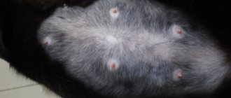
What do bumps on a cat’s nipples indicate, and how to deal with them?
A fluffy meowing lump brought into the house is not just a faithful friend, but also a considerable responsibility that falls on the shoulders of the owners. Just like people, cats get sick, and it often happens that the disease is so serious that you have to see a doctor and undergo a long course of treatment. Older animals are especially susceptible to disease. One of the symptoms indicating a dangerous illness is that the cat has a lump on the nipple, which causes concern among the owners. What disease does this sign indicate? How to properly respond to the appearance of a neoplasm?
Who is susceptible
Older cats that are not neutered or late neutered (after three years) are most susceptible to this disease. The greatest risk of detecting a mammary tumor is in a cat over 10 years of age.
This disease is rare in cats - 8 times less common than in cats. There is also a breed predisposition - Siamese cats have a much higher risk of developing mammary tumors, especially at a young age, and representatives of the Persian breed are also slightly more susceptible to the disease than others.
Preventive measures
A preventive measure that provides almost one hundred percent protection against this disease is sterilization of the cat in childhood, even before the onset of the first heat, and castration of the cat before the age of 1 year.
Regular prevention consists of the following:
- when playing with a cat or male cat, it is necessary to conduct an incidental examination of the mammary glands;
- the animal is subjected to an annual preventive examination at a veterinary institution after reaching 10 years of age;
- hormonal medications that regulate the cat’s sexual activity and delay the onset of pregnancy should be abandoned;
- It is equally important to follow the principles of proper and nutritious nutrition for your pet;
- and, finally, it is necessary to promptly treat inflammatory diseases of the genitourinary system of pets.
Breast tumors are a very serious disease. Any person caring for their pet is interested in the question: how long do cats with this disease live? If detected early, their lifespan may not be limited by the disease. In other cases, their average lifespan ranges from 2 to 20 months (if you count from detection).
You should never despair and lose hope for the recovery of your family friend. We must do everything possible and believe, according to the principle: as long as you believe, you live.
Stages of the disease
In cats, there are 4 stages of development of the malignant process (zero is distinguished separately):
- Stage zero - tumor less than 5 mm in size;
- The first stage is a tumor up to 2 cm in diameter without metastasis;
- The second stage is a tumor with a diameter of less than 3 cm, no metastases;
- The third stage is a tumor larger than 3 cm without metastases or less than 3 cm with metastases in regional lymph nodes;
- Stage four - a tumor with a diameter of more than 3 cm with damage to regional lymph nodes; tumor of any size with distant metastases; inflammatory carcinoma.
Prognosis and probable outcomes of the disease depend on its stage: cancer at the zero and first stages is curable in the vast majority of cases, at the second stage the prognosis is often favorable, in other cases it is rarely possible to completely cure a cat, but treatment is carried out in any case in order to prolong life animal.
However, in some cases at the fourth stage, the advisability of surgery and chemotherapy may be questionable; in such cases, maintenance therapy is carried out.
Life expectancy forecast
How long a cat with cancer will live depends on the stage of the disease, the age of the patient, as well as the conditions of keeping and feeding. Veterinary oncologists call the following numbers:
- If the tumor is up to 2 cm in size - about 3 years;
- if more than 3cm - about half a year.
However, if you undergo surgery and see a doctor annually (due to the high percentage of relapses), the chances of your pet having a longer and happier life are much greater.
Benign tumors
Fibroepithelial hyperplasia is a tumor characterized by rapid growth and proliferation of epithelial cells, an increase in the number of ducts in the mammary gland. The stroma can be either loose or dense, structured. The neoplasm tissue consists of the same cells as those of a healthy mammary gland.
These tumors contain progesterone-sensitive receptors. One of the mammary gland packages may be affected, but more often several lobes or even the entire mammary gland are involved in the process. With this disease, the mammary glands usually significantly increase in volume, which is noticed by the owners of the animal, and the cat is brought to the veterinary clinic.
Benign neoplasms - symptoms, treatment
It often happens that the anxiety of owners who discover lumps around the cat’s nipples is unfounded - the neoplasms do not pose any particular danger to the animals. Veterinarians warn that it is difficult and sometimes impossible to independently determine what exactly triggered the appearance of growths without special research, so in any case you cannot do without help. The following symptoms indicate benign neoplasms:
- the lump is motionless, when pressed with a finger it remains in place;
- when touched or pressed, the cat does not show concern;
- the tumor slowly increases in size and does not spread to other parts of the body;
- the growth is of an even shade, does not have a vascular network;
- the animal sleeps well, moves actively, does not lose weight or appetite.
Typically, conservative treatment is used in such cases, although this may depend on many factors. The veterinarian takes into account the pet’s age, general health, and the size of the tumors. Treatment is carried out at home, but under the mandatory supervision of a specialist who determines how effectively the disease responds to medication.
Malignant tumors
Carcinoma is the most common type of mammary tumor in cats. These tumors, as a rule, are very aggressive, characterized by rapid growth, and can metastasize to the lymph nodes, lungs and pleura already in the early stages of the disease.
Most often they do not contain estrogen and progesterone receptors. The most common carcinomas found in cats are tubulopapillary and solid carcinomas.
Sarcoma is a rare malignant tumor of the mammary gland in cats; it is the most aggressive, consists of poorly differentiated cells, and is characterized by very rapid growth.
What does pain indicate?
For most women, burning and mutual dull pain in the chest is a consequence of premenstrual syndrome. A burning sensation and pain in the chest is observed at the beginning of pregnancy.
Lumpiness and tenderness in the breasts occurs when milk accumulates in the mammary glands of a nursing mother. When cracks form in the nipples, there is a risk of infection and the development of mastitis.
Localized pain and burning in the chest can be the cause of the development of benign formations. This pain intensifies in a certain position of the body.
Burning and pain in one side of the chest may indicate the development of cancer. Tumor formations of the breast are most often located in the upper outer quadrant.
Establishing diagnosis
Various types of diagnostics are used to make a diagnosis and determine the best treatment regimen. Any appointment with a veterinarian for a cat suspected of having a mammary tumor begins with a history taking.
The specialist must find out the time of appearance and growth rate of the tumor, the number and specifics of the operations performed (first of all, castration is important), as well as everything related to the cat’s reproductive system: the number of births, if any, taking hormonal medications. The owner is asked about the cat’s recent condition, complaints, the presence of concomitant diseases, and the peculiarities of keeping the animal.
At the initial appointment at the veterinary clinic, the cat must also be examined. Breast tumors are usually easily detected by palpation; they look like a dense formation with clear boundaries, easily moving relative to the skin and muscles, but some tumors can involve surrounding tissues in the pathological process.
The diffuse form of mammary cancer is much more difficult to determine based on the initial examination: most often, when examining an animal, you can notice inflammation with unclear edges, which may be similar to other diseases of the mammary gland. Lymph nodes must be palpated and examined; their involvement in the pathological process is an important diagnostic and prognostic factor.
When the first two pairs of mammary gland packets are affected, the axillary lymph nodes react, the last pairs - the inguinal lymph nodes. A careful and detailed examination of the skin is also important - the presence of ulcers, skin metastases, and involvement of the skin lymphatic ducts significantly worsens the prognosis for such a disease.
What symptoms require immediate medical attention?
If the following signs appear, you should consult a specialist:
- Aching pain in the chest and mammary glands.
- Well palpable lumps in the chest.
- Stagnation of breast milk during lactation, changes in its color and smell.
- Formation of weeping wounds on the skin.
- Unpleasant-smelling nipple discharge.
- A sharp change in the size and shape of the mammary glands.
- Itching of the nipples and any change in their appearance.
You should not postpone a visit to the doctor if breast swelling appears after the installation of implants. Patients with chronic sexual diseases must undergo routine examinations. Regular visits to a mammologist are recommended for women with a family history. Trauma to the chest can lead to the formation of cysts or tumors.
Instrumental research methods
X-ray examination
The most common and routinely used method of instrumental diagnosis of breast tumors. The study is usually carried out immediately after the animal goes to the veterinary clinic, as well as subsequently to monitor the success of treatment.
X-ray allows you to assess the presence of large metastases and damage to other organs and tissues by the malignant process. At least two projections are always made (three are often recommended): one or two lateral (lateral, on the left and right side) ventrodorsal (direct).
Types and diagnosis of tumors
The tumor is a lump, the size of which varies from a small nodule to a tennis ball. It can be of two types:
- Benign (cyst, adenoma) - does not pose a threat to life, but can cause significant inconvenience to the pet if it reaches a large size. It usually has a regular shape, is separated from nearby tissues by a capsule, is located only on one of the glands and grows slowly, without penetrating into nearby tissues, but only moving them. But such compactions require constant monitoring, as they can develop into malignant formations.
- Malignant (carcinoma, sarcoma) is a deadly neoplasm that provokes the development of metastases and grows into neighboring tissues and organs. It usually has an irregular shape and a lumpy, nodular surface.
Most often, AMF occurs in unsterilized elderly individuals (over 7 years old), as well as in those who have suffered trauma to the mammary glands or suffer from hormonal imbalances. Moreover, benign formations account for 10-15% of cases, while the rest belong to the malignant category.
A neoplasm can be diagnosed using palpation, x-rays or ultrasound. But its type and nature can only be determined through cytological or histological examination of material taken from the tumor using a syringe. Additionally, a general blood test is taken and other studies are carried out to assess the animal’s health and determine further actions that will be appropriate in a particular case.
Only a specialist can determine breast cancer after conducting a series of laboratory tests. The most common diagnostic method is tumor biopsy. This procedure is carried out carefully to minimize the possibility of damage to healthy tissue. Experienced specialists also advise performing a biopsy of nearby lymph nodes, which are a common site for the development of metastases. If the disease is identified in a timely manner, you can try to cope with conservative treatment methods.
For an accurate diagnosis, a specialist may prescribe:
- ultrasound and x-ray (will help to see existing metastases in other organs);
- urine and blood analysis;
- tumor puncture.
Unprofessional taking of a biopsy can provoke the occurrence of metastases in the pet’s body.
Additional Methods
Often, a diagnostic method preferable to radiographic examination is computed tomography (CT). It allows you to detect a larger number of metastases (on average, CT is four times more accurate).
In some cases, a cat may be indicated for magnetic resonance imaging (especially if the first pair of mammary glands is affected and damage to the central nervous system is suspected).
Cytological examination
This study is based on an analysis of the cellular composition of the contents of tumors and adjacent tissues. The material for it is selected by puncturing the tumor with a needle in several places. Cytological examination is less invasive than, for example, histological examination, but its accuracy is much less, so it is not used routinely for all cats with mammary tumors, but only when there is an indication for its use.
Most often, it is used to differentiate breast tumors from tumors of surrounding tissues, such as skin, to detect relapse of breast cancer after treatment, and to determine the presence of metastases in certain areas, including enlarged lymph nodes. Cytological examination also makes it possible to differentiate inflammatory carcinoma (a tumor that invades the lymphatic vessels of the skin and causes inflammation) from other breast diseases that are not oncological (for example, purulent mastitis).
Which doctor should I contact for examination?
You should know that mastopathy and mastitis can cause the development of cancer. Regular examinations by a specialist will help avoid dangerous pathologies. If you have pain or lumps in the mammary glands, you should contact a mammologist. After an accurate diagnosis, he will prescribe appropriate treatment.
For preventive purposes, it is recommended to visit a mammologist annually. In case of complicated heredity, due to individual characteristics, in the presence of concomitant pathologies, additional visits to the doctor are required.
Treatment methods
Based on the diagnostic results, the type of tumor, stage of the disease, prognosis and further treatment plan are determined. The main method of treatment is surgery.
It is indicated for almost any cat with a mammary tumor, except animals with inflammatory carcinoma, diffuse cancer, and those at risk of metastasis in non-adjacent tissues.
Lumpectomy
This is the simplest and least invasive option of the operation, which consists of removing one tumor node. In this type of surgery, the skin is cut, and the tumor is separated from surrounding tissue and removed.
More often, surgery is used for diagnostic purposes—sending the tumor for histological examination—than for cancer treatment.
Simple mastectomy
This type of operation involves removing a section of glandular tissue along with the nipple and adjacent skin. It is not performed for excision of malignant tumors; it is used for skin lesions or damage to the mammary gland not related to cancer.
Unilateral mastectomy
After a diagnosis of mammary tumor in cats is made, this operation is performed most often. This type of operation involves the complete removal of the entire ridge of mammary glands along with the adjacent skin, subcutaneous fatty tissue and regional lymph nodes - inguinal, axillary (if they have metastases in the chest area, allowing them to be removed, or they are enlarged), additional axillary.
Sometimes the underlying deep pectoral muscle is also removed to prevent tumor invasion into it. If both ridges are affected, two operations are performed sequentially to completely remove the mammary gland. A radical operation such as a unilateral mastectomy often allows the cat to live for several years without recurrence.
Bilateral mastectomy
Indications for bilateral mastectomy occur much less frequently than for unilateral mastectomy. This surgical intervention is performed if the tumors located in both ridges are fused with each other and removal of one ridge will lead to damage to the tumor tissue.
Such an intervention is as invasive as possible - both ridges are removed immediately along with the skin, underlying fatty tissue and all regional lymph nodes. The volume of tissue removed is very large, lymphatic vessels are damaged; Due to the large area of skin removed, the tissues in the suture area are stretched, causing significant discomfort in the animal.
Chemotherapy
The main goal of chemotherapy is to prevent relapses of the disease and reduce the risk of further metastasis. The method is aimed at destroying small tumor cells and metastases and is usually used in the postoperative period (adjuvant chemotherapy), but can also be used without surgery, especially in diffuse forms of cancer, helping to transform it into a nodular form. Chemotherapy is definitely indicated at stages 3-4 of the disease, if the tumor is invasive (grows into surrounding tissues), inoperable, or is a sarcoma.
So far, there is no single protocol for chemotherapy and various combinations of drugs are used, most often including doxorubicin. Typically, the animal undergoes several treatment sessions, the frequency is once every three weeks. Before each administration of drugs, a general clinical (and sometimes biochemical) blood test is required.
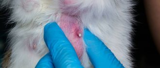
Symptoms of dangerous diseases requiring immediate veterinary intervention
The most dangerous diseases for pets are cancer and mastopathy, which have a common sign - the cat has a lump near the nipple. In the absence of timely treatment, diseases usually result in the death of the animal. Diagnosing the disease is difficult, but careful regular examination of your cat will help you recognize dangerous signs. The following symptoms should alert owners:
- swollen, dark red nipples;
- when you lightly press the nipple, the animal behaves restlessly and feels pain;
- the pet refuses to eat food;
- hair falls out near the affected area, weeping erosions appear;
- the animal quickly loses weight.
With the development of cancer, lumps near the nipples can spread to other parts of the body - head, back, limbs. Treatment in such cases may be useless, so it is better not to hesitate to visit a veterinarian.
Prevention
The main measure to prevent the occurrence of mammary tumors is timely castration of the cat. When surgery is performed before the first estrus, the risk of developing a mammary gland tumor is subsequently reduced by 25 times, and before the third estrus - by 7 times.
Castration performed at a later age does not in any way prevent the occurrence of a mammary gland tumor; it is also useless when removing a tumor, since it does not in any way affect the possibility of relapses (the exception is fibroepithelial hyperplasia, in which removal of the ovaries is the main method of treatment, removing from the body hormone-producing organ).
Back
Postoperative period: animal care
In order for the operation to produce positive results, it is necessary to properly organize postoperative care for the animal:
- Create a comfortable environment for your cat so that she can recover from surgery in complete peace.
- Feed her only those foods that are recommended by your veterinarian. Leave all the “goodies” and rewards for later.
- Treat the wound several times a day and only with the products prescribed by a specialist.
- Remember to give your cat medications in exactly prescribed doses and on schedule.
- To prevent the seam from coming apart, use a special tightening blanket.
If the animal's condition worsens or its stitches come apart or begin to rot, immediately take the animal to the veterinarian!
In the first few days after surgery, your cat will need to have blood and urine tests taken to understand how the postoperative period is going. In the following months, the doctor recommends that you come to the clinic again for tests to exclude a relapse.