Owners, when bringing a sick cat to a veterinary clinic, are almost constantly faced with the fact that veterinary specialists, in addition to a clinical examination of the sick cat, examine blood samples from the sick animal in order to make an accurate diagnosis of the disease. Modern effective treatment of a sick animal cannot be based only on examining the sick animal.
If not so long ago no one seriously took the possibility of conducting a study to assess the condition of an animal using a clinical and biochemical blood test in the laboratory, today every self-respecting veterinary specialist, and even more so a modern veterinary clinic, considers it the norm to do a blood test on their patients.
A blood test is one of the most informative ways to examine pets. By conducting a blood test, a veterinarian has the opportunity not only to confirm or refute the clinical diagnosis, but also to identify hidden pathological processes in the animal’s body (subclinical pathology) that have not yet produced characteristic symptoms.
The main types of research are general clinical and biochemical blood tests. Using the CBC, you can establish the basic composition of the blood. Biochemical research allows a veterinary specialist to more accurately judge the functional state of the animal. Before performing an operation or other intervention, a blood test is a mandatory norm.
How to properly prepare an animal for blood donation
- Before taking blood for analysis, the cat is not fed for 10-12 hours, and there must be free access to water.
- We limit the animal’s physical activity (refusal of games, etc.).
- Taking any medications is prohibited. If your cat regularly takes any medications as prescribed by your veterinarian, consult about stopping them.
- Before taking blood, therapeutic procedures, ultrasound, x-rays, and massage are unacceptable.
Why is regular testing necessary?
For some reason, people ignore blood tests for both themselves and their pets. Today we will talk about the second case.
Any veterinarian or entire veterinary clinic cannot begin to diagnose the problem without having recent blood tests on the animal. Every day, owners lose precious minutes of time that could be spent helping their pet.
Having a blood test in front of him, the veterinarian is able to immediately confirm the presence of a particular disease, or begin treatment of your friend’s maturing sore.
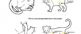
The procedure for taking blood for analysis
Blood is taken from the vein of the animal. To do this, the cat is placed on its side, and if the cat resists, it is secured in a special veterinary bag. Blood is taken from the front paw by shaving off a small area of fur. The injection site is treated with a disinfectant solution, a needle from a disposable syringe is inserted into the vein, blood in the amount of 2 ml is drawn into a test tube with heparin or sodium citrate.
What blood tests are performed in veterinary clinics?
In modern veterinary clinics, two laboratory blood tests are performed:
- General or clinical.
- Biochemical.
General blood test for a cat
A general blood test based on the number and condition of formed blood elements shows the health status of the cat’s body. When performing a general blood test, parasites such as dirofilaria (dirofilariasis), hemobartenella can be detected in a cat’s blood.
What indicators does the clinic’s veterinary specialist receive when conducting a general blood test:
- Hematocrit
- Hemoglobin.
- Average content and concentration of hemoglobin in an erythrocyte.
- Color indicator.
- ESR (erythrocyte sedimentation rate).
- Red blood cells.
- Leukocytes.
- Neutrophils.
- Lymphocytes.
- Eosinophils.
- Monocytes.
- Platelets.
- Basophils.
- Myelocytes.
Biochemical blood test for a cat
Biochemical blood tests allow veterinary specialists to identify subclinical (hidden) cat diseases. A biochemical blood test allows you to determine the functioning of the body’s enzymatic system and provide information about damage to a particular organ in a cat.
A biochemical blood test for a cat includes enzymatic, electrolyte, fat and substrate indicators.
Basic biochemical parameters:
- Glucose.
- Protein and albumin.
- Cholesterol.
- Bilirubin is direct and total.
- Alanine aminotransferase (ALT).
- Aspartate aminotransferase (AST).
- Lactate dehydrogenase.
- Gamma glutamyl transferase.
- Alkaline phosphatase.
- a –Amylase.
- Urea.
- Creatinine.
- Calcium.
- Magnesium.
- Creatine phosphokinase.
- Triglycerides.
- Phosphorus, inorganic.
- Electrolytes (potassium, calcium, sodium, iron, chlorine, phosphorus).
What tests are performed in veterinary clinics?
To ensure that your pet is always healthy and there is no threat of disease, it is recommended to carry out analysis regularly once a year. If the cat has been ill, then after six months it is recommended to donate blood again.
There are three different approaches to identifying diseases using analysis.
- An integrated approach involves studying the maximum amount of data. Prescribed only when previous studies have not made it possible to make an accurate diagnosis.
- The partial approach is used to exclude diseases with similar symptoms.
- Single is used to check the established diagnosis.
Regular visits to the veterinary clinic will save your pet from many diseases.
When diagnosing, the following types of blood tests can be used:
- general. Determines the overall picture of the pet’s health. Helps identify inflammatory processes, allergies and other diseases;
- biochemical analysis. Helps establish a diagnosis with inaccurate symptoms. Allows you to determine exactly where the problem is and how affected the internal organs are;
- hormonal analysis. There are a large number of endocrine (hormonal) diseases in animals, so analysis of the level of hormones in the blood is an integral step in the diagnosis of these diseases.
But the main ones are general and biochemical analysis.
Indicators of blood tests obtained and their characteristics
Each indicator of a blood test shows the functioning of individual organs or entire systems, while the veterinarian takes into account not only each data separately, but also the relationship to each other.
Hematocrit is a conditional indicator showing the ratio of all formed elements of blood to its volume, i.e. determines the thickness of blood. Shows how much blood can carry oxygen.
Hemoglobin is a protein contained in red blood cells that ensures the movement of oxygen and carbon dioxide throughout the animal’s body.
The average concentration of hemoglobin in an erythrocyte shows in percentage terms how much erythrocytes are saturated with hemoglobin.
The color (color) blood index shows how much hemoglobin is contained in red blood cells in relation to the normal value.
ESR is an indicator that determines the presence of an inflammatory process in the body.
Erythrocytes are red blood cells that take part in tissue gas exchange and maintaining acid-base balance. It’s bad when test results go beyond the norm, not only in the direction of decrease, but also of increase.
Leukocytes (white blood cells) - indicate the state of the animal’s immune system. Leukocytes include lymphocytes, neutrophils, monocytes, basophils and eosinophils. For a veterinarian, the relationship between these cells is of diagnostic importance.
- Neutrophils are responsible for destroying bacteria in the blood.
- lymphocytes - speak of a general indicator of immunity.
- monocytes - perform the function of destroying foreign substances in the blood,
- caught in the blood.
- eosinophils - perform the function of fighting allergens.
- basophils - together with other leukocytes, help the body recognize and identify foreign particles that have entered the blood.
Platelets are blood cells responsible for blood clotting. In addition to this function, they are responsible for the integrity of blood vessels. Both high and low levels of them are dangerous for the body.
Myelocytes are located in the bone marrow and normally should not be in the blood.
Leukocyte formula
This percentage of different types of white blood cells is determined by counting a stained blood smear under a microscope.
Neutrophils
A type of white blood cell responsible for creating a response to inflammation, destroying bacterial infections in the blood, dead and damaged cells. Form nonspecific immunity. The age of a neutrophil is estimated by the shape of the nucleus. An increase in the proportion of young cells may indicate the severity of the inflammatory reaction.
Norm: 60-75% for mature neutrophils (segmented) and up to 6% of young (band) neutrophils.
Lymphocytes
They are the main cells of the immune system. They form specific immunity by producing antibodies.
Norm: from 18 to 25%.
Eosinophils
They form antiparasitic immunity and participate in allergic reactions.
Norm: from 1 to 5%.
Basophils
Together with other leukocytes, they recognize foreign particles in the blood. Their presence in the blood may indicate an immediate acute allergic reaction (anaphylactic shock).
Norm: from 0 to 1%.
Blood chemistry
Glucose is an informative indicator indicating the functioning of a complex enzymatic system in the body, including individual organs (liver, pancreas, kidneys). Glucose metabolism in the body involves 8 different hormones and 4 complex enzymatic processes. A disorder is considered to be either high or low blood sugar levels in a cat.
Total protein in the blood reflects the correctness of amino acid metabolism in the body. Shows the total amount of all protein fractions - globulins and albumins. Proteins in an animal’s body take part in almost all life processes of the body. For specialists, both their increased and decreased amounts are important.
Albumin is the most basic blood protein produced by the liver. Albumin in a cat’s body performs a large number of functions (transporting nutrients, maintaining reserve reserves of amino acids for the body, maintaining osmotic blood pressure, etc.).
Cholesterol is a structural component that ensures the strength of cellular structures and takes part in the synthesis of many vital hormones. Veterinary experts judge lipid metabolism in a cat’s body based on cholesterol levels.
Bilirubin is a bile pigment found in the body in two forms - direct and indirect. Indirect bilirubin is formed in the blood as a result of the breakdown of red blood cells, and bound (direct) bilirubin is converted from indirect bilirubin in the liver. Bilirubin shows the functioning of the hepabiliary system (biliary and hepatic). Refers to “color” indicators i.e. when its content in the body is increased, the tissues turn yellow (jaundice).
Alanine aminotransferase (ALT, ALaT) and aspartate aminotransferase (AST, ACaT) are enzymes produced by liver cells, heart cells, red blood cells and skeletal muscles. It is an indicator of the functions of these organs or departments.
Lactate dehydrogenase (LDH) is an enzyme involved in the final step in the breakdown of glucose. Veterinary LDH specialists monitor the functioning of the cardiac and hepatic systems, and also judge the risks of tumor formation.
ɤ-glutamyltransferase (Gamma-GT) – in combination with other liver enzymes, provides insight into the functioning of the hepabilary system, pancreas and thyroid glands.
Alkaline phosphatase is determined to monitor liver function.
ɑ-Amylase – produced by the pancreas and parotid salivary gland. Their work is judged by its level, but always in conjunction with other indicators.
Urea is the result of protein processing, which is excreted by the kidneys. Some remains circulating in the blood. Using this indicator, you can check your kidney function.
Creatinine is a muscle byproduct excreted from the body by the renal system. The level fluctuates depending on the condition of the urinary excretory system.
Potassium, calcium, phosphorus and magnesium are always assessed in complex and in relation to each other.
Calcium is involved in the conduction of nerve impulses, especially through the heart muscle. By its level, you can determine problems in the functioning of the heart, muscle contractility and blood clotting.
Creatine phosphokinase is an enzyme that is found in large quantities in the skeletal muscle group. By its presence in the blood, one can judge the work of the heart muscle, as well as internal muscle injuries.
Triglycerides in the blood characterize the functioning of the cardiovascular system, as well as energy metabolism. Usually analyzed in conjunction with cholesterol levels.
Electrolytes are responsible for membrane electrical properties. Thanks to the electrical potential difference, cells pick up and execute commands from the brain. In pathologies, cells are literally “thrown out” from the nerve impulse conduction system.
What problems are most common?
What deviations from the norm occur most often? In cats, as in dogs, especially if the owner does not feed his pet properly, high or low cholesterol levels occur. What does each option mean?
If cholesterol exceeds the norm, this may indicate problems with the liver and lack of thyroid function. Low cholesterol may indicate a malignant neoplasm (in rare cases) or hepatopathy, that is, a certain list of liver diseases.
Pet owners also often encounter problems with glucose. Elevated glucose levels may mean your pet has pancreas or liver problems. Low glucose indicates possible starvation of the animal, an overdose of insulin or severe poisoning.
It is important to understand that these are just examples of what can happen. In other cases, there may be more rare diseases. We have mentioned the diseases that occur most often.
Norms of blood tests in cats
- General (clinical) blood test
- Blood chemistry
| The name of indicators | Units | Norm |
| Ø hematocrit | % (l/l) | 26-48 (0,26-0,48) |
| Ø hemoglobin | g/l | 80-150 |
| Ø average hemoglobin concentration in an erythrocyte | % | 31-36 |
| Ø average amount of hemoglobin in a red blood cell | pg | 14-19 |
| Ø color indicator; | 0,65-0,9 | |
| Ø ESR | mm/hour | 0-13 |
| Ø red blood cells | million/µl | 5-10 |
| Ø leukocytes | thousand/µl | 5,5-18,5 |
| Ø segmented neutrophils | % | 35-75 |
| Ø band neutrophils | % | 0-3 |
| Ø lymphocytes | % | 25-55 |
| Ø monocytes | % | 1-4 |
| Ø eosinophils | % | 0-4 |
| Ø platelets | million/l | 300-630 |
| Ø basophils | % | — |
| Ø myelocytes | % | — |
Norms of blood tests in cats
General (clinical) blood test
Blood chemistry
| The name of indicators | Units | Norm |
| Ø glucose | mmol/l | 3,2-6,4 |
| Ø protein | g/l | 54-77 |
| Ø albumin | g/l | 23-37 |
| Ø cholesterol | mmol/l | 1,3-3,7 |
| Ø direct bilirubin | µmol/l | 0-5,5 |
| Ø total bilirubin | µmol/l | 3-12 |
| Ø alanine aminotransferase (ALT) | U/l | 17 (19) -79 |
| Ø aspartate aminotransferase (AST) | U/l | 9-29 |
| Ø lactate dehydrogenase | U/l | 55-155 |
| Ø ɤ-glutamyltransferase | U/l | 5-50 |
| Ø alkaline phosphatase | U/l | 39-55 |
| Ø ɑ-Amylase | U/l | 780-1720 |
| Ø urea | mmol/l | 2-8 |
| Ø creatinine | mmol/l | 70-165 |
| Ø calcium | mmol/l | 2-2,7 |
| Ø magnesium | mmol/l | 0,72-1,2 |
| Ø creatine phosphokinase | U/l | 150-798 |
| Ø triglycerides | mmol/l | 0,38-1,1 |
| Ø inorganic phosphorus | mmol/l | 0,7-1,8 |
| Ø Electrolytes | ||
| Ø potassium (K+) | mmol/l | 3,8-5,4 |
| Ø calcium | mmol/l | 2-2,7 |
| Ø sodium (Na+) | mmol/l | 143-165 |
| Ø iron | mmol/l | 20-30 |
| Ø chlorine | mmol/l | 107-123 |
| Ø phosphorus | mmol/l | 1,1-2,3 |
Preparing for the examination
Before collecting biomaterial, the animal should not undergo an ultrasound procedure.
Blood tests in cats are carried out after certain preparation rules. Before surrendering, you should try to remove your pet from active games. It is not advisable to take medications on the eve of the test. Blood sampling from a cat is carried out in the morning strictly on an empty stomach. Before the procedure, ultrasound, X-ray examinations, and massage are prohibited.
The preparation rules are the same for both general analysis and biochemical analysis. Failure to comply will result in unreliable results.
Blood tests in cats (transcript)
All deviations in indicators are considered in complex and in relation to one data to another within the same results from the study of one blood sample. Only an experienced veterinarian should interpret blood tests (results).
General (clinical) blood test
| The name of indicators | Promotion | Decline |
| 1. Hematocrit |
|
|
| 2. Hemoglobin |
|
|
| 3. ESR |
|
|
| 4. Red blood cells |
|
|
| 5. Leukocytes |
|
|
| 6. Segmented neutrophils (mature) |
|
|
| 7. Band neutrophils (immature) |
| |
| 8. Lymphocytes |
|
|
| 9. Monocytes |
|
|
| 10. Eosinophils |
| |
| 11. Platelets |
|
- incoagulability of blood. |
| 12. Basophils | hemoblastoses | Normally absent |
| 13. Myelocytes |
| Normally none. |
Blood chemistry
| The name of indicators | Promotion | Decline |
| 1. Glucose |
|
|
| 2. Protein |
|
|
| 3. Albumin |
|
|
| 4. Cholesterol |
|
|
| 5. Direct bilirubin |
| |
| 6. Total bilirubin |
|
|
| 7. Alanine amino-transferase (ALT, ALaT) |
| |
| 8. Aspartate aminotransferase (AST, ASat) |
|
|
| 9. Lactate dehydrogenase |
| |
| 10. ɤ-glutamyl transferase |
| |
| 11. Alkaline phosphatase |
|
|
| 12. ɑ-Amylase |
|
|
| 13. Urea |
|
|
| 14. Creatinine |
|
|
| 15. Calcium |
|
|
| 16. Magnesium |
|
|
| 17. Creatine phosphokinase |
| |
| 18. Triglycerides |
|
|
| 19. Inorganic phosphorus |
|
|
| 20. Electrolytes | ||
| · potassium |
|
|
| · sodium |
|
|
| · iron |
|
|
| · chlorine |
|
|
| · phosphorus |
|
|