Causes and types of corneal damage
The cornea of the eye is very thin and practically unprotected, except for the movable eyelid. It can be damaged accidentally or intentionally, as a result of an accident or failure to follow safety precautions when wearing contact lenses and glasses.
The main causes of corneal damage are divided into external and internal. External ones occur due to foreign objects, substances, thermal or wave effects entering the eye. Internal - due to congenital or acquired diseases that affect the condition of the cornea and provoke a violation of its integrity.
External causes of corneal damage are considered the most numerous and varied. They are divided into several groups depending on the type of effect on the cornea:
- Mechanical injuries of the cornea
. They are sharp and dull. An acute violation of integrity is characterized by the fact that a foreign object penetrates through the layers of the cornea and reaches the vitreous body and the internal structures of the eyeball. Most often this is a scratch, puncture or cut of the cornea. Blunt eye injuries are expressed by a bruise of the cornea of the eye, which is not accompanied by radical dissection of the tissue. - Thermal damage to the cornea
is a burn of the outer shell by a hot object, excessively heated air, or steam. In rare cases, thermal burns of the eye shell are recorded due to its contact with objects that are too cold. - Chemical damage
. This cause of corneal damage is considered the second most common after mechanical trauma. It occurs when drops of acids or alkalis enter the eye. - Radiation injury to the cornea
, which occurs when unprotected visual contact with excessively bright light (so-called “bunnies”), magnetic, electrical or radiation waves.
Ophthalmologists call primary infections of the eye shell another cause of external corneal damage.
Internal processes leading to the development of pathology do not have a clear classification. These include:
- disruption of metabolic processes in the body or directly in the tissues of the eye, which cause thinning of the membrane, detachment of its outer layer from the membrane;
- dry mucous membrane due to high load on the organs of vision, wearing contact lenses, allergies or the functioning of the lacrimal glands, which provokes premature accelerated wear of the outer layer of the cornea;
- autoimmune thinning of the cornea, during which protective cells attack the tissue of the stratum corneum;
- a genetic anomaly affecting the formation of collagen in the body, as a result of which the cornea loses its firmness and elasticity and becomes brittle;
- instability of intraocular pressure, which causes a loss of strength of the cornea, provokes micro-tears that can become a gateway to infections;
- age-related changes.
They all have the same nature - they cause thinning or a decrease in the protective properties of the eye membrane. Such reasons often provoke erosion, detachment or rupture of the cornea.
Unlike external causes, which appear in one eye, internal ones can affect both eyes simultaneously or sequentially.
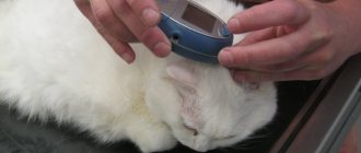
What eye injuries can a cat have?
Bruising in a pet's eye can occur as a result of exposure to sharp or blunt objects. If the damage was caused by blunt objects, then they are safer, because in this case the eyeball does not suffer. If the wound is caused by sharp objects, then the following may occur:
- displacement of the eye lens;
- severe opacities in the anterior chamber of the eye;
- damage to bone tissue near the eye socket;
- detachment of the eye cornea;
- profuse bleeding.
Most often, symptoms of eye injury are observed in the spring, when cats go into labor. In addition, such damage is often observed in pets that live near major highways. Small stones can jump out from under the wheels of passing cars and harm the animal.
However, the damage is not in all cases mechanical in nature. Sometimes injuries occur due to excessive care of the owner, who gives his pet strong veterinary drugs without first consulting a specialist. For example, unnecessary or inappropriate antibiotic therapy can cause complete loss of vision or hearing. If your pet's condition worsens, it should be shown to a veterinarian as soon as possible.
Main symptoms
There are many nerve receptors on the surface of the cornea, which is why the slightest impact on it is accompanied by severe discomfort. This protective reaction helps reduce the duration of traumatic contacts and protects against penetrating damage to the cornea.
Characteristic symptoms of injuries and damage to the outer shell of the eye are:
- intense lacrimation;
- intolerance to bright light;
- blepharospasm (uncontrolled tension of the eyelid muscles);
- pain that feels like sand in the eyes;
- partial loss of vision, inability to focus;
- redness of the mucous membranes of the eyelids, the appearance of a vascular pattern on the sclera.
With deep penetrating wounds of the eyes, when the destructive process affects the optic nerves, the clinical picture is complemented by intense headache, nausea, and dizziness.
3.Treatment of eye burns at home
After burning your eyes, rinse your eyes well with cool water. It's done like this
: Fill the sink with water. Place your face in the water and then open your eyes so that the water gets into all parts of the eye.
To manage pain and fever, try over-the-counter medications—acetaminophen (Tylenol), nonsteroidal anti-inflammatory drugs (ibuprofen-based), aspirin.
If you think that you managed to cope with the burn on your own and there was no serious damage to your eyes, still carefully monitor the condition of your eyes for several days. If the feeling of irritation, burning and redness persists, you should definitely consult a doctor. If pain increases, blurred vision or signs of eye infection appear, this should be done as quickly as possible. Treatment of an eye burn by a specialist will help preserve vision and eye health.
About our clinic Chistye Prudy metro station Medintercom page!
First aid
Correctly provided first aid is the guarantee that the damage will be stopped and eliminated without significant consequences for health. There is a clear algorithm for performing actions after receiving an injury or damage of another nature. First you need to stop contact of the cornea with the traumatic factor:
- If a foreign body gets into the eye, you must carefully remove it with clean hands, a sterile cloth or a stream of lukewarm water. It is not advisable to use tweezers, as they can further damage the membrane of the eye and mucous membrane. Cotton wool is also not suitable, as it leaves microscopic fibers on the surface tissues.
- If the eyes are damaged by chemicals, rinse them with running water for 10-15 minutes, spreading the eyelids with your hands.
- In case of a thermal burn, it is necessary to cool the eye by applying a cloth soaked in ice water to it.
In case of a penetrating wound, it is strictly forbidden to remove the foreign object yourself. As a result, vision will be lost forever.
After eliminating the source of injury, it is necessary to isolate the eye from the external environment. No matter how badly the cornea is damaged, only sterile gauze should be used to apply a bandage. It is applied as an application, without pressing it to the eye socket. You can secure the tissue with a plaster or a loose bandage made from a wide bandage. To get rid of reactive blinking, the remaining eye should also be closed.
After providing first aid, it is important to take the victim to the hospital as quickly as possible. Its treatment will be carried out by an ophthalmologist-therapist or an ophthalmologist-surgeon, depending on the extent of the damage.
Diagnostics
Trying to find the source of discomfort, the ophthalmologist examines the fundus, membranes of the eye, and the cavity of the upper and lower eyelids.
A slit lamp examination will help find a foreign body in the eye. For internal damage to the eye, an ophthalmoscope, x-ray or ultrasound of the eye is used.
If the grain of sand cannot be seen using a regular examination, then the ophthalmologist uses special eye drops with a dye. This helps to see the location of the speck and remove it in a timely manner.
1 Help with foreign body entry and removal from the eye
2 Help with foreign body entry and removal from the eye
3 Help with foreign body entry and removal from the eye
Treatment
The treatment regimen depends on how severe the damage to the cornea is. For superficial injuries of the eye shell, conservative therapy is used. On the first day, to prevent complications, the following is used:
- an antibiotic or antimicrobial drug in the form of an ointment, most often tetracycline;
- antiseptics (chlorhexidine and its analogues) in the form of solutions for instillation (rinsing) of the eye;
- non-steroidal anti-inflammatory drugs of systemic action in the form of tablets.
To speed up regeneration, eye drops with antioxidants (vitamins A and E), B vitamins, collagen, peptides and amino acids, and hyaluronic acid are used.
When the membrane of the eye is deeply destroyed, radical measures are resorted to, most often corneal plastic surgery (keratoplasty). After recovery, a course of antibiotic therapy is prescribed, and drugs with antihypoxic and anti-inflammatory effects are used to prevent neovascularization, scarring and trophic pathologies.
Penetrating lesions on the cornea can only be eliminated surgically. After removing foreign elements that have entered the eye, the displaced elements are repositioned: the iris, lens, etc. If the lens is damaged, it is replaced with a prosthesis. At the end of the operation, nylon purse-string sutures are placed on the eye capsule. They are left for at least 1.5 months. If the cornea is crushed and cannot be collected with sutures, artificial patches or an autograft (a piece of cornea from the surviving eye) is transplanted in its place.
After the operation, victims will undergo long-term rehabilitation, after which the doctor will assess the condition of the eye capsule and, if necessary, prescribe a correction: laser removal of scar defects on the cornea or vision correction.
Removing foreign bodies from the eyes
Removal of a foreign body from the eye should only be carried out by a specialist - this will avoid complications.
Removal of foreign bodies located on the surface of the eye
Grains of sand and debris from the surface of the eye can be removed quite simply with the help of jet rinsing and a swab treated with an antiseptic.
Removal of a foreign body from the conjunctival sac
It is performed after preliminary anesthesia using a tampon. If foreign pieces have been in the eye for some time and are covered with epithelium, they are removed with tweezers or an injection needle. After the foreign body is removed from the eye, bactericidal eye drops are used.
Removing a foreign body from the cornea
Wooden, glass and metal pieces that get on the cornea can cause infiltration and then suppuration within a few hours of their presence. The diagnosis is clarified using diaphanoscopy (examination with an electric light bulb) and biomicroscopy (slit lamp). Then an anesthetic is instilled into the eye, and the foreign body is removed with special instruments.
Removing a foreign body from the eye cavity
Fortunately, foreign bodies entering the eye cavity are quite rare. Such an injury can lead to vitreous opacification, dystrophy and retinal detachment. Foreign bodies inside the eye are removed surgically. A small piece of foreign body in the eye is removed using tweezers or a magnet through an incision in the sclera or cornea.
Helpful Tips:
- To prevent foreign particles from clogging your eyes, be sure to use safety glasses when carrying out agricultural or carpentry work;
- if a foreign object gets into your eye (wood shavings, metal, pieces of glass), do not try to blink and do not rub your eyelid, this will lead to even greater injury to your organ of vision;
- Pulling out a speck on your own can contribute to the appearance of an intraocular infection or the formation of a rough scar on the eye.
Highly professional ophthalmologists at the MedicCity clinic will provide the necessary assistance if a foreign body gets into the eye. We carry out all research and treatment procedures using expert-level equipment! We know how to make your eyes healthy!