Why is it developing?
Spondylosis occurs extremely rarely in kittens; it is often diagnosed in older cats. This disease is characterized by degenerative-dystrophic processes that develop as a result of aging, overload and injury to the spinal column. In addition, discopathy can also develop due to the genetic characteristics of a particular cat breed. For example, the Kurilian Bobtail has a short tail, due to which it has disturbances in the sacral spine. These signs can trigger discospondylitis and other problems associated with intervertebral discs. If a pet has been diagnosed with rickets, the possibility of impaired disc flexibility cannot be ruled out. Subsequent growth of the kitten can cause deformation and protrusion of the fibrocartilaginous formation.
Diseases of the nervous system of cats
Diseases of the central nervous system of cats are very diverse and can be caused either by direct damage to the nervous tissue itself or as a consequence of some systemic lesions. Such as hepatitis or hormonal disorders.
Aggression
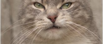
The most common and frequent symptom of many neurological diseases. A behavioral disorder that can be a completely normal condition during periods of “hunting,” pregnancy or lactation. This problem is solved by hormonal therapy or sterilization. In most cases, periods of aggression pass on their own without medical help. But the aggressiveness of cats may be a consequence of any disease, infection in particular. You should be especially wary if the cat's behavior has changed suddenly. This often speaks in favor of rabies. During this period, it is necessary to protect all family members from possible scratches and bites, and create a calm environment around the pet. If these measures do not help, you should immediately contact your veterinarian.
Types of discopathy
First type
This variety is characterized by an abrupt onset and rapid progression. This pathology is rarely diagnosed in cats. Mostly the first type of discopathy develops as a result of infectious processes that make the intervertebral discs hard and prevent healthy movements of the spinal column. During the course of discospondylitis, deformation of the layer between the vertebrae is observed; it gradually shifts and is severely destroyed. The symptoms of type 1 pathologies vary quite a bit. In some pets, only mild stiffness of movement is noticeable. Other cats move very poorly, some can be completely paralyzed.
Second type
Reduced activity and limitation in movement in older animals may be associated with the development of discopathy.
It is similar to the type of discopathy described above, however, unlike it, it begins with a smooth onset. The pathology occurs gradually, so it is difficult for owners to determine its onset. Often observed in elderly pets. The early stages of development are characterized by subtle limitations of movement and decreased activity. If at this stage the disease is not identified and appropriate therapy is not carried out, the cat’s posture will be impaired.
Third type
Often this type is a consequence of serious disorders in the spinal column. The main feature of type 3 pathology lies in the lightning-fast manifestation of symptoms against the background of good health of the pet. The core of the intervertebral disc is pressed into the spinal cord, resulting in a herniation. Cats experience severe pain, which causes them to stop moving. Often there is a lack of coordination and paralysis. Often the third type of discopathy leads to death due to respiratory arrest.
Discopathy: what is it?
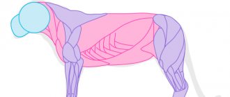
Intervertebral discs are a kind of “cushions” that consist of cartilage and fibrous tissue lying between the vertebrae. There are no intervertebral discs between the first cervical vertebrae. These “pillows” are designed to act as a lubricant and shock-absorbing element. As a result of their degeneration, the shock-absorbing function is reduced, which is fraught with the formation of hernias and compression of the spinal cord.
The term “discopathy” in veterinary medicine means a collective name for diseases characterized by changes in the intervertebral discs: displacement, prolapse into the spinal canal, compression of the spinal cord. It is these pathologies that lead to changes in the cat’s behavior, affecting its motor activity.
Many owners believe that discopathy in cats is almost a death sentence, but this is not true. The disease does not always lead to paralysis. If adequate treatment is started in a timely manner, the prognosis is favorable.
What symptoms indicate pathology?
The owner can suspect problems with the cat's spine by reducing the activity of the pet. Then symptoms such as:
- stiffness of movements;
- lameness.
Constant meowing due to pain and lameness are signs of the pathology.
A cat can limp on two or one paw at once, which is directly related to the segment of damage to the spinal column. The pet begins to meow for no reason and becomes restless. Such symptoms are caused by severe pain that is localized in the back area. If the owner tries to touch it, the cat may begin to hiss and scratch. As discospondylosis progresses, the animal becomes less oriented in space and its gait changes. The possibility of a convulsive syndrome cannot be excluded. If the lumbosacral spine is affected, the emptying process is disrupted.
Diagnosis of lumbosacral radicular syndrome
The use of modern diagnostic methods in dogs with cauda equina syndrome makes it possible to localize the area where the nerve damage occurs. Thus, from the collective concept of “cauda equina syndrome” (lumbosacral syndrome) it is possible to identify several separate pathologies. One place where nerve compression can occur is at the intervertebral foramina, through which the segmental nerve roots pass. Damage at this level leads to the development of radiculopathy (radicular syndrome). Radiculopathies are rarely diagnosed in veterinary medicine. This is due to several factors. Firstly, pathologies that exclusively affect the nerve root are relatively rare. Secondly, topical and visual diagnosis of such lesions can be difficult due to the rare manifestation of symptoms localizing the lesion. Thirdly, such lesions are often “masked” behind orthopedic pathologies in the pelvic limb.
Diagnostic measures
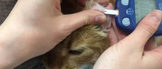
When a pet owner suspects that he or she has developed spondylosis, it is important to contact a veterinarian as soon as possible. The veterinarian will conduct a survey during which he will find out how long ago the cat’s spinal problems began and what symptoms were noticed. Then the doctor begins to examine and palpate the spinal column. Next, the animal is prescribed radiography, with the help of which it is possible to see changes and distinguish discopathy from traumatic spinal injuries. An x-ray image shows the following deviations:
- destruction or narrowing of intervertebral discs;
- degree of spinal cord damage.
If required, radiography is performed with the introduction of a contrast agent.
You can find out the location of a spinal disc lesion in a cat by performing an ultrasound examination.
Sometimes a cat is prescribed a magnetic resonance or computed tomography scan, but not all veterinary clinics are equipped with such expensive equipment. To confirm or refute the presence of an infectious process, a puncture of the cerebrospinal fluid is performed, which allows one to determine its composition. Bacterial culture is also required. To accurately detect the area of damage to the spinal column, ultrasound is used.
Causes of discospondylitis
Main causes of the disease:
- introduction of infection by hematogenous route from a focus in the body (for example, the primary focus may be chronic infections of the genitourinary system);
- Systemic infectious process (brucellosis, aspergillosis, etc.);
- Foreign body (grass seeds, plant needles and thorns, wood chips);
- Postoperative infection (non-sterile suture material);
- Some immunosuppressive conditions;
There is a breed predisposition (for example, German Shepherds, Bernese Mountain Dogs), but no gender predisposition has been identified.
How is the treatment carried out?
If an animal is diagnosed with discospondylitis or other spinal problems, it will need to be rested. Veterinarians recommend equipping the bed in a warm and secluded place, making it as soft as possible to reduce pain. When discopathy provokes urinary or fecal incontinence, the bed must be equipped with a waterproof diaper and constantly monitor the dryness of the material. Food and water bowls should be moved as close to the cat's resting area as possible.
Treatment is carried out with medications or surgery. If we talk about medications, then a sick pet is prescribed non-steroidal anti-inflammatory drugs and painkillers. The latter can be used before a visit to the veterinary clinic, which will improve the cat’s condition. Often they resort to the help of Papaverine with Diphenhydramine. In addition, a cat with spondylosis is prescribed sedative medications.
When the pathology has been detected in an advanced stage and conservative treatment does not bring the required therapeutic effect, the cat is prescribed surgical intervention. During the process, damaged intervertebral discs can be excised and replaced with artificial prostheses. After the operation, the cat experiences serious relief and can return to its normal lifestyle. If a pet has been paralyzed, in most situations, complete recovery of the body is not possible.
Radiography
X-rays in the lateral and ventrodorsal projections reveal the presence of endplate osteophytes. To visualize lateral endplate osteophytes, left and right oblique ventrodorsal alignments with an inclination of 15–30° may be useful. On radiographs taken in the correct lateral projections, you can try to assess the size and osteophytes of the lateral foramina (except for the last lumbar) lying near the center of the X-ray beam. Radiography does not make it possible to assess the condition of the L7-S1 intervertebral foramina, since there are no placements and projections in which other parts of the vertebrae or pelvic bones are not superimposed on the projection of the foramina. X-rays do not reliably assess the condition of the facet joints. To correctly diagnose narrowing of the foraminal openings, tomographic methods are needed.
Magnetic resonance imaging (MRI)
This is a method of visual diagnosis of soft tissues, the method of choice when examining the central nervous system. To date, diagnosing the central nervous system without MRI is not possible. When examining the lumbosacral region, MRI can reveal lesions such as inflammation (discospondylitis, myelitis); degenerative-dystrophic lesions of the intervertebral disc and the degree of their influence on the nerve endings (damage to the nerve roots during protrusion (Fig. 4) or extrusion (Fig. 5) of the intervertebral disc); neoplasms of vertebral bodies, nerve roots; consequences of injury (integrity of nerve endings and the degree of their traumatization). If it is necessary to assess dynamic compression, the study is carried out in stress settings. Based on the results of MRI diagnostics, the doctor can determine the patient’s treatment tactics and predict the outcome of the disease. MRI in no case excludes CT examination. In most cases of disease, the two methods significantly complement each other, which allows the doctor to make the only correct diagnosis and prescribe treatment.
Computed tomography (CT)
Often, native (without the introduction of a contrast agent) computed tomography is enough to understand the cause of the development of radicular syndrome. In medium to large dogs, it is possible to visualize the spinal conus and cauda equina nerves due to the surrounding epidural adipose tissue.
CT makes it possible to evaluate the shape and structure of bones, the presence of zones of hyperostosis or areas of destruction, narrowing of the spinal canal, the size of the intervertebral foramina, the presence and localization of osteophytes (Fig. 6), displacement of fragments during fractures (Fig. 7), displacement and changes in the section of the spinal cord and nerve roots. In most cases, it is possible to visualize extrusion and protrusion of the lumbar discs.
Stress CT allows one to evaluate vertebral instability and identify dynamic stenoses. Contrast enhances the capabilities of CT in cases where the cause of radicular syndrome is caused by soft tissue. Subarachnoid contrast-enhanced CT (CT myelography) allows visualization of the nerves of the cauda equina and assessment of its compression. Epidural contrast can be used to evaluate root compression in the lateral foramina.
Pictures and videos
Figure 1. “Spinal cord compression”
Figure 2. “Spinal cord compression in the cervical region”
Figure 3. “Stage of intervertebral hernia”
Video 1. “Destabilization of the lumbar vertebrae. Before the operation."
Video 2. “On limb failure in animals”
Neurological problems in the neck area in dogs and cats
This article will discuss pathologies in the cervical spine in domestic animals, which cause varying degrees of neurological disorders, often the leading symptom being pain in the neck.
These problems are less common than, for example, surgical diseases of the musculoskeletal system, but they do exist and must be correctly and timely diagnosed and treated professionally. After all, the success of further treatment depends on a quickly established diagnosis. The problem is that most neurological pathologies in the cervical spine have similar clinical signs, and there are quite a lot of such pathologies. Also, in the initial stages, neurological disorders manifest themselves as lameness, which is typical for diseases of the musculoskeletal system. All this prevents the timely provision of assistance to a sick animal.
Etiology. Pathologies of the cervical spine and spinal cord are divided into:
Degenerative (Wobbler syndrome), damage to the joints of the spine, herniated disc or discopathy;
Developmental anomalies and genetic predisposition (atlanto-axial instability, syringo-, hydromyelia, atlanto-occipital dysplasia, arachnoid cysts, vertebral deformity, spondylosis deformans;
Metabolic and feeding disorders (secondary hyperparathyroidism), vitamin A hypervitaminosis in cats;
Neoplasia (tumors of various nature and localization);
Idiopathic pathologies;
Inflammation and infection (discospondylitis, epidural empyema);
Traumatic (fractures, spondylolisthesis, disc damage, gunshot wounds);
Toxic (botulism, tetanus);
Vascular disorders (fibrocartilaginous embolism, hematoma in the spinal canal, necrosis of epidural tissue, myelomalacia).
Clinical signs. They are very diverse and depend on the degree of involvement of nervous structures in the pathological process. The only sign that all these pathologies have in common is the presence of a neurological deficit in the thoracic and pelvic limbs or one of them. In the acute course of the process, the development of tetraplegia and tetraparesis (paresis of all four limbs) is possible. Also, the following signs can most often be observed (one or more of the above symptoms are noted simultaneously):
the animal is lame on one or both thoracic limbs,
the animal is constrained, less active,
holds his head in a forced position - head down,
impaired coordination of voluntary movement and balance in a standing position (ataxia),
A painful reaction is often expressed when examining the animal, usually when the neck is extended.
owners complain of a changed position of the limb and a change in gait. In this case, the limb(s) are supported on the volar (front surface of the paw) surface.
All these clinical signs should prompt the veterinarian to think about a possible pathology in the cervical spine and conduct additional studies to confirm the diagnosis.
Diagnostics. It is impossible to make an accurate diagnosis of a neurological problem in the cervical spine by collecting only anamnesis and conducting a neurological examination of the animal. After a neurological examination, it is only possible to assume the localization of the process. A very small number of neurological pathologies of the neck can be diagnosed using an x-ray. Therefore, more modern and informative diagnostic methods are required:
Therapeutic techniques
If any of the above symptoms appear, you should immediately take your pet to a veterinarian. A sick cat should be given complete rest, especially carefully isolated from small children. There is no need to play or tease the cat, as any sudden movement is fraught with sudden paralysis. At first, if the symptoms are not too threatening, drug treatment is practiced, the purpose of which is to stop the inflammatory process. But in most cases, this only manages to smooth out the main signs. There is no talk of a complete cure without surgery.
It is highly advisable to prescribe sedatives , which will naturally limit the animal’s activity and eliminate the likelihood of sudden movements. Drug pain suppression is recommended only for cats that feel pain and retain tactile sensation. In other cases, it is not recommended to prescribe painkillers, as they can blur the picture of the course of the disease.
In veterinary practice, there have been many cases where cats, after four to six weeks of complete rest, treated with non-steroidal anti-inflammatory drugs, fully recovered. However, there are also many relapses, especially in the case of animals that have already reached the age of five or seven years.
If no significant improvements are observed throughout the course of drug treatment, or if there is a marked tendency towards worsening the course of the disease, surgery is strongly recommended. The sooner this is done, the greater the chance of a complete recovery of the animal.
What is the essence of surgical intervention for this pathology? In most cases, what is called “surgical decompression” is used. This is a method in which the “prolapsed” part of the spinal disc is partially excised, restoring the outer lining of the organ. Decompression is the only effective treatment when paralysis or para/tetraplegia occurs.
Important! If a cat suddenly loses tactile sensitivity, the operation should ideally be performed within two hours, since after this period the nerve tissue of the spinal cord begins to die. When more than two days have passed since the loss of sensitivity, euthanasia is recommended, since the chance of success of the operation is less than 4%.