If, while petting your cat, you feel a certain growth on the back of her neck, know that you cannot ignore it. But there’s no need to be alarmed prematurely either. Perhaps it's just a lipoma. Lipoma in a cat is one of the most common benign neoplasms in animals, consisting of adipose tissue. It is formed in the subcutaneous tissue and in those internal organs that have a fatty layer. It can penetrate deep into the body, spreading muscles and vascular bundles, to the periosteum. Lipoma usually progresses slowly. It can be removed through a simple surgical operation.
What is lipoma
A lipoma is a soft growth under the epithelium that usually does not cause pain to the cat. But if, as it grows, it pushes aside and compresses muscles and nerve endings, then pain and symptoms of impaired blood flow may appear in the affected areas. It is localized on any part of the body where there is adipose tissue, but more often on the withers or on the stomach. A lipoma may also contain connective tissue, in which case its consistency is denser.
Having discovered such a growth in his pet, the owner must contact a veterinary clinic for an in-person examination, diagnosis and further recommendations for treatment. With these actions he prevents the following unpleasant moments:
- having increased in size, the wen can interfere with the animal’s walking or the functioning of some internal organs;
- Vital vessels and airways in the neck (vertebral artery, trachea) may be compressed;
- sometimes an oncological disease is hidden under the guise of a harmless lipoma;
- lipoma has the ability to degenerate into a malignant tumor (liposarcoma).
The main reasons for the appearance of lumps on the stomach of cats
The causes of a lump under the skin in a cat can be harmless. These include:
- Small balls that form after an injection. They do not pose any danger, since without any treatment they resolve on their own over time.
- Consequences of a wasp or other insect bite.
- Mats on wool. Of course, it’s difficult to mistake them for a bump, but this option also happens.
However, there are more serious types of formations. If after the appearance of one bump new ones begin to appear, then this can be a very bad sign. It is likely that this is a malignant formation. But there is no need to rush with premature species.
What is a wen and what does it look like?
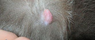
A fatty tumor is a neoplasm that is located between the skin and muscles in loose adipose tissue and can. It feels like the consistency of dough or silicone, i.e. soft and elastic. There is normal mobility and painlessness. In some cases, a lipoma can grow into the muscles - in such cases, the mobility of the tumor decreases and its density increases. Wen, as a rule, grow very slowly, but are capable of growing to significant sizes. Wen can appear anywhere there is loose fatty tissue. At first, the lipoma looks like a small, loose tubercle (bump), inactive in a relatively limited area. Depending on the conditions and condition of the body, the tumor begins to increase. Sometimes it grows to such a size that it involuntarily begins to attract attention to itself and provokes discomfort in the animal. This is explained by the fact that during the growth process the neoplasm can affect the nerve endings in the surrounding tissues.
Wen appears in dogs and cats for two main reasons:
- violation of fat metabolism;
- genetic predisposition.
The main metabolic disorders that provoke the appearance of wen include insufficiently correct functioning of the body's enzymatic system. This means that the animal’s body lacks the required amount or type of enzyme to fully break down accumulated fat into water and internal energy. Those.
Lipomas appear not because too much fat accumulates in the body, but because it is not properly and fully broken down, resulting in the formation of intermediate substances that provoke tumor growth. It is because of this reason that the risk group consists of old animals and overweight animals.
Lipomas are most often found in dogs on the back, sides and tops of the limbs; in cats - on the stomach, in the chest area and along the upper surface of the hind legs.
The diagnosis of lipoma (or wen) is usually made on the basis of fairly specific clinical manifestations. It is also advisable to obtain a tumor sample for additional studies - histology and cytology. Specimens are obtained by aspiration, biopsy, or exploratory surgery. For large lipomas, ultrasound may be indicated.
In any case, no matter where this neoplasm appears, it is absolutely necessary to show your pet to a specialist. It is always necessary to exclude purulent abscesses, cysts and malignant tumors.
Wen in cats is rare. Such skin growths are more common in dogs. In felines, lipid metabolism occurs very quickly, so fatty tumors rarely appear. But there are cases when cats experience metabolic disorders. This becomes In veterinary medicine, such neoplasms are called lipomas. How dangerous are they and how to get rid of them? We will consider these questions in the article.
Can lipomas reappear after surgery? In 70% of cases, no recurrence of tumors is observed. Veterinary experts have found that removing the wen promotes the overall health of the body and prolongs the life of the pet by several years.
The situation becomes somewhat more complicated if the cat suffers from multiple lipomas (lipomatosis). In this case, after the operation it is necessary to review the pet’s diet. You may need to follow a lifelong fat-restricted diet, as well as take medications that normalize lipid metabolism. This will help prevent the recurrence of lipomas.
Wen is found not only in humans, but is characteristic of the Sphynx and other breeds of the cat family. Lipoma in cats, as a rule, is not accompanied by pain and is a cosmetic defect. Such compactions belong to benign tumors consisting of connective tissue. Neoplasms are localized between the epidermis and the muscle corset of the pet. A lipoma can form in the area of the shoulder blades, on the paws or back; less commonly, multiple fatty lumps are found throughout the body.
Methods for treating wen
Wen (lipoma) is always a benign tumor that rarely causes any inconvenience to the animal.
If this neoplasm:
- small sizes,
- painless,
- has no growth dynamics,
- does not affect the life of the pet,
- has no signs of inflammation,
- does not open on its own,
then it is customary to leave it in the format in which it is. You should not smear the wen with anything, pierce it, apply compresses, etc. – there is always a risk of provoking increased tumor growth and/or its degeneration into cancer.
In all other cases, the lipoma can only be removed surgically. Especially when the lipoma is located in a place that is subject to constant mechanical stress (armpit, for example).
All lipomas are divided into:
- non-infiltrative;
- infiltrative (spilled).
The first group of tumors has a clear, limited contour, making them very easy to remove. The second group is characterized by germination into neighboring tissues and is difficult to remove (since damaged neighboring areas also have to be cut out). Also, it is this group of wen that most often degenerates into an oncological (malignant) form.
It is strictly forbidden to independently open (pierce) any seals on the body of pets, because... a non-specialist will never determine for sure what is hidden under one or another subcutaneous lump, and whether it is a wen and benign!
- Before the operation, the animal must be prepared. For at least 12 hours (ideally 24 hours), animals - be it a cat or a dog - are not fed, but water is given upon request. This contributes to a faster effect of general anesthesia and prevents narcotic vomiting. Local novocaine or ledocaine anesthesia is also used (ledocaine is contraindicated in cats due to individual sensitivity and numerous severe side effects, including death). If the wen ruptures or there are signs of bleeding from it, the operation is performed urgently and without fasting preparation.
- Removal of the wen usually occurs through a small incision. If infiltration (growth into surrounding tissues and muscles) was observed, then the fatty tumor will be removed along with the affected areas. If the tumor is large, a drainage must be formed - a place for the drainage of exudate, which will certainly form in the postoperative period. The postoperative wound will also be washed through the drainage.
- Fatty tumors that are located on the border with the main nerve ganglia or branches deserve special attention - operations in such cases are carried out with extreme caution so as not to catch them and not to disrupt the innervation (the passage of nerve impulses).
- If there are any signs of purulent inflammation of a postoperative wound, you should immediately contact a veterinary clinic for help, and not necessarily the doctor who operated (although this is recommended!). It is also necessary to monitor the wound dressing and comply with all the specialist’s requirements.
- The likelihood of recurrence after surgery is minimal. Exceptions are infiltrated lipomas, which are very difficult to remove completely, so cases of the return of wen with this form of neoplasm are quite possible. If there is a genetic predisposition to the formation of lipomas, they may reappear not only in the place where surgical removal was performed, but also in any other place on the animal’s body.
- Depending on the outcome of the operation, the cat/dog may be prescribed a course of antibiotics, painkillers and sedatives.
- If, after a biopsy, a malignant degeneration of the wen is determined, then chemotherapy is prescribed. Typically, a chemotherapy course consists of the following drugs: prednisone, vincristine, cyclophosphamide and doxorubicin. In 71% of all cases, therapy has a positive effect. Schemes and dosages are calculated strictly by a veterinary specialist, based on this specific situation.
We recommend reading: Solvimin Selenium for productive animals - instructions for use
Types of lipomas
Wen in cats can be single in appearance, or can be multiple in nature, especially in the presence of concomitant diseases (deviations in the functioning of the endocrine system). This multiple formation of wen is called lipomatosis.
According to the nature of germination, a lipoma can be simple or infiltrating:
- A simple lipoma has clear boundaries and, as it grows, does not affect the surrounding muscles, so it can easily be surgically removed.
- Infiltrating lipoma grows into the thickness of the muscles and vascular bundles and does not have a clear boundary. It is this type of neoplasm that tends to degenerate.
Varieties
Such formations can be multiple in animals.
Different types of lipoma look different in cats. In pets there is a single lump or multiple formations are possible. The latter is likely in case of secondary diseases in a pet, especially from the endocrine system. If there are several lipomas on the skin at once, then in veterinary medicine the pathology is called lipomatosis. The table shows the types of tumor, taking into account the nature of its germination:
| Type | a brief description of |
| Simple | The boundaries of the lipoma are clearly defined |
| When the wen grows, it does not affect nearby muscles | |
| Easy to remove surgically | |
| Infiltrating | Grows into thick layers of muscle and vascular bundles |
| Without clear contours | |
| It is possible that a lipoma in a cat may degenerate into a cancerous tumor. |
What to do if there is a lump under the skin
If a cat has a lump under the skin, you need to contact a veterinarian who will conduct the necessary tests, establish a diagnosis and prescribe treatment.
Neoplasms are diagnosed using:
- medical examination;
- urine and blood tests;
- Ultrasound;
- endoscopy;
- biopsies.
If oncology or tumors that cause discomfort in the animal are detected, radical therapy is prescribed. In other cases, conservative treatment is acceptable.
If the lump bleeds
Bleeding tumors are life-threatening for your pet, so you cannot do without the help of a veterinarian. To prevent contamination of the wound, the lump is treated with chlorhexidine and bandaged with a sterile bandage. If the formation bleeds, it should not be wetted, cauterized with alcohol-based products, or lubricated with cream without a doctor’s prescription.
Lump with pus
Seals with purulent contents require caution. They should not be pierced or squeezed out to avoid infection and injury to internal organs. If the tumor bursts on its own, the fluid that has accumulated under the skin may break out. The wound is treated with an antiseptic that does not contain alcohol.
A common reason for the appearance of pus-filled bumps is the final stage of cancer. In cats, the mammary glands are affected, and in males, the testes are affected. If the disease is advanced, the veterinarian will prescribe painkillers.
What not to do
Most often, the owner, while stroking the pet, accidentally discovers a wen on the cat. What to do in this case? First of all, you should not try to fight lipoma on your own. Self-treatment can lead to disastrous results.
Under no circumstances should you squeeze or pierce the wen. Any trauma to the tumor can lead to its suppuration or malignancy. It is impossible to remove lipoma at home by any means.
If you find a wen in a cat, you should immediately show your pet to a veterinarian. Only a specialist can distinguish a lipoma from malignant neoplasms and provide qualified treatment.
Reasons for appearance
Conclusions about the causes of the disease in each specific case can only be made by a specialist after a comprehensive examination of the animal. But based on the experience of previous cases, we can say that the most common causes of lipomatosis are the following:
- complicated heredity;
- metabolic disease;
- hormonal imbalances in the body;
- physical inactivity;
- endocrinological diseases;
- elderly age.
It is believed that the growth of adipose tissue in the body is promoted by enzymatic deficiency, namely, a deficiency in the body of the enzyme that breaks down fat - lipase. This statement is supported by the fact that not only obese animals are susceptible to this disease, but also those whose layer of fat in the subcutaneous tissue is small.
What is the problem?
Its structure helps to distinguish a lipoma in a cat from a malignant or other dangerous tumor. The wen is soft to the touch and forms under the skin. Its main difference is the absence of pain. It can form on any part of a pet’s body where there is adipose tissue, but in most cases a lipoma is observed in a cat on the withers or abdomen. Veterinarians note that the main source due to which a benign tumor appears is a disrupted metabolic process. There are other factors that lead to lipoma:
- heredity;
- hormonal imbalance in the cat’s body;
- weakening of muscles due to low activity of the animal;
- diseases of the endocrine system;
- elderly age.
Pathological growth of adipose tissue is influenced by enzymatic deficiency. If a cat's lipase level is reduced, the risk of lipoma increases. This is precisely what explains the fact that a benign neoplasm occurs not only in obese felines, but also in thin breeds of cats.
How long do cats with tumors live?
If the lump is benign, then it will not affect the pet's life expectancy. Such neoplasms either do not require treatment or are easily treated.
Oncological diseases are diverse, and the development of tumor processes is unpredictable, so it is impossible to accurately determine how long a cat will live.
It is important that the doctor assess the situation and prescribe treatment based on the individual characteristics of the animal. There are situations when surgery and other emergency methods are contraindicated due to the presence of chronic diseases. In this case, removing the tumor will not prolong life, but will complicate it.
Sometimes we are talking about several months or days of life without pain. The question may also arise about euthanizing your pet. But there are also frequent cases of complete recovery without relapse.
Diagnosis and preparation for surgery
As we have already said, the biopsy plays the “main violin” here (since any lipoma looks the same). In this case, a piece of tissue is taken from the very depths of the tumor, which is subsequently subjected to cytological examination. In cases where the suspected lipoma is located in hard-to-reach areas, ultrasound and X-ray examinations are also used, which make it possible to determine the prospects for surgical intervention. It is important to understand that this category of tumors (even if they are benign) should be taken seriously and several samples must be taken at once in order to insure against sudden surprises during surgery. Alas, surgery is the only suitable treatment for lipoma. It will not be possible to overcome it by other methods.
Clinical picture and diagnosis
Single subcutaneous lipomas, typically located, can be easily palpated during examination as soft, painless formations that are easily displaced upon palpation. If the wen is large and located deeper (along the spine or nerve endings), then when pressure is applied, the animal may feel discomfort and pain.
If a lipoma has formed in any organ, you can find out about its presence by indirect signs of its compression. To clarify the diagnosis, modern diagnostic methods are used (magnetic resonance imaging or computed tomography). These methods are effective in preparing an animal for surgery, as they make it possible to determine the exact size of the tumor and its location.
The most informative diagnostic method is a histological examination of the affected tissue to exclude other pathological processes (vascular aneurysm, parasitic invasion or cancer). Typically, a piece of tissue for cytological examination is taken before surgery. But, if the tumor is located in a hard-to-reach place, then an accurate diagnosis can only be made after its removal.
Diagnostic methods
If you suspect a lipoma, you must promptly contact a veterinarian and treat the animal. Before selecting treatment, diagnostic examinations are performed to determine the structure of the tumor and its nature. Diagnostics is also necessary before surgical removal of a lipoma in a cat. To confirm the diagnosis, the following manipulations are performed:
- magnetic resonance and computed tomography;
- histology of damaged tissues to exclude vascular aneurysm, helminthic invasion and oncology.
Histological examination of a tumor in a cat is performed only after removal of the lipoma, especially if it is located in a hard-to-reach place.
Establishing diagnosis
Due to the fact that lipomas do not cause pain, owners often do not pay attention to them. However, after detecting a formation in a cat, it is important to consult a doctor to determine the correct diagnosis. This is explained by the fact that only in the clinic will they be able to determine whether it is a wen or other types of malignant tumor. And also prevent the growth of the tumor. Wen is not an infectious disease and therefore cannot be transmitted to people or other pets.
The veterinarian carries out a number of preliminary measures:
- signs of a wen in a cat are determined by external examination;
- X-ray, ultrasound; puncture biopsy.
© shutterstock
When palpating a cat, single wen can be easily felt. They are soft and easy to move. An x-ray will determine the presence of metastases. In cases where wen is located in hard-to-reach areas, ultrasound will help determine the need for surgery. Using a biopsy, the type of cells that make up an organ or formation in it is determined. A puncture biopsy will clarify the nature of the tumor: benign or malignant. Cytological examination is carried out before surgery.
Difference from dangerous tumors
How to distinguish a tumor from a wen in a cat? This question often interests animal owners. It should be noted that lipoma is also a tumor, but benign. It can be differentiated from dangerous malignant neoplasms by the following characteristics:
- A cancerous tumor usually has a bumpy surface and indistinct edges. Lipoma looks like a smooth lump with clear boundaries.
- Malignant neoplasms grow rapidly. Non-infiltrative wen increases in size very slowly.
- Lipoma has a soft consistency because it is made of fat. The structure of dangerous tumors is usually hard and elastic.
It is important to remember that infiltrative lipomas can be very similar to malignant tumors. Such wen are characterized by rapid growth. Therefore, it is very difficult to make a diagnosis yourself at home. It is possible to distinguish a lipoma from dangerous formations only with the help of laboratory tests.
Treatment of the disease
Based on the diagnostic results, the doctor can make two decisions:
- constant monitoring of the dynamics of the tumor;
- performing surgery to remove a lipoma.
Surgery to remove a simple lipoma is usually simple and is done under local anesthesia. A large tumor that is difficult to reach may require general anesthesia.
Preparation for the operation should include the following:
- it is necessary to inform the surgeon about what medications the animal has taken recently;
- you should not feed the cat on the last day before surgery;
- You need to give her enough fluids.
In the case of a burst wen, the operation to remove it is transferred from the category of planned to the category of urgent.
This operation usually does not last long, after which the patient recovers quite quickly. The doctor makes an incision and removes the capsule containing the wen, separating it from healthy tissue. The resulting cavity is carefully stitched. Healing usually goes well, and after 7-10 days the stitches are removed.
If the surgeon has to deal with an infiltrating lipoma that has grown into muscle tissue, then it takes more time to free the affected muscles. This increases the wound surface, and, accordingly, rehabilitation lasts longer. Sometimes large nerves in the back are involved in the pathological process. In this case, special professionalism is required from the doctor so as not to damage them without impairing the sensitivity and motor function of the body.
The postoperative period requires maintenance therapy:
- prescribing sedatives, antibiotics and anti-inflammatory drugs;
- prescribing IVs with an electrolytic solution to combat dehydration;
- home monitoring of the animal's condition;
- A protective collar on the cat's neck will help protect the post-operative area from licking and infection.
Deterioration in the animal’s well-being requires urgent consultation with a veterinarian.
Treatment
If the lipoma is small, grows slowly and does not cause any particular inconvenience to the animal, then dynamic observation is recommended. In this case, it is necessary to regularly show the cat to the veterinarian. The doctor will monitor changes in the size of the tumor.
If the wen poses a danger to the health of the animal, then it is necessary to remove the tumor. It is important to remember that it is impossible to cure lipoma with medications or folk remedies. The only way to get rid of a fatty tumor is through surgery.
Surgery to remove a lipoma requires preparation. The day before surgery, you must stop feeding the cat. During this period, the animal can only be given clean water. You should also inform the surgeon about all medications your pet is taking.
Fat pads are usually removed under local anesthesia. However, for operations on very large tumors, general anesthesia is used. For non-infiltrating lipomas, surgery is easy. The doctor makes a small incision in the animal's skin, removes the tumor and stitches the wound.
Infiltrative wen requires more complex surgical intervention. It is necessary to remove not only the lipoma itself, but also part of the muscle tissue adjacent to the tumor. When suppuration occurs, it is necessary to drain the tumor cavity.
Clinic
Older cats are most often affected. These animals suffer much less often than their canine counterparts. If any formation appears on the skin of an animal, you should immediately seek qualified medical help to exclude a cancerous tumor and carry out treatment in a timely manner.
The symptoms are always the same; swelling appears on some part of the body. She can remain in this position for a long period of time. Over time it grows, the sizes may vary. The pet's general condition does not suffer, it may cause discomfort when walking or lying down, depending on the location of the wen.
Veterinarians, having carried out an examination and diagnosis, remove the tumor, followed by a histological examination of the cells.
Rehabilitation
The duration of the rehabilitation period after surgery depends on the nature of the lipoma. Restoring your pet’s health after removal of large infiltrative lipomas can take a long time. During this period, the cat needs special care.
It is very important to monitor the condition of the seams. At the slightest suspicion of inflammation, the animal should be taken to a veterinarian. After the operation, you need to put a special protective collar on the cat so that the pet does not lick the wound.
It is also necessary to ensure that the postoperative bandage fits snugly to the body. If the bandages are loose or dirty, then it is necessary to bandage them.
After removing a wen on a cat’s abdomen, a special bandage should be used. This will help protect the postoperative suture from infection.
Disease prognosis
The prognosis of the disease, subject to surgical intervention, is favorable, especially if the size of the wen is small and the cat is in good physical condition. Relapses of the disease are rare. Almost all cases of relapse relate to infiltrating variants of the disease. Recently, veterinarians have been practicing radiotherapy for such forms of the disease, which has significantly increased the percentage of cases of complete recovery.
Lipoma or wen are diseases for which self-medication is unacceptable. At best it will be ineffective, and at worst it will be harmful. Therefore, the only possible way out in such a situation is to visit a veterinary clinic.
What is the outcome of the pathology?
If the size of the benign neoplasm is small, the nerve fibers are not pinched and the operation is performed on time, then there is no threat to the life and health of the cat. In rare cases, relapses of the pathology are possible, which are more difficult to treat and are more typical for the infiltrating form of the disease in a cat. If you delay therapeutic actions or self-medicate, there is a high probability of developing complications. The most dangerous consequence is the degeneration of the neoplasm into cancer. Recently, veterinarians have been trying to eliminate lipomas using radiotherapy, which is less traumatic, more effective and less likely to lead to relapses.
Diagnosis of pathology in cats
An owner who is concerned about the condition of his pet should bring him to a competent specialist for examination. The veterinarian will conduct a number of mandatory studies:
- First, the doctor must carefully examine the animal.
- Next, he will ask the cat's owner questions about her health.
- Afterwards, general laboratory tests are carried out, which include blood and urine tests.
- If a specialist cannot diagnose a disease based on the test results, he will prescribe an ultrasound scan for the animal.
In addition to ultrasound, CT is also prescribed. These diagnostic methods allow the veterinarian to determine the degree of infiltrative neoplasm.
In rare cases, it becomes necessary to conduct a study of the cat's immune system. This diagnosis helps the specialist make the correct diagnosis. After all, lipoma symptoms often appear in various immunodeficiency states of pets, as well as in leukemia.
Diagnostics
The veterinarian examines the animal and palpates the tumor. A biopsy is performed to determine the nature of the tumor. Using a puncture needle, the contents of the tumor are taken for analysis. The biomaterial is sent for cytological examination. This allows you to accurately determine the benign or malignant nature of the tumor and determine its structure.
For infiltrative lipomas, a CT or MRI examination is additionally prescribed. This allows you to determine the degree of germination of the wen into the vessels and muscles.
Dangerous symptoms
The larger the formation, the more discomfort it causes the animal. Wen often remains undetected in cats for a long time, since there is no obvious clinical picture. The earlier a lipoma is recognized, the easier it is to treat and restore your pet. Externally, this is a soft neoplasm, upon palpation of which the pet does not feel pain. If you try to move it to the side, it moves easily. Unpleasant symptoms are observed in the case of large growth of the lipoma, as a result of which there is a high probability of compression of the nerve endings. Often a lipoma is localized on an internal organ.
Is it dangerous
Lipoma is a painless formation. However, a large wen can cause a lot of inconvenience to an animal:
- A lipoma in the neck can put pressure on blood vessels and the trachea. This leads to breathing problems and brain hypoxia.
- Wen on the paws can make walking difficult.
- A large tumor on the body can put pressure on internal organs and disrupt their function.
A cat's fatty tissue grows very slowly. But under unfavorable conditions, it can degenerate into a malignant neoplasm - liposarcoma. Most often this happens when the lipoma is traumatized. Therefore, the area of skin with a wen should be protected from rough mechanical influences and friction.
First aid and treatment for cats
As a rule, first aid and treatment of a cat in such cases is the prerogative of a veterinarian. We can advise the owners themselves the following:
- It is necessary to immediately provide the animal with complete rest; if necessary, it should be isolated from other pets and small children.
- If the lump has formed suddenly, you can apply cold in the first half hour to an hour. Do not use warming or cooling ointments.
- To make paw care and treatment easier, you can clip the hair around the coat.
- If pus or other exudate is released from the neoplasm, it must be regularly removed from the skin.
Surgery and care for a recovering cat
First, a simple incision is made. As a rule, small lipomas are removed without any special frills, and therefore the size of the postoperative wound is small. In cases where the weight of the tumor reaches several kilos (the size of such a “miracle” is visible in the photo), massive drainage has to be done. It is worse when it is necessary to operate on an infiltrative lipoma, since in this case it is also necessary to excise the affected muscles, which significantly increases the postoperative recovery period of the animal. Liposarcomas require aggressive resection of almost all tissues that were in direct contact with the tumor. You should also know that hip lipomas are often associated with the sciatic nerve, and therefore increased caution is required during surgery.
In the postoperative period, sedatives and non-steroidal anti-inflammatory drugs are often prescribed. For some time, the cat will be unable to move independently, and therefore must be kept on a drip to avoid dehydration. At home, you should constantly monitor the condition of the surgical field, and at the slightest sign of infection (inflammation, pus), immediately seek help from a veterinarian. Always make sure that the animal does not take off the tight bandage or blanket to lick the cut.