Urolithiasis (UCD) is a pathology associated with metabolic disorders. It is often found in domestic cats, is accompanied by severe pain, worsens the quality of life and even reduces its duration. Therefore, if your pet has urolithiasis, you need to take immediate action - even if you suspect it, you should consult a doctor.
According to doctors, along with medications, therapeutic nutrition plays an important role in the treatment and prevention of diseases of the genitourinary system. It allows you to reduce unpleasant symptoms and reduce the amount of insoluble sediment in the urine, due to which the animal experiences severe pain when visiting the toilet.
Let's look at why KSD can develop, how to organize nutrition, and what cat food helps cope with the disease.
Features of ICD in cats
Among all diseases of the genitourinary system, cats most often suffer from renal failure and urolithiasis. If an animal develops urolithiasis, an insoluble sediment begins to be deposited in the urethra, bladder and renal pelvis - uroliths. These are crystals consisting of mineral and organic substances that form calculi - hard particles similar to sand or stones. This process is called urolithiasis. The accumulation and movement of stones in urolithiasis leads to blockage of the urinary tract, causing the cat to experience pain.
Uroliths are divided into:
- oxalates - dense round formations from oxalic acid salts are formed, most often, in urine with a high level of acidity;
- struvites - loose or solid formations of magnesium, ammonium or phosphate nature;
- urates - solid formations with sharp traumatic outgrowths on the surface, formed from uric acid salts;
- cystines - consist of sulfur and cystine and are extremely rare.
The most common type of urolithiasis is the struvite type. Such formations occur in 60% of cases, mainly in cats under six years of age. In second place are oxalates; they accumulate more often in animals older than seven years. Urates and cystines are rare; these types of urolithiasis are treated primarily with medications.
Classification of stones
The classification can be based on the shape, location, chemical composition and size of the stones. Stones formed in the urinary system can have the following forms:
- round;
- oblong;
- flat;
- combined (from different configurations);
- coral-shaped.
If the systematization is based on the chemical composition inherent in stones, then the following types can be distinguished:
- protein;
- from inorganic compounds (we are talking about carbonates, phosphates, oxalates);
- magnesium;
- from derivatives of uric acid;
- polymineral.
The size range of stones can range from one millimeter to ten-centimeter “giants”; the weight of some specimens reaches a kilogram or more. Doctors performing operations to extract substances have to deal with stones of various appearances.
| Stone type | Color | Surface |
| Uric acid (urate) | Yellow-brown | Smooth |
| Oxalate | Brown-black | Uneven, with thorns |
| Phosphate | Whitish gray | With pores |
If we consider stones according to their location, then doctors identify substances that have settled in the renal pelvis, in the bladder area, in the ureter, in the kidneys.
Causes of pathology development and methods of combating
Urolithiasis and other renal pathologies are caused by various reasons:
- improperly organized cat nutrition, excess salt in the diet;
- small quantity of water or its poor quality;
- inflammatory, systemic diseases - cystitis, urethritis and others;
- sedentary lifestyle, high-calorie foods and, as a result, obesity;
- genetic predisposition - Persian, Siamese and Russian blue cats suffer from urinary tract pathologies more often than others, which is due to the peculiarities of crossing during breeding.
In cats, the risk of developing urolithiasis is higher than in cats, since their urethral lumen is narrower. Sterilized animals are also at risk, as they are less active and quickly gain excess weight, which leads to obesity.
Therapeutic nutrition, plenty of clean water, active movement and weight control are decisive factors in the fight against urolithiasis. Cat food selected for the specific situation helps reduce the amount of insoluble sediment in the urine and, together with plenty of drinking, removes stones from the animal’s body. It is important that the pet does not overcool, otherwise inflammatory processes may develop in the genitourinary system.
Genesis of formal type
The basis of the stone is its matrix, which has a protein structure. The matrix is formed as a result of metabolic disorders, infection of the body, various inflammatory diseases, and atypical blood circulation in the kidneys. Insoluble compounds (calculi) are deposited in the components of the urinary system, which accumulate and increase over time. The kidneys and other organs that remove urine from the body are in conditions of insufficient blood flow, which reduces their nutrition and leads to malfunctions.
Rules for feeding cats with ICD
Therapeutic nutrition can be organized in two ways: transfer the animal to dietary natural food or give it special food for cats. In any case, if you suspect urolithiasis, you should immediately visit a veterinarian. He will prescribe tests, since the type of stones can only be determined in the laboratory. The composition of uroliths determines what can be given to a cat and what cannot.
If it is determined that the type of stones in urolithiasis is the struvite type, you need to create a diet so that the natural mixture or cat food acidifies the urine. Then it will neutralize alkaline uroliths. It is important to reduce the caloric intake of the diet, since struvite urolithiasis often provokes obesity. When feeding your cat natural food, you should give preference to boiled meat and fish, egg whites, and liver.
If the stones are acidic (calcium oxalates), the cat needs an alkalizing diet. It should contain less calcium, so dairy and offal products are excluded from the diet. Vegetables, boiled meat, rolled oats, and rice are healthy.
For urolithiasis of all types, nutrition is selected individually. Veterinarians recommend feeding cats five to six times a day, in small portions, and forming the basis of a natural diet of lean meat and fish, meat broths, boiled vegetables - carrots, cauliflower, broccoli, potatoes, beets.
If you have urolithiasis or other kidney diseases, you should not give your animal:
- sausages, lard, smoked meats;
- offal (except liver in some cases) and bone marrow;
- cream and milk with a fat content of more than 1.5%;
- pasta. bakery products;
- eggs (yolk);
- legumes, peas and mushrooms;
- spicy and salty.
How to deal with urolithiasis during pregnancy
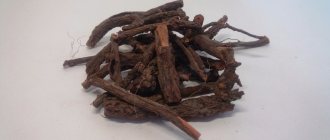
Treatment of the disease in pregnant women is carried out with great care so as not to harm either the fetus or the expectant mother. Most often, herbal preparations are prescribed (cyston, cystenal, canephron, phytolysin). Physiotherapy during pregnancy is not suitable for combating stones. Sticking to a diet, drinking enough clean water (if there is no swelling), not sitting still - this is what doctors recommend to expectant mothers. High-level specialists of the Global Clinic Center will do everything possible to eliminate discomfort and negative manifestations of the disease in pregnant women. Traditional methods As an additional remedy, but not the main treatment, you can use:
- use of herbs;
- drinking mineral water (selected depending on the type of stone);
- inclusion of healthy foods in food - watermelon, cabbage, strawberries, cranberries.
All traditional recipes require the approval of your doctor.
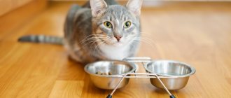
What foods are suitable for cats with urinary tract diseases?
Due to strict restrictions in diet composition, natural nutrition is difficult to organize. The solution is specially developed ready-made diets - wet canned food (pouches) or dry food for cats. They must be of high quality - premium or, preferably, super-premium. Budget cat food does not contain the best protein, a lot of salt, and chemical additives. They worsen the animal’s condition and can lead to dangerous disorders of the gastrointestinal tract and kidneys.
Therapeutic food for cats with pathologies of the genitourinary system is selected taking into account the type of stones. If it is oxalates, natural food and/or wet canned food can be given. For struvite-type urolithiasis, you can choose special dry cat food.
Rules for choosing diets:
- There should be a note on cat food that it is suitable for animals with diseases of the genitourinary system and what types of uroliths it affects;
- It is advisable that cat food contain vitamin A and tocopherol - they improve the condition of urolithiasis;
- the composition should contain a minimum of calcium, phosphorus and vitamin C - ascorbic acid provokes the accumulation of uroliths;
- The percentage of protein in cat food should be reduced - because of this, animals may be reluctant to eat it, and you need to gradually accustom your pet to the new diet.
Prevention
In order not to provoke the appearance of urolithiasis, preventive measures should be observed:
- Lead an active lifestyle: moderate exercise, giving up bad habits, frequent walks in the fresh air.
- Resist stress and nervous overload (vitamin complex, self-discipline, yoga).
- Try to avoid overeating and consuming high-calorie, canned, salty, smoked and fatty foods.
- Increase the amount of fluid you drink.
- Include fruits and vegetables in your diet.
Nutrition: enemy and ally
Malnutrition in cats is one of the provoking factors for the development of urolithiasis. Therefore, when choosing food, you need to consider the following nuances:
- A number of feeds contain excess amounts of phosphorus, magnesium, and calcium. These elements contribute to the formation of crystals in urine.
- The food has a significant effect on the acidity of urine (pH). The cat’s body is “tuned” to digest animal food, which makes the urine slightly acidic, and therefore prevents the formation of stones. In such an environment, crystals dissolve. If the diet is dominated by products of plant origin, which is often found in cheap brands, the pH of the urine shifts towards slightly alkaline. And this contributes to the formation of struvite - one of the most common types of stones.
The likelihood of developing the disease increases if the cat drinks little. In this case, the urine becomes too concentrated and stones form faster.
Modern approach to the treatment of urate nephrolithiasis
The problem of treating urolithiasis remains one of the most pressing in modern urology. Urate urolithiasis is one of the types of urolithiasis, the frequency of which has increased significantly in recent years - from 5–10% in the 50s of the last century to 20–30% currently, which is associated with the increasing impact of a number of environmental factors leading to the accumulation of excess lead in the body, as well as increased alcohol consumption.
The etiopathogenesis of urate lithiasis is the most studied and is associated with complex physicochemical processes occurring both in the body as a whole and at the level of the urinary system of a congenital or acquired nature. The biochemical basis for disorders of purine metabolism are hyperuricemia and hyperuricuria, leading to the formation of stones consisting of uric acid, as well as sodium, ammonium and calcium (very rarely) salts of this acid. The process of stone formation goes through a number of stages - from saturation and supersaturation of urine with salts and further to the phases of enucleation, crystallization and crystal growth to clinically significant sizes, when mechanisms for inhibiting crystal growth are ineffective or absent.
Hyperuricuria creates the preconditions for crystallization of uric acid, mainly in the area of the terminal nephron and at the apex of the renal papilla, similar to Randall's plaques. Uric acid crystals can also lead to the development of aseptic necrosis of the tubular epithelium, which, when rejected, under conditions of hyperuricemia, can become the core of a future calculus. Hyperuricemia leads to the accumulation of uric acid crystals in the interstitial tissue of the kidney and causes interstitial nephritis or early changes in small renal vessels with the development of arterial hypertension.
The genesis of urate nephrolithiasis has both common causes for stone formation and a characteristic feature unique to it, which is that the formation of urate stone requires high acidity of urine, since uric acid dissolves only in slightly acidic and alkaline environments. When the pH of the urine is above 6.5, crystallization of uric acid does not occur, and it is released in a dissolved state; a decrease in the pH of the urine below 5.5 leads to a supersaturation of the urine with uric acid crystals, which precipitate and serve as a framework for the formation of stones.
An increase in the concentration of uric acid in the blood is associated with excess nutrition (especially with an increase in the proportion of food rich in protein in the diet), prolonged fasting, physical inactivity, frequent consumption of alcohol and caffeine, and the use of certain medications: laxatives and diuretics, antibiotics, corticosteroid hormones. In some cancers and malignant blood diseases, hyperuricemia can also develop.
Attempts to explain the development of urolithiasis by any one reason have led to nothing, therefore, in each specific case, it is necessary, before prescribing treatment, to conduct an examination in order to find out all the possible causes of the development of the disease in a given patient.
The examination of patients involves the collection of anamnestic data, laboratory (with mandatory testing of the level of calcium and uric acid in the blood, excretion of calcium, oxalates, phosphates, uric acid), ultrasound, X-ray (survey and excretory urography) examination, as well as bacteriological analysis of urine. Additional information can be obtained from helical computed tomography with a bolus of radiocontrast or magnetic resonance imaging. When preparing for surgery, it is also necessary to consult a therapist, an anesthesiologist, and, if indicated, other specialists. After spontaneous passage or removal of the stone in one way or another, a study of the chemical composition of the stones is carried out.
The examination results reveal various variants of purine metabolism disorders: an increase in both the level of uric acid in the blood and its daily excretion in an average of 25% of patients; increased levels of uric acid in the blood with normal daily excretion in 20% of patients; normal levels of uric acid in the blood with increased daily excretion in 15% of patients.
In other patients, the level of uric acid in the blood and its daily excretion may be normal, despite the urate composition of the stones.
More than 50% of patients with urate nephrolithiasis belong to the age category of 50 years and older and have various concomitant diseases (obesity, coronary artery disease, arterial hypertension, diabetes mellitus, gout, etc.), which influence the choice of treatment method and necessitate the need for them additional preoperative preparation.
Modern medicine has a whole arsenal of conservative, surgical and combined methods for treating urate urolithiasis. The choice of treatment method is determined by the number of stones, the location of the stones, their size and shape, the duration of the disease, the presence of concomitant urinary tract infection, the functional capacity of the kidney, the presence of concomitant diseases, the general condition of the patient, the anatomy of the upper urinary tract and other features. It is important to note that treatment is carried out for both emergency and planned indications.
Open surgical interventions (pyelolithotomy, nephrolithotomy), although they have not lost their importance to date for nephrolithiasis, are not of decisive importance and are used in 3–5% of patients. They should be considered as a forced measure and used in specific circumstances and when other treatment methods are impossible. Basically, these operations are performed in emergency situations in acute obstructive pyelonephritis, caused by blockage of the urinary tract with large kidney stones, or when destructive forms of this disease occur. In a planned manner, open operations are indicated in the presence of secondary kidney stones in combination with various anomalies of the upper urinary tract, which cannot be corrected otherwise than by resorting to surgical correction (stricture, periureteritis, etc.).
In recent decades, laparoscopic stone removal and retroperitoneal pyelolithotomy (endoscopic stone removal using percutaneous retroperitoneal access) have competed with open operations for nephrolithiasis.
The main interventions for the purpose of removing urate kidney stones are extracorporeal lithotripsy (ESLT), percutaneous nephrolithotripsy (PCNL), which can be combined and combined with litholytic therapy.
The introduction of percutaneous nephrostomy into practice has revived interest in local litholysis, which refers to the direct application of a dissolving drug to the stone. An optimal litholytic effect can be achieved with uric acid and struvite stones. The method can be combined with percutaneous stone removal or EBRT.
Percutaneous X-ray endoscopic surgery in the “era of DLT” is used to solve complex clinical cases of urate nephrolithiasis: unsuccessful DLT, in the presence of contraindications for DLT, and also as an independent or combined with DLT (“sandwich therapy”) method in the treatment of large, multiple stones , stones in abnormal, repeatedly operated kidneys, in a single kidney, bilateral stones, as well as when removing coral stones.
EBRT is successfully used for kidney and ureteral stones up to 2.5 cm in size. However, if for relatively small stones (up to 1.5 cm maximum linear size) it is indicated as monotherapy, then for larger stones it must be combined with kidney catheterization, installation internal stent or, less commonly, percutaneous puncture nephrostomy (PPNS).
With DLT, only the destruction of the stone occurs. The most responsible period is the period of spontaneous passage of fragments after fragmentation, when periods of disturbance in the passage of urine from the kidney exposed to shock waves are observed. The main methods of kidney drainage used in the postoperative period in case of DLT for ureteral obstruction or attack of pyelonephritis are also ultrasound-guided PPNS, installation of an internal stent, and renal catheterization.
Currently, DLT is the method of choice in the treatment of patients with various clinical forms of urate nephrolithiasis that has not responded to litholysis. Alternative treatment methods (endoscopic or open surgery) should be used only in case of contraindications or prognostic ineffectiveness. The importance of EBRT is especially increasing in older patients with urate nephrolithiasis, since a comprehensive assessment of the invasiveness, clinical effectiveness and impact on the quality of life of all modern treatment methods in the elderly allows us to consider EBRT as the method of choice for stones up to 2.5 cm in size. Introduction of EBRT , undoubtedly, has significantly changed the approach to the removal of urinary stones. However, it is specifically for urate stones that the implementation of this method has so far been associated with certain difficulties, since visualization, and therefore the conduct of an EBRT session as monotherapy for urate stones, is possible only under ultrasound guidance. Since ultrasound guidance and crushing of urate stones are limited to their localization in the kidney, pelvic and prevesical sections of the ureter, after crushing, endoscopic contact lithotripsy of fragments and “stone paths” that caused occlusion in the middle sections of the ureter is most often used, due to the fact that their visualization Ultrasound and DLT are not possible. That is why, more and more often, works are appearing whose authors recommend carrying out DLT and litholytic therapy against the background of installing an internal stent in the preoperative period.
External shock wave lithotripsy for urate lithiasis should be performed under ultrasound guidance. As an alternative method, especially in case of ureteral obstruction in the postoperative period of DLT, X-ray guidance can be used using the following techniques: intravenous administration of a radiocontrast agent before DLT, provided that the renal pelvis is “filled” with a contrast agent, indicating the location of the “stone path” in the ureter ; retrograde insertion of a catheter to a stone in the ureter or pelvis with the introduction of a radiopaque substance into the renal cavity system; antegrade administration of contrast through nephrostomy drainage in the absence of an internal or external stent or ureteral catheter.
Our experience in the use of DLT for the treatment of patients with urate nephrolithiasis indicates that for stones with a maximum linear size of more than 1.5 cm (especially more than 2 cm), DLT gives better results in conditions of kidney drainage, which is preferably carried out by installing an internal stent, since this significantly reduces the incidence of postoperative complications. DLT of uric acid stones in the form of monotherapy can be carried out for relatively small stone sizes - up to 1.5 cm. Litholytic therapy with citrate mixtures, carried out for 1 month and which did not allow dissolution of the stone during subsequent DLT, significantly improves treatment results (the number of complications and time frame for the kidney to be freed from stone fragments). The administration of citrate mixtures can be recommended as preoperative preparation for large urate kidney stones - more than 1.5 cm.
However, the “gold standard” for the treatment of urate stones should be recognized as oral administration of citrate mixtures, which provide dose-dependent alkalinization of urine without changing the acid-base balance of the blood. With concomitant hyperuricemia, treatment must be supplemented with uricostatics to reduce the level of uric acid in the blood. The daily dose of citrate mixtures is selected individually within the range of 6–18 g, evenly distributed throughout the day in 2–3 doses to maintain urine pH at 6.2–6.8, and it does not affect the level of potassium, sodium, oxygen, carbon dioxide and bicarbonate in the blood. The mechanism of action of citrate mixtures is based on reducing the processes of crystallization in the urine and the binding of calcium ions from the gastrointestinal tract to the urinary tract, where this effect is maximally manifested due to the highest concentration of citrate. Thus, the complex effect of citrate on the physicochemical state of urine leads to an increase in the solubility of urates, calcified oxalates, complex magnesium-ammonium phosphates and some other salts, helping to inhibit stone formation and dissolve already formed stones.
There is sufficient data in domestic and foreign literature on the effectiveness of litholytic therapy for urate stones. It should be noted that in the treatment of large urate stones, the effectiveness of litholytic therapy of urate stones increases significantly after their fragmentation by DLT, since after fragmentation the area of contact of urine with the surface of the stone increases hundreds of times.
In addition, for lithokinetic purposes, conservative therapy for urate lithiasis may include the prescription of antispasmodics, anti-inflammatory and antibacterial drugs.
Litholytic therapy for urate nephrolithiasis should be comprehensive and aimed at reducing the content of uric acid in the extracellular space. For this purpose, drugs that have a uricostatic effect and diuretics, including official ones, are used. Patients are prescribed a diet with limited protein-rich foods. Litholysis of urate stones is carried out under ultrasound control. It is important to note that only uric acid stones are reliably soluble during litholytic therapy. Stones consisting of uric acid salts: sodium urate, potassium urate - dissolve less well. Stones consisting of ammonium urate are practically insoluble, so it is advisable to additionally prescribe potassium preparations, which promote the conversion of uric acid salts into potassium urate salts (the solubility of these urate salts is higher).
Citrate therapy in patients with urate nephrolithiasis is carried out provided that the patient follows a diet with limited protein-rich foods, such as meat products, their derivatives and by-products, fatty fish, mushrooms, legumes, lentils; strong alcoholic drinks, red wine, dark beer, chocolate, strong tea and coffee, pickles, smoked meats, raspberries, figs, alkaline mineral waters (Borjomi), sorrel, spinach, celery, peppers, radishes, cauliflower. Patients with impaired purine metabolism are additionally prescribed uricostatics (allopurinol), in the presence of metabolic disorders in the form of oxaluria + hyperuricuria, magnesium oxide is used, in the presence of metabolic disorders in the form of hypercalciuria + hyperuricuria, hypothiazide is added in normal dosages.
In general, when litholytic therapy is carried out correctly by patients, it is possible to dissolve up to 60% of all stones, and in 40% of cases it is necessary to resort to alternative methods of removing stones from the kidneys, and primarily to DLT, the absolute indications for which are: urate stone, in which it is possible to achieve the optimal pH value or there is no effect from long-term therapy; a calculus that caused occlusion of the ureteropelvic segment and an attack of acute pyelonephritis; kidney stone, which disrupts the passage of urine and causes pronounced dilatation of the collecting system; frequent gross hematuria; pain that renders the patient unable to work.
A relative contraindication to DLT of urate stones is the combination of urolithiasis with the active stage of chronic pyelonephritis and constant aching pain in the lumbar region. Relative contraindications include clinical situations such as intolerance to an internal catheter such as a stent (dysuria, hematuria, vesicorenal reflux).
Thus, the currently existing methods of treating urate nephrolithiasis can basically solve the problem of removing urate kidney stones without changing the fundamental principles and principles of treatment, achieving results with a significantly lower risk for the affected organ and the patient’s body as a whole.
It should be noted that patients with urate nephrolithiasis with impaired purine metabolism after stone removal require clinical observation and therapy aimed at preventing relapse of the disease.
Thus, urate urolithiasis is one of the most complex types of urolithiasis; With this disease, successful use of conservative therapy (litholysis) is possible. Indications for surgical treatment have been developed. EBRT in the form of monotherapy is the most favorable method of surgical treatment of urate stones up to 1.5 cm; EBRT in patients with urate nephrolithiasis with stones larger than 1.5 cm gives optimal results in conditions of kidney drainage, which is preferably carried out by installing an internal stent, as this significantly reduces the incidence of postoperative complications. For urate stones larger than 1.5 cm, litholytic therapy with citrate mixtures as preoperative preparation for 1 month before DLT also increases its effectiveness; after DLT of urate stones of any size, litholytic therapy significantly increases the effectiveness of treatment of urate lithiasis, increasing the area of contact of urine with the surface of the stone; the combined use of percutaneous surgery and radiotherapy also maximizes the release of the kidney from stone fragments in the most severe forms of this disease (large stones and coral nephrolithiasis), thereby increasing the effectiveness of treatment. Open surgical interventions are currently rarely performed and are performed mainly for emergency reasons or when other treatment methods are ineffective.
Literature
- Alyaev Yu. G., Kuzmicheva G. M., Rapoport L. M., Rudenko V. I. Modern aspects of citrate therapy in patients with urolithiasis // Medical class. 2004. No. 4. P. 20-24.
- Alyaev Yu. G., Rapoport L. M., Rudenko V. I. Citrate therapy to prepare for extracorporeal lithotripsy // Materials of the plenum of the board of the Russian Society of Urologists. M., 2003. P. 59-60.
- Alyaev Yu. G., Rapoport L. M., Rudenko V. I. et al. 1st International Consultation on Stone Disease. aris, 3-4 July 2001. Book of abstracts; 63:63.
- Baibarin K. A. Surgical methods of treating nephrolithiasis in gerontological patients: Dis. ...cand. honey. Sci. M., 2004.
- Beshliev D. A. Dangers, errors, complications of extracorporeal lithotripsy, their treatment and prevention: Dis. ...Dr. med. Sci. M., 2003. 356 p.
- Bugaeva N.V., Balkarov I.M. Arterial hypertension and disorders of purine metabolism // Ter. arch. 1996. No. 1. P. 36-39.
- Dzeranov N.K., Grishkova N.V., Boyko T.F., Golovanov S.A. Conditions for performing external lithotripsy with different physicochemical composition of urinary stones // Urology and Nephrology. 1994. No. 6. P. 10-13.
- Dutov V.V. Modern aspects of the treatment of some forms of urolithiasis: Dis. ...Dr. med. Sci. M., 2000.
- Kaprin A.D., Ivanenko K.V., Ivanov S.A. Treatment of patients with urate nephrolithiasis with disorders of purine metabolism. M., 2003.
- Martov A. G. X-ray endoscopy and external shock wave lithotripsy in the combined treatment of nephroureterolithiasis: method. recommendations. M., 1994. pp. 11-14.
- Nephrology: A Guide for Doctors /ed. I. E. Tareeva. M., 1995. T. 2.
- Pytel Yu. A., Zolotarev I. I. Urate nephrolithiasis. M., 1995. 176 p.
- Pytel Yu. A., Chakaleva I. I., Shemyakin F. M. About some biochemical changes in the body during citrate therapy of patients with urate nephrolithiasis // Urology and Nephrology. 1972. No. 6. P. 28-33.
- Pytel Yu. A., Zolotarev I. I. On the issue of the use of conservative therapy in patients with urate nephrolithiasis // 4th Lithuanian Conf. urologists. Kaunas, 1987. pp. 66-68.
- Rudenko V.I. Urolithiasis. Current issues of diagnosis and choice of treatment method: Dis. ...Dr. med. Sci. 2004.
- Sergienko N. F., Shaplygin L. V., Kuchits S. F. Citrate therapy in the treatment of urate nephrolithiasis // Urology and nephrology. 1999. No. 2. P. 34-36.
- Buck AC The epidemiology, formation, composition and medical management of idiophatic stone disease//Curr. Opin. Urol. 1993; 3: 316-322.
- Grases F., Ramis M., Villacampa Al., Costa-Bauza A. Uric acid urolithiasis and crystallization inhibitors // Urol. Int. 1999; 62(4): 201-204.
- Hoffman N., McGee SM, Hulbert JC Resolution of ephedrine stones with dissolution therapy//Urology/2003/may: 61(5): 1035.
N. K. Dzeranov , Doctor of Medical Sciences, Professor, Academician of the Moscow Aviation Institute D. A. Beshliev , Doctor of Medical Sciences R. I. Bagirov K. A. Baibarin , Candidate of Medical Sciences RMAPO, Moscow
Instructions for use
A portion of such food should not exceed the permissible dose authorized by the doctor.
Royal Canin Urinari Feline cat food is considered medicinal, so it can be given to your pet only on the recommendation of a veterinarian in the dosage prescribed by the doctor. It is prohibited to increase or increase the dose on your own, as well as to cancel the diet. For a low-weight cat, the norm should be added by 15-20 g, and for a pet with normal weight, the amount of food is shown in the table:
| Cat weight (kg) | Daily value (g) |
| Dry | |
| 2—3 | 33—44 |
| 4—5 | 54—63 |
| 6—7 | 72—80 |
| 8—9 | 88—96 |
| 10 and above | 103 |
| Wet | |
| 2—3 | 115—175 |
| 4—5 | 230—290 |
| 6—7 | 350—405 |
| 8—9 | 465—520 |
| 10 and above | 580 |
Classification of chemical types of stones
- Inorganic stones
- At pH0 – calcium oxalate (vivelite)
- At pH 5- calcium phosphate (carbonatopatite)
- At pH 0 magnesium ammonium phosphate (struvite)
- Organic stones
- At pH5-6.0 – uric acid its salts urate
- Cystine and xanthine stones are associated with inborn errors of metabolism at the level of the body as a whole.
Diagnostics:
- Ultrasound can detect stones in the CL, ureteropelvic and vesicoureteral segments. Diagnose dilatation of the upper urinary tract.
- Survey urography - allows you to identify shadows suspicious of stones in the projection of the kidneys, ureters and bladder, and determine their radiopacity.
- Multislice CT allows you to determine the calculus, its linear dimensions, volume, density, and assess the anatomical and functional state of the urinary tract. Currently, it is the “gold standard” in the diagnosis of ICD.
Treatment
Medication for hepatic colic - pain relief with non-steroidal anti-inflammatory drugs (NSAIDs). They have a better analgesic effect compared to opiates. The following drugs are used: to relieve pain, diclofenac, indomethacin, ipobrufen, tramadol.
Taking alpha blockers daily also reduces the likelihood of a recurrent episode of hepatic colic.
If the analgesic effect is not achieved with drugs, they resort to surgical methods of drainage of the upper urinary tract or disintegration of the stone (Ureter stenting or PPNS-percutaneous puncture nephrostomy)
Surgical treatment: ESWL, remote shock wave lithotripsy, CLT (contact lithotripsy, ureteroscopy), percutaneous nephrolitholapaxy.
Silence is not always golden?
For a long time, the stone may not show itself in any way. But this is not dangerous only in those rare cases when stones can be considered “clinically insignificant” - they are not only “silent”, but also do not impair kidney function for many years. However, everything is for the time being, so such patients need to periodically visit a urologist.
A kidney stone makes itself felt with an attack of renal colic. When it happens depends not at all on its size, as many people think, but on where exactly the stone settled, whether it blocked the outflow of urine and other factors.
Existing assortment
Today, many dry food manufacturers produce products that help prevent the occurrence of urolithiasis. All of them belong to the premium or super premium class. Typically, such diets are prescribed to increase urine production, relieve signs of inflammation and dissolve existing sediment.
Almost every well-known global manufacturer has such lines. Products marked “Urinari” are available in the range of such products as “Purina”, “Eukanuba” and “Hills”.
Cat Chow Urinary Tract Health
This food is intended for the prevention of urinary tract diseases in adult cats. It contains an optimal ratio of minerals and a reduced concentration of magnesium. Regular consumption of this food allows you to maintain an optimal pH value and minimize the risk of stone formation.
This Urinari line contains processed meat products, cereals and vegetables. This food also contains chicory root, soy flour, chopped spinach, rosemary, animal and fish oil.
Description and composition
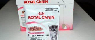
This type of food can be purchased for the animal in bags.
Royal Canin cat food "Urinari" is produced in the form of dry kibble, packaged in packages of 400 g, 1.5, 3.5, and 7 kg. Wet food is sold in 100 gram packs and looks like pieces of meat in sauce or pate in tins weighing 195 g. It belongs to the super-premium class due to the content of natural ingredients. The composition reduces the amount of magnesium, calcium and phosphorus, which contribute to the formation of crystals. The increased sodium content encourages the cat to drink more water, which has a beneficial effect on the condition of the kidneys and promotes the elimination of substances that provoke urolithiasis.
Healthy Ingredients
Therapeutic food "Royal Canin Urinari" has the composition shown in the table:
| Component | Benefit |
| Dried poultry meat | Source of essential amino acids and protein |
| Fish fat | Contains Omega-3 and Omega-6 fatty acids essential for skin and coat |
| Has an anti-inflammatory effect | |
| Cystine | Natural antioxidant |
| Has anti-inflammatory, immunomodulatory and regenerating properties | |
| Marigold extract | Relieves inflammation and spasms |
| Strengthen immunity | |
| Prevents the formation of blood clots | |
| Taurine | Promotes the formation of bile acids |
| Essential for healthy mental development | |
| Fructooligosaccharides | Maintains the level of beneficial intestinal microflora |
| Arachidonic acid | Improves the functioning of the digestive system |
| Reduces the risk of joint inflammation | |
| Normalizes hormone levels | |
| Biotin | Regulates blood supply to tissues |
| Improves coat growth and structure | |
| Linoleic acid | Necessary for normal function of the kidneys, blood vessels, and nerves |
| Iodine | Vital for thyroid function |
| Iron | Participates in hemoglobin synthesis |
| Zinc | Helps absorb B vitamins |
| Has regenerating properties | |
| Selenium | Protects organ cells from oxidation |
| Neutralizes the effects of heavy metals | |
| Vitamins B, C, E, D3 | Vital for the normal functioning of all organs and systems |
Urinari cat food – rating
Among the foods labeled “Urinari for cats,” almost all have a therapeutic effect, but only a few brands are popular with veterinarians:
- Proplan Urinari food for cats
contains a special St/Ox complex, which prevents the formation of oxalate and struvite uroliths and dissolves existing ones. - Purina Urinari for cats
is suitable for pets with struvite formations in the urinary tract. The presence of oxalates is a contraindication to its use. - Hill's Urinari for cats
is the most prescribed medicinal food. It is available in several variations. Only your pet’s attending physician can select the correct variety. - Monge Urinari for cats
is a super premium food that contains substances both to combat urolithiasis and to maintain the intestinal microflora and the beauty of the animal’s coat.
Foods labeled Urinari for cats are widely used in the treatment of urolithiasis and cystitis in pets. Their use helps reduce both acute attacks of the disease and prevent possible relapses in the future. It is important to remember that such foods belong to medical nutrition, so they should be prescribed strictly by the attending physician with regular monitoring of the animal’s tests.