Quick transition Treatment of atrophic rhinosinusitis
Atrophic rhinitis is a clinical syndrome of progressive atrophy of the nasal mucosa.
It is characterized by paradoxical nasal congestion and thick, unpleasant nasal discharge. Most patients are diagnosed with concomitant sinusitis, and therefore this disease is more correctly called atrophic rhinosinusitis .
Atrophic rhinosinusitis can be divided into two forms: primary and secondary.
Nose injuries
The crusts on your cat's nose may be brownish or black. This symptom is characteristic of a fungal infection, an inflammatory process in the nasopharynx, or an allergic reaction. The most harmless cause of crust formation is considered to be household injuries.
The fact is that a healthy cat's nose is slightly moist. This prevents the normal restoration of damaged skin, so wounds can take a long time to heal. If the owner is absolutely sure of the nature of the crusts, it is necessary to help the pet’s body recover as quickly as possible. To do this, it is recommended to treat your nose with an antiseptic several times a day. Preventing re-infection of the wound significantly accelerates regeneration and the crusts begin to disappear a few days after the start of treatment.
A dark crust on the nose, which is accompanied by discomfort and is a consequence of skin damage, should not be a cause for concern. In this case, no treatment is required; the crust will fall off on its own when the tissues are completely restored.
Treatment with an antiseptic is only necessary if weeping erosions or crusts appear at the site of the wound, blocking the respiratory tract.
Diagnosis and treatment methods
The therapeutic course for spots on the nose is selected by the veterinarian based on the diagnostic results. The main thing in making a diagnosis is a laboratory test to determine the pathogenic microflora that caused the rash on the face. Blood and urine tests may be required. For black spots, local treatment is prescribed, but for advanced cases of the disease, an integrated approach is required using medications from different groups.
Among the antiseptics, Miramistin is suitable for animals.
When rashes on a cat’s nose are associated with an allergic reaction, drugs with antihistamines come to the rescue. For daily treatment, antiseptics are used, such as:
- "Miramistin";
- "Chlorhexidine."
When spots on a cat’s nose are caused by a dermatological disease, antibacterial agents that suppress the activity of pathogenic microorganisms are necessarily used for treatment. It is equally important to adjust your pet’s diet by eliminating fatty foods and adding more fresh food. Omega-3, 6 and vitamin supplements help strengthen the cat’s immune system and prevent the appearance of other spots. It is possible to use folk remedies, such as a decoction of chamomile or celandine, which is used to treat the cat’s nose.
Infectious diseases
If a cat has black crusts on his nose that do not heal for a long time, the cause should be sought in infectious diseases. Associated symptoms of infectious diseases:
- Nasal discharge.
- Conjunctivitis.
- Otitis.
- Frequent sneezing.
- Decreased appetite.
- Lethargy.
- Increased sleepiness.
In this case, the animal may refuse to eat and be lethargic. Particularly severe forms of infectious diseases are accompanied by vomiting, diarrhea, and dehydration.
Cat infections are very dangerous, especially if the animal does not have all the necessary vaccinations. Black crusts on the nose are one of the most harmless symptoms; in severe cases, infectious diseases can lead to the death of the pet. You cannot try to cure the cat yourself; you should take the animal to a veterinary clinic. This may require long-term hospital treatment.
We recommend the article: Why do sores appear on a cat’s face?
Sensation of speck in the eye
Table of contents
A real speck or a symptom: how to determine?
Is it possible to remove a foreign body yourself? Can I wash my eyes? The feeling of a speck in the eye as a symptom of disease When should you see a doctor? Prevention of injuries and eye diseases Gilan solution for dry eye syndrome The eyeball is sensitive to any external influence. Its most vulnerable part is the cornea, which is not surrounded by hard tissues and bones. A thin layer of tear film performs a protective function. In its absence, microcracks appear on the surface of the cornea, which cause irritation and foreign body sensation. A similar reaction is caused by mechanical trauma or, indeed, by the contact of large dust particles with the cornea, which can complicate the diagnosis of the disease.
A real speck or a symptom: how to determine?
Without special equipment, it is difficult to determine what caused the eye irritation. An indirect sign that a speck has entered the eye is a sudden feeling of pain and discomfort when blinking.
Eye injuries
There are several types of eye injuries: wound, blunt trauma, chemical burn. Damage can be either superficial or penetrating, with or without infection.
Foreign body entry
When a foreign body enters the eye, the degree of injury to the eye can vary: from irritation of the cornea to severe conjunctivitis, keratitis, clouding and rupture of the cornea. The longer a foreign body remains in the eye, the higher the risk of infection and purulent inflammation of the external and internal structures of the eyeball.
As a rule, we are talking about microparticles of debris, decorative cosmetics, hair, and animal fur getting into the eye. If safety precautions are not followed at work, small fragments of building materials get into the eye: pieces of metal or wood shavings, concrete, plastic.
Is it possible to remove a foreign body yourself?
What to do if a foreign body gets into your eye? Should I try to remove it myself or contact a specialist? Specks, tissue fibers, eyelashes, as a rule, are removed on their own with rapid blinking. Due to irritation, increased production of tears begins, which washes away the foreign body from the cornea without damaging it.
If the foreign body is in a hard-to-reach place, for example, in the corner of the eye or stuck deep under the eyelid, then it is wiser to consult an ophthalmologist.
Can I wash my eyes?
If the feeling of a speck in the eye does not go away, then intuitively there is a desire to rinse the eyes. It is not recommended to do this at home, since there is a high risk of deeper injury to the cornea and infection.
Bruises
For bruises, it is recommended to put a bandage on the eye and apply ice on top. This will help reduce pain. Some types of eye drops have an analgesic effect.
Burns
In case of a chemical burn - that is, if cleaning agents, glue, paint, other industrial or household liquids or fumes get into the eyes - as first aid, you can carefully rinse your eyes with running water, opening both eyelids with your fingers, and tilting your head down and to the side - so that water can flow freely. Afterwards you need to apply a sterile bandage and be sure to consult a doctor.
The feeling of a speck in the eye as a symptom of disease
The feeling that something is bothering the eye occurs with some eye diseases, for example, conjunctivitis or dry eye syndrome. In fact, there is no foreign body in the eye, and the feeling of discomfort appears due to irritation of the cornea, which occurs as a result of its insufficient hydration and oxygen supply.
If, while working at a computer, in the open air, it stings in the eye, there is a stinging and burning sensation, a feeling of dryness appears, or, on the contrary, lacrimation increases, we can talk about the development of dry eye syndrome (DES).
The disease is characterized by a decrease in the quality and speed of tear production and can lead to serious consequences, including loss of vision. Violation of the visual apparatus is provoked by several factors:
- work in difficult conditions, in offices, in hazardous industries;
- use of contact lenses with low gas permeability;
- unfavorable environment;
- extreme weather conditions;
- eye diseases;
- eye surgery;
- age over 45 years.
When should you see a doctor?
Dry eye syndrome (xerophthalmia) is dangerous due to its consequences. Without adequate treatment, the disease becomes chronic, which not only reduces the overall quality of life, but also leads to irreversible structural changes in the eye. You should consult a doctor if several symptoms appear at once: a feeling as if there is a speck stuck in the eye, rapid eye fatigue, decreased visual acuity or clarity.
What should you not do if you feel a speck in your eye?
If you have a feeling of “sand” in your eyes, do not rub them with your hands or rinse them with water. When an infection gets on the irritated surface of the cornea, inflammation quickly develops, which can affect the internal structures of the eye.
Possible complications
Dry eye syndrome is difficult to treat once the process is started. A common complication of the disease is corneal erosion, a chronic impairment of the quality of tear fluid, which leads to the inability to wear contact lenses, drive a car, or perform certain jobs.
Prevention of eye injuries and diseases
Ophthalmologists note that people with strong immune systems are least susceptible to dry eye syndrome. Maintaining hygiene when wearing contact lenses, limiting the time you use gadgets and working at the computer, and maintaining general health reduce the risk of dry eyes. To reduce the negative impact of the environment, it is recommended to use moisturizing drops based on natural ingredients.
Gilan solution for dry eye syndrome
The drug "Gilan" contains hyaluronic acid, which is a natural source of hydration of the cornea. It belongs to the group of tear substitutes that show effectiveness in the treatment of dry eye syndrome. Gilan solution is compatible with all types of contact lenses, protects the eyes from drying out, and quickly relieves pain and irritation in case of corneal injuries.
Dermatological diseases
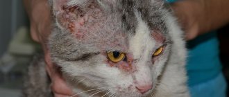
Crusts on a cat’s nose can be the result of skin pathologies and dermatitis. These diseases are caused by fungi and bacteria. To accurately determine the cause of the formation of crusts, it is necessary to undergo a series of tests, including a nasal microflora smear and skin scraping from the affected area. The doctor will also illuminate the animal with a special lamp, which allows you to quickly determine the presence of fungal infections.
Treatment depends on the type of disease. For fungal infections of the epidermis, antimycotic drugs are used. Treatment is carried out using ointments and solutions for treating the nose. In case of a bacterial infection, the crusts should be treated with antibacterial drugs, for example, Levomekol ointment.
For dermatological diseases, antiseptic treatment of the affected area is mandatory. This is necessary to prevent secondary infection. For this purpose, a solution of chlorhexidine or miramistin is used.
Primary atrophic rhinosinusitis
Primary atrophic rhinosinusitis is more often diagnosed in patients from low socioeconomic status groups living in geographic areas with warm climates. Areas of high prevalence include southern Saudi Arabia, China, Africa, India, the Mediterranean and the Philippines.
Atrophic rhinosinusitis is more common in women. It is diagnosed more often at young ages than at older ages.
Clinical picture
Patients experience a constant sensation of an unpleasant odor in the nose (this disorder of smell is called cacosmia). At the same time, there is also an unpleasant odor from the mouth, noticeable to others. This is where the term “ozena” (“stench”) comes from, which is sometimes used as a synonym for severe primary atrophic rhinosinusitis.
Other symptoms include lack of sense of smell (anosmia), nosebleeds, nasal pain, sleep disturbances, and suffocation due to excessive crusting.
Patients complain of nasal congestion even with excessively wide nasal passages, which is better characterized by the concept of “lack of sensation of breathing.” Nasal congestion occurs due to improper passage of air through the nasal cavity, lack of resistance and sensation of air flow due to loss of tissue containing sensory receptors. The same phenomenon is observed in patients with perforation of the nasal septum.
The disease is characterized by the replacement of normal pseudostratified columnar epithelium with squamous epithelium. This tissue lacks cilia and mucus-producing goblet cells.
The most common culprits of the bacterial process in the nose are Klebsiella ozaenae, Proteus, Escherichia coli, Staphylococcus aureus, Streptococcus pneumoniae.
ENT examination
During rhinoscopy (examination of the nose), the doctor sees a shiny, thin, pale, and sometimes ulcerated mucous membrane, covered with thick yellow, brown or green crusts, sometimes bloody, covered with a purulent coating. Resorption (destruction) of the underlying cartilage and bone leads to an increase in the volume of the nasal cavity. Some patients are diagnosed with perforation of the nasal septum and secondary saddle deformation of the external nose (recession of the nasal dorsum).
Predisposing factors to the development of primary atrophic rhinosinusitis are completely unknown. It is discussed that these could be endocrine, vascular, infectious and autoimmune diseases, as well as occupational hazards (working in dusty, contaminated areas with chemical or industrial dust).
Computed tomography of the nose and paranasal sinuses
Possible finds include:
- atrophy of the mucous membrane of the lower and middle turbinates and bone resorption;
- resorption of cells of the ethmoidal and uncinate labyrinth;
- an increase in the volume of the nasal cavity with destruction of its lateral walls;
- thickening of the mucous membrane of the paranasal sinuses;
- hypoplasia (reduction) of the maxillary sinuses with a decrease in their pneumatization (airiness).
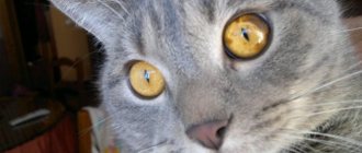
Scabs on a cat's nose photo
Sunlight has no effect on lentigo
It is known that in humans, freckles become more intense in color in the spring and summer. Lentigines are not exactly freckles (although they are similar in “behavior” and appearance) and they do not depend on sunlight.
However, direct sunlight must be treated with care. Lentigines may darken slightly due to tanning, and the skin around them may become damaged due to burns. Since the nature of lentigines is still not understood, many veterinarians assume that the bulging growths can develop into melanoma (skin cancer).
Important! It is lentigo that often causes the first symptoms of skin cancer – dark, raised spots – to be missed.
Wounds and allergies
Sores and crusts on a cat's nose and around the lips may be the result of an allergic reaction. Typically, such symptoms appear in response to food allergens, but damage to the skin of the nose can be caused by contact with household chemicals.
For treatment, special antihistamine drops are used. You can buy them at a veterinary pharmacy. If the allergic reaction is moderate, it is enough to eliminate the irritant for the symptoms to subside. No specific treatment is required; it is enough to avoid contact with the allergen in the future and treat the crusts with an antiseptic to prevent infection.
This is simple lentigo
Another possible cause is simply your cat's nose being discolored. Lentigo is the development of small areas of hyperpigmentation on your cat's skin and it is a common occurrence around your cat's nose and mouth, if a black spot is adjacent to your cat's nose and looks like part of the skin, congratulations, your cat essentially has a new freckle.
Lentigo is not dangerous and does not affect your cat's health in any way. Once you notice one lentigo spot, you are likely to see more. The spots look like freckles or age spots; your cat will likely get more, but don't expect any side effects from the spots.
Allergic reactions
Another common cause of sores on the nose can be considered allergic reactions, regardless of their origin. You can guess the nature of the lesions by the presence of the following signs:
- Sores of allergic etiology are almost always accompanied by the development of severe itching. At the same time, the animal constantly strives to scratch its nose with its paws, or by “wiping” furniture and other interior items.
- Allergies are characterized by mild swelling and swelling of the affected tissues, which is caused by local migration of leukocytes to the site of penetration of allergens.
First aid is provided as follows:
- You can give your cat ½ tablet of diphenhydramine. Submission frequency: up to two times a day, three days in a row.
- The sores themselves are treated with the drugs already described above.
- Until the root cause of the allergy is identified, it is better to change the food to a hypoallergenic one; we recommend feeding your pet from a glass or metal bowl (allergies to plastic can also occur).
Non-communicable causes
There are the following types of non-infectious diseases in which crusts form on the nose:
- allergic conditions;
- injuries;
- violation of conditions of detention.
Allergic conditions
All allergens that cause a runny nose lead to the release of exudate, which dries out and crusts form. The nasal mirror is injured by hard scabs, ichor is released, secondary microflora is added and the pet’s condition worsens.
The development of a hypersensitive reaction is influenced by the following unfavorable factors:
- increased sensitivity to feed mixture ingredients;
- response to pollen from flowering plants;
- irritating effects of perfume odors, tobacco smoke, household chemicals;
- irritating effects of toys, tray filler, bedding made of synthetic materials;
- reaction to human hair or the fur of other pets;
- side effects of medications.
Treatment consists of eliminating the causes. Caused an inappropriate reaction. They stop taking medications that can cause an allergic response and switch to ready-made hypoallergenic food.
Remove objects that emit odors from the room and stop smoking in the presence of the pet. Change the tray filler and bedding. In some situations, the use of antihistamines is required.
Injuries
Such crusts heal on their own
. Crusts are formed if a cat scratches its nose in a fight or through contact with sharp or prickly objects. When you hit the nose, slight bleeding may occur; the released liquid dries, forming crusts. If there is no suppuration under the scabs, observe the healing process of skin defects. Otherwise, use an antiseptic wound-healing ointment.
Be sure to read:
A cat sneezes: causes, symptoms, what to do, treatment methods, what should alert you
Violation of containment conditions.
When the central heating is turned on in winter or during hot weather, the air dries out and the skin on the cat's nose cracks. The ichor is released, which dries and forms crusts. If in hot weather the cat is exposed to a stream of cold air from an air conditioner, hypothermia develops and rhinitis occurs, in which snot is released from the nose. They dry out and form crusts.
Frequent bathing in combination with drafts in the apartment also leads to hypothermia and rhinitis. Treatment consists of introducing immunomodulatory or antimicrobial drops into the nasal cavity.