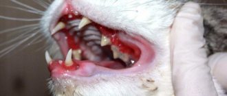
When kittens change their milk teeth, owners should pay special attention to their little pet. In some pets, a similar process occurs at the age of 5-6 months, while in others, teeth change for about a year. In rare cases, in lop-eared kittens and representatives of other breeds, tooth replacement occurs painlessly. But often owners notice that when the baby gets new front teeth, the gums itch, there is pain and other unpleasant symptoms.
Owners are advised to contact a veterinarian, who, if necessary, will prescribe medications for the pet to facilitate the process of teething, and suggest other methods to eliminate discomfort.
Why do new teeth emerge?
Just like in humans, kittens 4 months and older gradually replace their milky bone tissue with permanent teeth. Many owners get scared when they notice that the cat is missing one tooth or is constantly scratching its lips and gums. There is no need to worry, since such a process is completely natural and necessary. It is only important to ensure that the animal grows a new set of teeth normally and does not have double teeth, in which two teeth immediately fall out in place of one tooth. Contacting a veterinarian is also necessary in other cases:
- the resulting wounds fester and heal for a long time;
- inflammatory reaction in the gum area.
Possible complications when changing teeth
Changing teeth in a kitten is a natural process. It can go smoothly, or it can get complicated.
Mild inflammation or redness of the gums is normal. However, it is worth observing all changes and, at the slightest suspicion of deviations, contact a veterinarian.
Symptoms that indicate possible problems:
- suppuration forms on the wound from a lost tooth;
- the kitten is in a bad mood, lethargic, restless;
- refusal to eat for more than a day;
- gums are very inflamed;
- formation of wounds near grown molars;
- The permanent teeth have grown, but the baby teeth have not yet fallen out.
The appearance of any of these signs should be a reason to contact a veterinarian.
Other problems
It happens that the entire row of permanent teeth has formed, but the baby teeth have not fallen out. This situation causes jaw injuries, crooked bites, and the development of periodontal disease. A qualified doctor will help you deal with this problem.
A very common problem when changing teeth is improper growth of fangs. There are several variants of this pathology, so the problem is corrected individually in each case. But this cannot be ignored, since improper tooth growth worsens the animal’s quality of life and creates digestive problems.
Each case of abnormal formation of fangs in a cat is considered individually
Age at which change occurs
The eruption of fangs in kittens occurs two weeks after birth, but some babies are already born with them.
Sometimes milk teeth in cats are observed immediately when they are born, but more often they appear 1-2 weeks after the kitten is born. The first two canines in Scottish cats and representatives of other breeds are observed by 4 weeks of age. Baby teeth in boys and girls can be replaced in different ways, but on average the process of complete replacement ends in six months. In this case, the permanent dentition should have 30 teeth. The table shows the age at which a kitten's baby teeth fall out and permanent teeth grow.
| View | Quantity | Period of eruption, month | Function | |
| Lower jaw | Upper | |||
| Incisors | 6 | 3,5—4,5 | Grasping | |
| Fangs | 2 | 5 | Tearing food | |
| Molars | 4—5 | Chopping | ||
| Premolars | 6 | 4 | 4,5—6 | |
Scheme for replacing baby teeth with permanent ones in healthy kittens
Formation of teeth in a kitten
The structure of the teeth of cats is typical for their species, and zoologists consider the structure of the dental system as its morphological feature. The structure of the dental system reflects the nature of the diet, and the cat is an undisputed predator - all its dental crowns are conical in shape and are designed to tear hunted game into pieces. The teeth located on the jaw form the upper and lower arcades, respectively. To form a breed characteristic—the appearance of a cat’s face—the correct formation of dental arcades is necessary. Teeth are necessary for capturing, holding and tearing prey; the cat uses them to intimidate the enemy, in fights, for grooming, and for carrying small objects, including kittens, in its mouth.
The formation of tooth buds in cats occurs during embryonic development in the form of a dental plate - this is a fold of epithelium containing tooth buds. The inner layer of cells of the dental plate - anameloblasts, form tooth enamel; The cells of the outer layer are odontoblasts, they form dentin. The connective tissue surrounding the tooth germ forms cement.
The development of teeth is associated with their eruption, when the teeth come out through the mucous membrane of the gums.
Teeth are made up of tissues:
- dentin is the modified bone tissue that makes up most of the tooth. Dentin forms the tooth cavity, where the pulp containing blood vessels and nerve fibers is located;
- enamel is a very durable white tissue that covers the surface of the tooth from the outside, where it comes into contact with the external environment;
- cement - covers the parts of the tooth located in its alveolus - the cell of the jaw where the tooth is located. Cementum is represented by bone tissue of a layered structure.
The following parts can be distinguished in the appearance of a tooth:
- tooth crown - part of the tooth that protrudes freely into the oral cavity and has a chewing surface;
- tooth root - located in the alveolus; the cementum of the tooth root and the periosteum of the alveoli are connected by the periodontium;
- neck of the tooth - connects the crown of the tooth with its root, here the coating of the tooth changes, the enamel of the crown passes into the cement of the tooth root. The neck of the tooth is normally located in the gum.
Based on function and form, they are distinguished:
- incisors - small, flat-shaped teeth located on the jaws in front and having one root. The incisor crown has three projections that wear away over time; The cat uses the incisors for grasping and holding, as well as grooming; the roots of the incisors are shallowly embedded in the jaw, so these teeth are often lost with age;
- fangs - the cat’s fangs are dagger-shaped, they are sharp and long, their roots are immersed very deeply in the bone tissue of the jaws; used to kill prey; the cat uses its fangs to separate the cervical vertebrae of the victim - this is a characteristic hunting feature of small cats; fangs are used for tearing food; each fang has only one root, the fangs are located next to the incisors;
- premolars - small molars; Kittens also have them; premolars are needed for grinding food and have a chopping effect; the number of premolars is different on the upper and lower jaws: there are 6 on top, 4 on the bottom. They have 2 or 3 roots, so before removing a premolar, the veterinarian prescribes an x-ray to clarify the number of roots and not leave one of them in the jaw; located next to the fangs;
- molars - large molars - do not have their milk equivalent, they grow immediately in the form of permanent teeth; molars are also designed for cutting and crushing food; located at the edges of the dental arcades; The upper molars have one root each, the lower molars have 2.
During the teething period, cats try everything, it is especially dangerous when they chew wires
When and in what quantity do baby teeth appear?
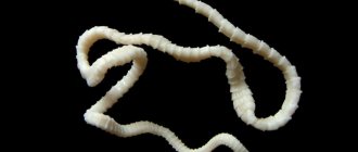
At birth, a kitten has no teeth at all, but starting from the 2nd week of life, milk teeth begin to appear. A cat's primary set of teeth is incomplete because molars, the large molars, are missing. They are called milk teeth because they appear when the kitten feeds on milk, and also because of the specific color of the tooth enamel - milky white, translucent. Baby teeth are smaller, sharper and more fragile compared to permanent teeth. In total, the milk set contains 26 teeth: 12 incisors, 4 canines, 10 premolars.
Timing for the eruption of baby teeth:
- incisors: the first primary incisor erupts at 2–3 weeks; the second - at 2.5–4 weeks; third - at 3–4 weeks;
- fangs: milk fangs erupt at 3–4 weeks of a kitten’s life;
- premolars: eruption in 4 to 8 weeks.
It is generally accepted that the age of a kitten can be accurately determined by its teeth, but this is not entirely true, since individual and breed characteristics of the timing of teething can create a large error in the accuracy of this method.
The practical benefit of knowing the average timing of the eruption of baby teeth is expressed in the ability to control their development, since the development of teeth is also an indicator of the development of the kitten.
The eruption of baby teeth usually goes unnoticed by the kitten’s owner, because the cat is still too small.
By 2 months the kitten has a full set of baby teeth.
At what age does teeth change?
The replacement of baby teeth with permanent ones occurs when the kitten is 3–6 months old. The permanent set includes 30 teeth: 12 incisors, 4 canines, 10 premolars, 4 molars.
Main symptoms of new teeth erupting
Sometimes kittens develop milk teeth within the first day after birth, and when new ones emerge, no unpleasant signs or discomfort are noted. If the process of changing teeth occurs painlessly, then there is no need to do anything. But when the baby develops restlessness and unpleasant symptoms, the help of a veterinarian may be required. Contacting a veterinary clinic is necessary if a kitten’s fang has fallen out and the following symptoms are recorded:
If unpleasant symptoms occur during the change of baby teeth, you should consult a specialist.
- Disgusting odor from the mouth. A healthy cat teething without an unpleasant odor from the mouth, and the presence of such an odor signals an inflammatory reaction and the progression of periodontal disease.
- Poor sleep and refusal to eat. Similar symptoms when changing teeth in a kitten are the result of severe pain. The pet is constantly restless and apathetic. It is necessary to try to feed the kitten at least once a day when teething, so that additional health problems do not arise.
- Copious secretion of saliva. The consistency of the liquid changes, it becomes thick and constantly hangs out of the kitten’s mouth, as a result of which the fur on the chest is constantly wet. Such signs indicate inflamed oral mucosa and stomatitis.
It is necessary to contact a veterinarian if, after 2 years of age, the cat’s primary canines have not been renewed or permanent teeth have begun to fall out. Help is also required if the change of dentition is accompanied by double eruption of some teeth. This problem can be solved by pulling out baby teeth. If the problem is not solved in time and the double dentition is not removed, then there is a high risk of severe inflammation, purulent process and the development of other complications.
Correction of canine position disorders in dogs and cats
F. Genne
Violation of the position of the canines is a change in the bite, which affects their functional state.
Incorrect canine alignment can be caused by a misalignment of the primary teeth or an abnormality in the size of the mandible.
The categories of these deviations from the norm are determined by the orientation of the canines in space.
The first part of the article presents a classification of violations of the position of the canines, while the second discusses the issue of ways to eliminate defects.
Classification
Abnormalities in the position of the teeth In the transverse projection An anomaly in the position of the fangs is much more often noted in the transverse projection. It is mainly found at the level of the lower canines, the position of which has a pronounced direction towards the tongue.
This anomaly, called canine convergence, refers to linguoversion (severe inclination of the canines towards the tongue) or linguoposition (severe displacement of dental implants towards the tongue). The etiology of these dental disorders is not well understood. If in small breeds of animals the persistence of primary canines is the main factor of these disorders, then in others this cause of defect is encountered much less frequently. Also, the lower canines may have a pronounced inclination in the lateral direction (vestibular version), which is also called divergence.
As for the upper canines, violation of their position is much less common. We can observe their deviation towards the palate (due to the persistence of baby teeth).
In lateral projection In dogs and cats, rostral deviation (rostroversion) of the upper canines can also be observed. This is most often observed in small breeds of dogs (Yorkshire terrier, poodle, dwarf dachshund, etc.) due to the persistence of primary canines. In addition, depending on the breed, the persistence of primary canines causes a more or less pronounced disturbance in the position of the permanent canines. This feature is typical for Italian and Scottish Italian greyhounds (photo 1). In any case, particularly in these breeds, it is very difficult to establish whether the persistence of the primary canine is the cause of this type of disorder or whether it provokes rostroversion of the primary canine.
| Photo 1. Rostroversion of the upper canine in an Italian greyhound. A persistent milk tooth was recently removed (the wound caudal to the tooth), but a fragment remained. |
In cats of brachycephalic breeds (Persians, exotics), these disorders are of a specific nature. If there is rostrodeviation of the upper canines in association with the persistence of primary teeth, then it should be borne in mind that one of the reasons for the intensity of this pathology is the maxillary-facial modification that we observe in brachycephalic breeds. As for the lower canines, the violation of their position is caused by the rostral deviation of the upper canine or a skeletal anomaly, characteristic of brachycephals.
Vertical projection A fairly common anomaly in the position of the canines in the vertical projection is egression (progression of one or more teeth that do not have antagonists), and it is observed mainly in small breeds of dogs. Egression is also noted with malocclusion.
Violation of the position of the teeth associated with changes in the structure of the skeleton In the transverse projection As for the violation of the position of the main teeth at the level of the lower canines (“convergent canines”), it is recommended to introduce the term mandibular endognathium (narrowing of the lower jaw). This skeletal anomaly in transverse view corresponds to a pronounced "internal" displacement of the position of the canines in relation to the maxilla.
In the lateral projection In the case of upper or lower prognathism (or brachygnathism), a violation of the position of the upper canines in relation to the lower canines is noted, due to the shortening of one of the jaws.
These two anomalies are regrouped because they are predominantly volumetric anomalies (transverse and sagittal projection), in which the mandible is narrower or shorter (micromandibularia). In this case, the lower canines have a more internal and caudal position.
Guide to action
Reason for consultation and medical history There are two reasons for consulting a doctor:
- mainly rostral deviation or divergence of the lower canines (the aesthetic factor plays a role here);
- indications for treatment in case of anomaly of the lower canines in the transverse projection (convergent canines) or in the case of rostroversion of the upper canines or micromandibulia.
The main causative factors that must be taken into account are: breed, how long ago the anomaly occurred, the state of occlusion due to the replacement or spontaneous loss of primary teeth, the presence of primary teeth, as well as the presence of anomalies during the growth period.
Determining the severity of the violation The main significance is the violation of the position of the lower canines. Due to their size, changes in the position of these teeth, mainly in the transverse projection, negatively affect the condition of the gums or palate. Sometimes the disease develops before the change of baby teeth, but becomes more pronounced from the moment the permanent teeth appear (from the age of five months). Treatment should be carried out before the age of one year (photo 5, 6).
| Photo 5. Damage to the palate due to linguoversion of the lower canine in a puppy aged six months. It should be noted that the canines have not yet fully erupted. | Photo 6. Occlusion of the maxillary teeth in this dog indicates damage to the palate, which is localized in the space between the teeth. Effective orthodontic treatment was carried out (Fig. 1). The lips fit tightly to the upper canine, which ensures fixation of the removable appliance. |
| Figure 1. Treatment options for palatal injuries caused by mandibular canine malalignment. 1) – damage to the palate is noted in the sagittal plane in relation to the space between the angle of the canine and the first third of its width. Orthodontic treatment is required. 2) – damage to the palate, located in the sagittal projection at the level of the second third of the width of the canine. It is necessary to amputate the crown. 3) – damage to the palate, localized in the sagittal projection at the level of the third third of the width of the canine. It is necessary to carry out amputation of the tooth crown or orthodontic treatment aimed at repositioning the tooth caudal to the upper canine, the tooth should be located between this canine and the first premolar |
Rostral deviation of the upper canines provokes a more or less complete closure of the space between the teeth (forming the shape of an angle) into which the lower canines lie. Consequently, the lower canine, subject as a result of the above changes to a certain degree of lateral (vestibuloversion) and/or rostral (rostroversion) deviation, provokes injury to the upper lip or covers it.
Making a diagnosis At the beginning of the consultation, it is necessary to carry out a differential diagnosis, if possible, between skeletal anomalies and a violation of the position of the tooth.
An external clinical examination of the patient allows one to assess the degree of symmetry of the skull and facial surface of the head, as well as the position and shape of the main elements of the skeleton.
An oral cavity examination allows you to evaluate:
• occlusion of incisors, canines and premolars; • position of premolars (overfilling, rotation or displacement); • the shape of the mandible (straightness and parallelism in terms of occlusion or curvature with an open bite or gaping at the premolar level).
Before starting treatment, it is necessary to very carefully and completely examine the oral cavity under general anesthesia, which also makes it possible to conduct radiographic examinations inside the oral cavity. Radiography makes it possible to identify remaining fragments of the roots of primary teeth (photo 2), as well as to assess the stage of development of the roots of the corresponding teeth.
| Photo 2. Rostroversion of the upper canine in an Italian greyhound at the age of six months. The tooth is immature: the pulp canal is very wide, the root wall is thin, and the apex is unformed. It should be noted the persistence of a fragment of the root of a primary tooth (arrow). This persistence distorts the position of the main tooth. |
Treatment
Lower canines Lingual deviation of the lower canines Lingual deviation of the lower canines is most often accompanied by disturbances in the position of the teeth (linguoversion or linguoposition), the orthodontic treatment of which is quite well developed.
If baby teeth are still present, it is recommended to remove them immediately with care so as not to damage the root. In case of doubt, X-rays are used.
The decision for orthodontic treatment can be made when the lower canines have grown to half their height, which corresponds to the animal's age of approximately six months. The therapeutic option (choice), depending on the situation, is to install an active appliance between the lower canines or a passive one, which is also called functional, used on the upper jaw (at an angle).
If treatment is carried out before the full growth of the canines (at the age of 7 months), it is recommended to use:
• active flexible, usually hinged apparatus, so as not to change the direction of divergent growth of the lower canines; • functional appliance (angled), removable, designed for the upper jaw, which does not put pressure on the lower canines.
In addition, the transverse growth of the maxilla should not be inhibited during this period, which requires the use of an inclined hinged or “telescopic” appliance that meets these requirements (photo 7, 8). In adults it can be used with a fixed inclination.
| Photo 7. Installation of a functional removable appliance (inclined). The device is held passively using the lips. | Photo 8. The result of orthodontic correction after removal of the apparatus. |
Lingual and caudal deviation of the lower canines • Orthodontic treatment If the anomaly seen in the sagittal projection (caudal deviation) is not so pronounced, then a functional appliance (at an angle) can be used to correct the tooth(s) while simultaneously performing rostroversion (moving forward) or vestibuloversion (lateral displacement). During the examination, we can determine the boundary of the first third of the length of the upper canine (photo 6). The lower canines, which extend beyond its limits when the jaws are clenched (Figure 1, Photo 8), should be treated primarily in a different way: with the help of an active, more complex orthodontic apparatus (Photos 9, 10) or by amputation of the crown.
| Photo 9. Damage to the palate in the sagittal plane, at the level of the second third of the width of the canine. The most adapted treatment is crown amputation | Photo 10. Linguoversion and distoposition (more caudal position) of the lower canine. |
• Crown amputation Endodontic therapy is a priority when orthodontic treatment is contraindicated or undesirable. It allows you to avoid injury to the palate caused by the fangs if they are incorrectly positioned (photo 4, 8). This method of treatment is resorted to when the animals are over seven months old, because this corresponds to the period of formation of secondary, that is, more durable dentin at the crown level. This treatment is based on amputation of the upper part of the crown (approximately half). The tooth is then subjected to partial devitalization by eliminating the pulp (pulpotomy or partial pulpectomy). As a result of the manipulation, the formation of secondary dentin in the tooth crown is suspended. And vice versa, in its radicular (root) part the pulp is preserved: for this purpose we place a biological coating (“pulp combing”), which allows us to maintain its vital activity (normal root maturation) and in particular the closure of the apex (apexogenesis) and thickening of the root part of the wall tooth (secondary dentinogenesis). X-ray control for an objective assessment of its physiological formation is carried out after four or six months (photo 11, 12).
| Photo 11. Correction carried out by installing the maxillary and mandibular active apparatus. | Photo 12. Amputation of the crown with pulpotomy and “combing” of the pulp in a puppy at the age of seven months. The root is not yet ripe. Pulp brushing material and obturation are recognized by detecting a radiopaque mass. |
Rostral and vestibular deviation of the lower canines In case of deviation of one or two lower canines outward (lateral) and forward (rostral), the upper lip is captured, which is the reason for consultation. If the deviation is mild, then the canine(s) may rest against the upper lip.
This positional imbalance, either unilateral or bilateral, is common in brachycephalic cat breeds in association with skeletal disharmony. In this case, orthodontic treatment is not recommended, since the disorder occurs not at the level of the teeth, but at the skeleton. In cases where a violation of the position of the teeth is not associated with a skeletal abnormality, it is often unilateral (one-sided) in nature. The lower canine may be in a state of rostroversion due to that of the upper canine or an incorrect inclination of the tooth.
Orthodontic correction of canine alignment disorders involves the use of an active device that has an elastic module. Anchoring should be performed at least at the level of the lower premolars and molars to ensure secure anchorage.
Upper canines Rostral deviation In cases of severe rostroversion of the upper canines, the therapeutic choice is focused on tooth extraction or orthodontic treatment.
Treatment of this tooth pathology cannot be neglected, since the lack of its functional activity predisposes to the development of periodontal disease.
Due to the fact that this anomaly is mainly found in small breeds of dogs, this already indicates a high predisposition to the development of periodontal disease in them. Orthodontic treatment involves moving the upper canine back. This manipulation, due to the large volume of the root and, therefore, reliable fixation of the canine, requires a delicate approach.
Another difficulty is that the lower canine is in the path of the upper canine when it is displaced back and, if the anomaly is pronounced, then the visible part of the crown of the upper canine is very limited in size (photo 1). Finally, to ensure stability of the anchoring, multiple teeth must be involved. In this case, the active apparatus may consist of several buckles and/or elastic modules (photo 13, 14). The force of the elastic modules is adjusted using an orthodontic dynamometer.
| Photo 13. Radiographic follow-up performed 6 months later in a dog that had previously undergone crown amputation. Root maturation (closing of the apex and thickening of the root wall) indicates a favorable outcome of treatment, which preserved the viability of the pulp. | Photo 14. Maxillary active appliance, supplemented with an exiting buckle, designed for caudal displacement of the upper canine in a Chihuahua. |
An approximate standard when working with a cat is the force it produces, which is approximately 150 g (photo 15). The found normal occlusion of the upper canine relative to the caudal aspect of the lower canine may reduce the phase of natural retention provided by the interdigitation (cohesion) of these teeth.
| Photo 15. Active device, including elastic modules, used to distalize (caudally shift or lower) the upper canine in a cat. Simultaneous anchoring of the upper and lower jaws. |
Root maturation is regularly monitored using radiographic examination, which makes it possible to ensure the quality of orthodontic treatment, which does not have a negative effect on the pulp and root of the tooth (photo 3, 4).
| Photo 3. Orthodontic treatment of an Italian greyhound suffering from rostroversion. It should be noted that the apex of the tooth has not yet matured. | Photo 4. Control carried out after treatment. The appliance is removed after the tooth has been restored to normal occlusion, and attention should be paid to root maturation with closure of the canine apex, indicating that pulp vitality is preserved. |
Hygiene of the vestibule of the oral cavity and teeth is very important, and during the treatment period it must be carried out regularly. For this purpose, chlorhexidine gel is used, which is used daily by applying it to the teeth and gums.
Deviation of the palate, causing a violation of the position of the upper canines Deviation of the palate, causing a violation of the position of the upper canines, is an extremely rare anomaly in brachycephalic breeds of cats. It occurs due to the persistence of primary fangs. These teeth often cause aggression.
Malocclusion can be corrected very early orthodontically from the moment the canine reaches one third of its height. In this case, an active mini-device in the form of an elastic buckle is used, which makes it possible to tilt the upper canine in the lateral direction (photo 16). Treatment does not always allow achieving complete egression of the tooth.
| Photo 16. Tensioning the elastic chain using a dynamometer. The tension is adjusted to a weight value of approximately 150 g. | Photo 17. Maxillary active appliance, complemented by a buckle, designed to bring the upper canine laterally (to the position of vestibuloversion), arriving at the moment in a state of palatoversion. |
Conclusion
Violations of the position of the canines are common and are the cause of a violation of their occlusion, which is responsible for the disruption of their functional activity, which should not be neglected.
The clinician must offer adequate treatment. Scientific knowledge and its practical application in the treatment of animals are constantly evolving in veterinary medicine. Lack of treatment or tooth extraction is currently not the only solution to this problem. The veterinarian, depending on the wishes of the animal owner and his capabilities, either independently chooses the method of treatment or consults with his colleague.
SVM 4/2005
Rate material
Like Like Congratulations Sympathy Outrageous Funny Thoughtful No words
Tags
Odontostomatology Orthodontic system Bite
Care and feeding
Changes in teeth in cats can make themselves felt as early as six months later, so owners should know how to properly care for their pet so that the process goes faster and less painfully. It is important to monitor the behavior of British cats and representatives of other breeds, preventing them from chewing furniture, shoes and other objects in the house. As a relief, the kitten is capable of biting the owner's fingers or toes, but this should not be allowed, since the habit can become a serious problem in the future. During the period of teeth change, it is worth observing a special diet, adhering to the following recommendations:
To ensure a sufficient amount of calcium for the animal’s body, it is recommended to include cottage cheese in its diet.
- Avoid very soft food, since the kitten may inadvertently swallow lost teeth, resulting in damage to the esophageal mucosa. It is recommended to introduce food of coarse consistency and large size into the diet.
- The meat is given in large pieces, with preference given to beef, turkey, and chicken. You first need to scald or boil it.
- Add cottage cheese and other fermented milk products, which contain a lot of calcium, to your daily diet.
- It is possible to add drops or small tablets of phosphorus to the main diet. It is possible to purchase such drugs at a veterinary pharmacy, and many of these drugs contain a small analgesic substance that eliminates pain when changing teeth.
- Twice a week the kitten is fed boiled sea fish, for example, hake.
- The diet is replenished with foods filled with vitamins A and D.
- It is recommended to let the kitten chew on special bones, which are previously purchased at a pet store. They consist of vitamin supplements, thanks to which you can not only scratch your gums and strengthen your growing teeth, but also get the necessary vitamins.
What to do and how to relieve the kitten’s discomfort?
It is recommended to buy special chewing toys for the kitten during the change of teeth.
When pets change their teeth, they often experience pain that does not allow them to live their previous lives. If a cat’s fang or molars are protruding, then to alleviate the discomfort, owners are advised to purchase special rodents or bones from the pet store, on which the animal can scratch its gums. For this reason, as soon as a kitten’s tooth falls out, it is worth removing all electrical appliances, shoes and other items that the baby can damage. If a cat experiences increased salivation when changing teeth, then no measures should be taken. In most cases, this symptom does not cause discomfort to the kitten. If his face is constantly wet, then periodically it can be wiped with a soft cloth or dry cloth.
When a kitten's teeth grow, its body may be constantly in a state of stress. Owners are advised to treat the baby with special attention, surround him with love, care and attention. Frequent contact with your pet and various games will distract him from changing teeth and reduce discomfort. During this process, the pet often rubs against the corners of furniture or the owner’s legs. You shouldn’t forbid him to do this, because with such actions he is trying to calm himself down and scratch his gums. It is also important to pay attention if these signs are not observed, as they may indicate a problem, so it is recommended to take the kitten to the vet.