Victoria Rashidovna Khazimulina
veterinarian Petstory
Liver diseases in cats are very dangerous. Cats are true carnivores, and the metabolism of proteins and fats is especially important for them. When the liver becomes inflamed, metabolic processes are disrupted, which disables other body systems. In addition, hepatitis is accompanied by cholecystitis. And due to the special structure of the excretory ducts of the gallbladder and pancreas, the disease can also affect the pancreas and duodenum, which will provoke a disease such as triaditis. In this article we will talk about an inflammatory disease, namely hepatitis in cats.
- Improper feeding
Hepatitis (hepatitis; hepat- + -itis) is inflammation of the liver. That is, liver diseases accompanied by an inflammatory process.
Causes of hepatitis
Causes of hepatitis in cats:
- Infections. Infectious hepatitis in cats develops as a complication of viral, fungal and bacterial infections (leptospirosis, panleukopenia, toxoplasmosis and chronic viral infections). Viral hepatitis does not exist as an independent disease in cats, unlike infectious adenoviral hepatitis in dogs.
- Autoimmune diseases.
- Toxins. Toxic hepatitis in cats can occur as a result of acute poisoning with poisons, chemicals, low-quality food (for example, if storage conditions are not met and the food is contaminated with fungal flora), medications, etc. All toxic substances that enter the animal’s body pass through the bloodstream through the liver, whose cells cleanse the blood of harmful components.
- Diseases accompanied by abnormal accumulation of copper in the liver.
- Due to the action of certain medications.
Features and causes of the disease
This disease in a cat can cause symptoms that are very similar to many other viruses. Therefore, it is important to show the cat to a good specialist who can competently conduct an examination and make a diagnosis. Improper treatment of this disease leads to a deterioration in the general condition of the cat’s body and the progression of hepatitis.
Hepatitis can be divided into two types depending on the reasons that led to the development of the disease in the cat:
- Toxic - occurs after severe poisoning of the body due to an overdose of drugs, under the influence of various poisons and chemicals. Since the liver filters out all dangerous substances, this greatly affects the functionality and health of this organ. If the poison penetrates the body for a long time, then therapy cannot give a positive result. Very often, hepatitis develops after treatment of helminths with toxic drugs. Therefore, timely prevention of parasites is one of the most important methods of preventing hepatitis. Helminths can also destroy the liver from the inside and affect it with the products of their vital activity, thereby provoking the development of the disease.
- Infectious – occurs as a result of diseases caused by viruses, fungi and bacteria. Most often, this type of hepatitis affects old and young cats that have not been vaccinated against diseases such as distemper, leptospirosis and enteritis. In this case, hepatitis is a complication, the symptoms of which complement the symptoms of the underlying disease.
It is worth noting that animal hepatitis does not pose any danger to humans. The course of this disease in humans differs in signs and symptoms from the disease in cats.
Clinical symptoms of hepatitis
Symptoms of hepatitis in cats are nonspecific; not all symptoms may appear, since their presence depends on the stage of development of the inflammatory process in the liver:
- lethargy;
- loss of appetite;
- weight loss;
- vomit;
- increased urination and thirst (polydipsia/polyuria);
- icteric staining of the gums and other mucous membranes;
- accumulation of free fluid in the abdominal cavity (ascites);
- neurological symptoms due to increased concentrations of ammonia in the blood (hepatic encephalopathy).
Most often, two types of hepatitis occur in cats: cholangiohepatitis
(acute and chronic) and
lymphocytic portal hepatitis
.
Prevention
It is impossible to protect a cat from hepatitis, but following the general rules of feeding and maintenance reduces the risks. You cannot feed your pet fatty, fried, salted and smoked foods, sweets - ice cream, cookies, yoghurt. It is important to treat your cat for parasites in a timely manner, vaccinate it, and prevent mice from being hunted where neighbors can poison them.
Moderate physical activity is indicated to prevent illness and relapse - overload and a sedentary lifestyle are harmful. Drafts, hypothermia and overheating should not be allowed. In old age, feline hepatitis can be prevented by regular preventive examinations.
Cholangiohepatitis
Cholangiohepatitis is an inflammation of the liver and bile ducts.
The wide prevalence of cholangiohepatitis in cats is associated with the peculiarities of their anatomy: the pancreatic duct and the gallbladder ducts connect before emptying into the duodenum. Therefore, inflammation of the small intestine or pancreatitis (inflammation of the pancreas) also leads to inflammation of the bile ducts (cholangitis).
Cholangiohepatitis can manifest itself in acute and chronic forms.
The acute form occurs more often in young cats. It begins with a sudden refusal to feed and lethargy. Vomiting appears, body temperature often rises, and the abdominal area is painful. With acute hepatitis, dehydration quickly occurs. After this, the so-called “jaundice” or icterus (yellowish tint of the skin and mucous membranes), which is noticeable on the sclera of the eyes and gums. During this period, the activity of liver enzymes, bilirubin and the number of leukocytes in the animal’s blood increases.
The chronic form of cholangiohepatitis is more common than the acute form, and older cats are more prone to it. Symptoms with this course appear and disappear in periods, with periods of exacerbation often associated with stress.
Depending on the type of cells that are found during microscopy of liver samples, chronic cholangiohepatitis may have different names. If lymphocytes predominate, it is called lymphocytic cholangiohepatitis; if neutrophils - then neutrophilic; if other defense cells (macrophages, plasmacytes) – then granulomatous.
All forms of cholangiohepatitis can eventually lead to liver atrophy (cirrhosis).
The causes of acute cholangiohepatitis are often bacterial infections that pass to the liver from the small intestine (duodenum) and pancreas. In addition, acute cholangiohepatitis can be caused by coronavirus infection, intoxication, or feeding with low-quality or unbalanced feed.
Among the causes of chronic cholangiohepatitis, genetic predisposition comes first; it can also be due to an autoimmune disease, helminthiasis, cystoisosporosis, or malnutrition.
Chronic liver diseases in small animals
Introduction
The liver is an important organ responsible for the breakdown of nutrients and the synthesis of many molecules such as albumin, blood clotting factors, cholesterol, glucose and many others. The liver has a phenomenal ability to regenerate. For example, in humans, half a liver can be transplanted from a living donor to a recipient, and after 6 weeks both the transplanted liver and the remainder of the donor's liver will have reached normal volume. Despite the liver's ability to regenerate, liver disease can lead to death, as the body cannot live without the liver, and exogenous support of liver function in dogs and cats is currently not possible, even for a short period of time. Liver disease is common in dogs and cats. Acute liver damage can be caused by infectious diseases or intoxications. Chronic liver diseases are more common than acute lesions. The most common chronic liver diseases are chronic hepatitis or cholangitis, hepatopathies associated with copper accumulation, iatrogenic liver diseases and vascular liver lesions, which, with the exception of vascular liver lesions, will be discussed in more detail below.
Chronic hepatitis and cholangitis
Chronic hepatitis is a heterogeneous group of chronic liver diseases in dogs that is associated with inflammatory infiltration of the liver. Similarly, cholangitis is a heterogeneous group of liver and bile duct diseases in cats that also result in inflammatory infiltration of the liver and bile ducts. There are many different etiologies for chronic hepatitis/cholangitis, including infections (eg, canine infectious hepatitis or leptospirosis), drug effects (eg, anticonvulsants), genetic predisposition (eg, Doberman Pinschers, Bedlington Terriers, Cocker Spaniels, West Highland Whites) terriers, etc.). However, most cases of chronic hepatitis/cholangitis are idiopathic.
Clinical picture
Clinical signs in dogs and cats with chronic hepatitis/cholangitis are often nonspecific. The most common are drowsiness, anorexia, weight loss and vomiting. Cats with cholangitis may also present with elevated body temperature and abdominal pain. If liver function is insufficient, other signs are possible, such as diarrhea, polydipsia, polyuria, jaundice, ascites and hemorrhagic diathesis.
The most common screening finding in dogs and cats with chronic hepatitis/cholangitis is elevated levels of liver enzymes such as alanine aminotransferase (ALT) and serum alkaline phosphatase (SAP). In more severe cases, hypoalbuminemia, hyperbilirubinemia, hypocholesterolemia, and decreased blood urea nitrogen (BUN) levels may also occur. About half of all cats with cholangitis have hyperglobulinemia, but it is not a common finding in dogs with chronic hepatitis.
Concentrations of bile acids on an empty stomach and after meals may be elevated depending on the severity of the pathological process.
In patients with liver failure, coagulation parameters may be abnormal, either due to impaired synthesis of coagulation factors or disseminated intravascular coagulation.
X-rays of the abdomen are usually normal. However, in patients with late-stage cirrhosis, the liver may appear smaller.
Ultrasound examination of the abdominal organs often reveals changes
echogenicity of the liver and uneven edges of the liver. In more severe cases, ascites and acquired intrahepatic shunts may be visualized.
Diagnostics
Diagnosis of chronic hepatitis/cholangitis is based on histopathology. Cytological evaluation of fine needle aspirate is not sufficient to make the diagnosis of chronic hepatitis/cholangitis. Biopsies for histopathological evaluation can be obtained by cutting needle biopsy, laparoscopy or exploratory laparotomy.
Cutting needle biopsy is less invasive but is only suitable for obtaining small biopsies. In contrast, laparoscopy provides significantly larger specimen sizes, as well as the ability to visually inspect the liver surface and directly detect bleeding.
One study suggested that laparoscopy biopsy was superior to needle biopsy. However, more studies are needed to make a definitive assessment.
Regardless of the method by which the biopsy was performed, one of the biopsy specimens should be sent for bacteriological cultivation.
Treatment
Treatment of chronic hepatitis/cholangitis may be aimed at the underlying cause of the disease, i.e. liver inflammation, or may be supportive or symptomatic in nature in patients with liver failure.
Therapeutic measures directed at the underlying cause may include antibiotic therapy in dogs with leptospirosis or in cats with suppurative cholangitis. In other cases, they may represent a change in anticonvulsant drug in dogs with chronic hepatitis due to anticonvulsant treatment. In patients in whom an infectious etiology has been excluded, anti-inflammatory therapy can be started.
There are no controlled studies evaluating the benefit of corticosteroid therapy in dogs with chronic hepatitis. However, a large retrospective study found a beneficial effect of corticosteroid treatment in dogs with chronic hepatitis. The author prefers to use corticosteroids in cases where the bacterial culture from a liver biopsy is negative and the patient does not respond to attempts at empirical treatment with antibiotics. If corticosteroids are used, start the patient on a high dose of prednisone or prednisolone 2 mg/kg twice daily for 5 days, then 1 mg/kg twice daily for 6 weeks, after which the dose should be tapered slowly to no. If the side effects of corticosteroids become very severe, you may consider using other anti-inflammatory agents such as azathioprine.
Patients being treated with anti-inflammatory drugs should be closely monitored as some patients may not benefit from corticosteroids and their condition may even worsen.
Maintenance therapy may include ursodeoxycholic acid, S-adenosyl-L-methionine (SAME), or other antioxidants. Although there are no controlled studies performed in dogs or cats, human trials have shown a beneficial effect of ursodeoxycholic acid in patients with chronic hepatitis. Recently, the antioxidant S-adenosyl-L-methionine (SAME) has been proposed as an adjuvant therapy in dogs and cats with chronic hepatitis/cholangitis. Initial studies have shown that SAME replenishes glutathione, but clinical data are still limited. Other agents, such as vitamin E, silymarin, or milk thistle extract, have been suggested by some authors, but there is little data regarding their effectiveness. Several agents are believed to have antifibrotic properties, including colchicine, prednisolone, azathioprine, and others. Unfortunately, none of these agents have been demonstrated to be effective in dogs and cats with chronic hepatitis/cholangitis.
Patients with liver failure should be fed low protein diets. In addition, oral lactulose and neomycin can be used to treat hepatic encephalopathy.
Hepatopathies associated with copper accumulation
Copper toxicosis (CT) is characterized by excessive accumulation of copper in hepatocytes and is an inherited liver disease that occurs primarily in Bedlington Terriers, but also in West Highland White Terriers, Skye Terriers, Doberman Pinschers, and recently has been found in Labrador Retrievers . A similar disease is very rare in Siamese cats.
Hereditary prerequisites
It has been shown that in Bedlington Terriers, copper toxicosis is inherited in an autosomal recessive manner. Linkage studies revealed the linkage of copper toxicosis with the marker CO4107. A genetic test for copper toxicosis is now available for Bedlington Terriers (www.vetgen.com). This test can be used to identify dogs predisposed to this disease that would benefit from treatment to prevent copper accumulation in order to prevent the development of clinical disease. The test is also a scientific tool that will help dog breeders develop breeding strategies.
Pathophysiology
Copper toxicosis in Bedlington Terriers is similar to Wilson's disease in humans. Copper is normally stored in hepatocytes. In dogs with copper toxicosis, liver copper levels increase until a critical threshold level is exceeded. Tissue damage occurs, the extent of which depends on the copper content of the liver and varies from acute liver necrosis to cirrhosis. Dogs with copper toxicosis may also experience severe hemolytic crisis after stressful events such as childbirth. Excess copper accumulated in the liver can be released into the bloodstream, leading to hemolytic anemia, acute renal failure, and disseminated intravascular coagulation (DIC).
Clinical signs
Anorexia, vomiting and diarrhea are common clinical signs. Chronically ill dogs may also develop jaundice, ascites, and hepatic encephalopathy. Any dog with copper toxicosis may develop a stress-related acute hemolytic crisis.
Diagnostics
For diagnosis, it is necessary to examine liver biopsies stained with a special dye to detect copper. Measurement of liver copper concentration is preferred, and copper levels greater than 850 μg/g dry weight are considered indicative of a copper storage disorder.
Treatment
Dogs with copper toxicosis have impaired copper storage and excretion. Therefore, the main therapeutic approach is to prevent excess copper accumulation. To ensure a low copper intake, protein sources such as crustaceans, liver, kidney and heart must be eliminated from the diet. Many commercial pet foods also contain high levels of copper, and foods should be carefully examined before feeding them to your pet over a long period of time.
Zinc has the ability to block copper absorption in the intestine and can therefore be used as a dietary supplement (100 mg zinc orally twice daily for 3 months, then 50 mg orally twice daily) in conjunction with a low-copper diet. Early dietary changes in dogs with a genetic defect associated with copper toxicosis may prevent or delay the onset of clinical signs.
However, if copper storage disorder has already developed, copper complexing agents such as D-penicillamine (10-15 mg/kg orally twice daily) should be given to try to reduce liver copper levels. .
Anticonvulsants
Anticonvulsants appear to be the most common cause of iatrogenic liver disease in dogs, but are a rare cause in cats because cats are placed on anticonvulsant therapy at significantly lower rates than dogs. Phenobarbital, primidone, or phenytoin may cause chronic liver disease.
Clinical signs in dogs with anticonvulsant-induced liver disease are similar to those in dogs with other chronic liver diseases, such as anorexia, lethargy, weight loss, jaundice, ascites, and bleeding diathesis. In addition, dogs may experience ataxia, sedation, and a decrease in the frequency of seizures.
The diagnosis of anticonvulsant-induced liver disease may be suspected in patients with a history of anticonvulsant drug use and clinical signs of liver disease and/or elevated serum liver enzymes. It should be noted that anticonvulsant treatment with phenobarbital, primidone, and phenytoin increases liver enzyme activity but does not cause an increase in serum liver enzyme activity, which is indicative of liver damage. However, acute increases in liver enzyme activity, clinical signs of liver disease, and markers of liver disease such as serum bile acid concentrations or serum albumin concentrations may be helpful in making the diagnosis. Ultrasound examination of the abdominal cavity is also useful to detect changes in the liver parenchyma.
Treatment of liver disease caused by anticonvulsants involves switching the patient to another anticonvulsant drug, such as potassium bromide, and possibly the supportive and symptomatic therapy described above.
Diazepam
Diazepam has been used as an appetite stimulant in cats. However, some cats have developed fatal liver necrosis. If clinical or biochemical signs of liver damage appear in cats receiving diazepam treatment, treatment should be stopped immediately and supportive or symptomatic therapy initiated if necessary.
Carprofen
Carprofen is a non-steroidal anti-inflammatory drug used to treat chronic arthritis in dogs. Carprofen is associated with idiosyncratic liver damage in dogs. The risk has been reported to be increased in Labrador Retrievers.
Treatment includes discontinuation of carprofen and, if necessary, supportive and symptomatic therapy.
Many other drugs can cause liver damage. However, in most cases, these lesions are considered idiosyncratic reactions, they develop acutely, and they are impossible to predict.
Complications of hepatitis
- Hepatic lipidosis.
Cats do not tolerate periods of not eating well (anorexia). At this time, their liver often begins to store fat, which leads to lipidosis; the functional liver tissue is irreversibly replaced by fatty tissue. Cats with a lack of appetite due to cholangiohepatitis are at risk. - Hepatic encephalopathy.
Due to increased levels of ammonia and other unwanted components in the blood, brain damage occurs. - Portal hypertension
and the formation of free fluid in the abdominal cavity (ascites). - Sometimes chronic cholangiohepatitis progresses to cancer. In humans, there has been a proven connection between chronic stimulation of lymphocytes and the occurrence of malignant lymphoma. Therefore, it is likely that chronic lymphocytic cholangiohepatitis in cats can provoke lymphoma and malignant abnormalities of lymphocytes.
Can Hepatitis A be treated at home?
In fact, it is possible if the disease is mild, which is extremely rare, and the patient is given the following recommendations:
- Follow a diet and do not violate bed rest.
- Take medications if necessary.
- If the condition worsens, call an ambulance.
To treat a viral infection at home, you will have to issue a waiver of hospitalization or ask the doctor to transfer the patient to outpatient treatment.
Children, elderly people, alcoholics, pregnant women, people with a reduced immune status are required to be hospitalized. Due to the high likelihood of complications.
Diagnosis of hepatitis
- General clinical examination of the animal.
- General clinical and biochemical blood test. The presence of hepatitis or chronic inflammation of the intestines and pancreas is indicated by a high level of GGT, an increase in ALT and alkaline phosphatase with normal levels of thyroid hormones. The level of bilirubin and globulins also increases, cobalamin and folic acid decrease.
- Serological studies. Used for suspected viral infections (feline leukemia, feline immunodeficiency, feline viral peritonitis), as well as toxoplasmosis.
- X-ray examination.
- Ultrasound of the abdominal cavity (is important in diagnosing cholangiohepatitis or obstruction (blockage) of the bile duct).
- Liver biopsy. A needle is inserted through the abdominal wall into the animal's liver and material is collected for further research. The best way to diagnose hepatitis is by examining small pieces of the animal's liver. They are obtained by exploratory laparotomy (surgically) or by biopsy. Both procedures pose a certain risk and must be carried out with extreme caution, because in severe cases of the disease, the affected organs may bleed during puncture, and anesthesia poses a risk to the sick animal.
- Bacterial culture of liver and bile. If liver and bile samples can be obtained for pathological examination, it may be possible to test them for the presence of bacteria.
Liver functions
The liver performs a number of vital functions in the body, so inhibition of the action of hepatocytes negatively affects the functioning of the entire body. This large gland is located in the upper right quadrant of the abdominal cavity under the diaphragm and has a lobular and segmental structure. Each segment includes a section of liver tissue, with an independent circulatory system, innervation and bile outflow system.
The functions of the liver are very diverse, necessary and unique, but the most important are detoxification and protection. The liver neutralizes and removes numerous toxins. These are pathogens, allergens, metabolic products, as well as excess synthesis of hormones and vitamins.
The liver can be called the biological powerhouse of the body. Here, the transformation of energy sources (amino acids and glycerol) into glucose occurs, and energy reserves are regularly replenished (regulation of carbohydrate metabolism).
The liver is involved in the process of hematopoiesis, synthesizes proteins, is a depot (storage) for vitamins (A, D, B12), microelements (iron, copper, cobalt) and participates in their metabolism. Blood supplies are replenished and stored here.
Another important function of the liver is participation in digestion and synthesis of necessary enzymes (bilirubin, bile), regulation of fat metabolism.
Treatment
- stabilization of the body's condition in critical cases (intravenous therapy with electrolyte solutions). If necessary, artificial or parenteral nutrition and antiemetics are prescribed;
- antibiotics;
- choleretics and hydrocholeretics (substances that help the passage of bile from the liver to the intestines). They are prescribed to prevent bile stagnation, because this is one of the main phenomena of cholangiohepatitis;
- anti-inflammatory drugs;
- immunosuppressants (prescribed for lymphocytic portal hepatitis);
- vitamins K, E, B12. With cholangiohepatitis, the ability to absorb these vitamins through the intestines is reduced. In some cases, the use of taurine, folic acid and L-carnitine is also indicated.
Diet
The cat should be switched to a diet containing a reduced amount of sodium, carbohydrates and an increased amount of protein. It is not advisable to feed your animal food containing sucrose or fructose.
To avoid hepatic encephalopathy, if the level of ammonia in the blood of a particular animal is elevated, the amount of protein in its diet should be limited, because Protein is the main source of ammonia in the body.
It is very important to feed your cat small meals multiple times throughout the day.
Incubation period of the disease
It begins from the moment the virus cells enter the body and continues until the first clinical signs of the disease appear.
General information about the incubation period:
- the average duration is from 14 to 28 days, but can reach 50;
- a short incubation period is observed in persons with reduced immunity, it is a week;
- from the moment this period begins, a person is considered a carrier of the virus and poses a danger to others.
Incubation ends when the first symptoms of the disease appear; from that moment on, the patient is not contagious and does not pose a danger to others, since he cannot infect them.
During the incubation period, the body fights viral agents, its immune system actively produces antibodies. But it cannot defeat the virus, since it is integrated into the RNA chain and “forces” the human body to produce affected cells in excess.
But once you have been ill, the virus remains in the blood, immunity to it is developed and the disease is no longer dangerous for humans.
You can become infected with hepatitis A once; once the virus is defeated, there is no need to fear a relapse.
Prognosis for hepatitis in cats
The liver is an organ that recovers well in the event of a sudden crisis, provided that some of its cells (hepatocytes) remain intact. Therefore, a cat with an acute form of hepatitis is not difficult to cure. However, with chronic hepatitis the situation is different. Diet and proper treatment help improve the animal’s condition, although the chronic form of the disease is not completely cured. The animal can live a normal life if the conditions of detention and therapeutic feeding are observed, although the disease can worsen with any stress.
The prognosis is less favorable for cats with advanced stages of hepatitis, when many systems are susceptible to inflammation and the disease has approached the early stages of leukemia (lymphoma).
(c) Veterinary center for the treatment and rehabilitation of animals “Zoostatus”. Varshavskoe highway, 125 building 1. tel.
8 (499) 372-27-37
Symptoms of the disease
The acute form of hepatitis is characterized by a rapid increase in symptoms. At the initial stage, hepatitis is indicated only by uncharacteristic changes in behavior and some deterioration in health. It is difficult to conclude from them that the animal’s liver is suffering. Cough, watery eyes, lethargy are more likely to indicate a cold.
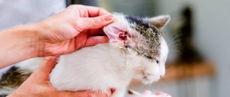
In cats, symptoms of hepatitis appear when the disease is already at an advanced stage. First, hepatic jaundice develops against the background of hyperbilirubinemia (high levels of bilirubin in the blood). The first signs of jaundice are yellowing of the inner surface of the ears, oral mucosa and conjunctiva. At the same time, the urine darkens, and the feces become colorless due to the lack of strecobilin pigment. Jaundice is accompanied by other symptoms:
- dyspepsia in the form of vomiting, loose stools;
- increased thirst due to dehydration;
- loss of appetite and weight loss;
- temperature increase;
- liver enlargement.