Among the diseases that cats suffer from, a significant proportion are those caused by various parasites. One of the most common diseases in this group is demodicosis. The causative agent of demodicosis in cats is the subcutaneous mite. This parasite can cause a lot of anxiety and problems for your mustachioed pet. The Murkoshi team will talk about what it is, how the disease progresses and how to remove this parasite in this article.
1) Pathogens of demodicosis and methods of infection 2) Risk groups 3) Types of demodicosis 4) Symptoms of subcutaneous mites 5) Treatment of a localized form 6) Treatment of a generalized form 7) Prevention of demodicosis
Pathogens of demodicosis and methods of infection
The causative agent of the disease is the subcutaneous mite - demodex. Actually, there are two types of subcutaneous mites (even three, but the third is currently poorly studied and not described): Demodex cati and Demodex gatoi. The first species (Demodex cati) always lives on the cat's body, in the hair follicles (the places where the hairs of the fur grow), and usually does not cause any trouble. This species is not at all contagious. But in some cases, such as illness, stress, decreased immunity of the kitten, this subcutaneous mite on the body multiplies excessively, accordingly, causing toothache and other inconveniences.
The second species (Demodex gatoi) is an infectious parasite. This subcutaneous mite is transmitted from a sick cat to another (but it is not dangerous for humans). It lives and reproduces in the stratum corneum of the skin (the surface layer consisting of keratinized cells). If this parasite gets on a cat's body, it can lead to illness. Although the body of healthy animals with strong immunity in most cases copes with subcutaneous mites on their own. Demodicosis occurs if the immune system is weakened for some reason.
When people and animals have a common enemy
Both man and his four-legged beloved creature are the object of desire of a harmful parasite. But since they have different types of ticks, mutual infection is excluded. True, there are isolated cases when people carry a “feline” type of infection in their bodies. Or cats are carriers of the “human” virus. Then, without affecting the intermediate link, that is, the foreign body, the tick infects the organism of an object “native” to its species.
The parasite can only be seen with multiple magnification, for example under a microscope, since it is very small, the female is up to 0.5 mm, the male is even smaller. The biological life cycle - eggs - larvae - adult creatures lasts up to 25 days. Demodex is unpretentious in its requirements. It quietly spends its life in one follicle or gland, without making passages under the skin, without sucking blood from the owner. If suddenly the tick’s “house” becomes clogged, it will become a favorable environment for the development of the disease. Several individuals (up to 15-17 pieces) accumulate together and provoke a purulent process. Such massive infections cause lesions over large areas of the skin.
Demodex is also present in apparently healthy skin. But the host’s strong immunity copes with the waste products of the harmful guest. However, once the immune system slightly weakens its protective barriers, the situation will change dramatically. What causes a decrease in immunity:
- poor nutrition, lacking vitamins;
- unsuitable living conditions;
- helminthic infestations;
- infections;
- stressful situations;
- antibiotic treatment.
The parasite quickly activates and manifests itself in all its nasty essence.
At-risk groups
Theoretically, all cats that have been in contact with sick animals can become infected with subcutaneous mites. But the following groups are most often susceptible to demodicosis:
— offspring of cats with demodicosis; — animals with weakened immunity and autoimmune diseases; — cats in the postoperative period; - animals in the recovery period after infectious diseases; — kittens and adult cats with rickets; - animals exhausted after starvation; - pets who have suffered severe stress; - some breeds: Siamese, Burmese, Devon Rex.
Prevention
- Avoid contact between a healthy cat and a cat with demodicosis.
- Timely detection of the disease and contact a specialist.
- Timely preventive treatment of cats against ectoparasites that live on the animal’s skin.
- Treating premises, cages, and places of residence of cats infected or suspected of demodicosis with acaricidal preparations, the action of which is directed against ticks.
- It is recommended to destroy bedding, feeders, equipment (rags, brushes, etc.) from a sick animal.
Varieties of demodicosis
Veterinarians distinguish two forms of demodicosis - localized and generalized. When localized, one area of the animal’s body (sometimes several) is affected, and there are no signs of the disease on the cat’s paws. With generalized disease, many areas of the body are affected at once, and the paws also suffer. The generalized form, of course, is much more difficult to tolerate and more difficult to treat. If a cat gets sick with a generalized type of demodicosis, then there is a high probability that she will pass the disease on by inheritance. Therefore, such cats must be sterilized.
Preventive measures
Prevention of demodicosis consists of several basic measures:
- regular and timely vaccination against fleas and ticks (special preventive drops and collars are sold in every pet pharmacy);
- selection of the correct diet (you need a balanced diet rich in proteins);
- providing the cat with all the necessary vitamins and minerals (additional vitamins can be prescribed by a veterinarian);
- providing comfortable and safe conditions for the cat (compliance with sanitary standards, protecting the animal from stress, etc.).
The pet's immune system will not fail if the cat is kept according to all the rules. She should eat well and live in a clean and dry house. A cat should not be left alone for a long time, because many animals suffer from loneliness, become depressed, and eventually get sick. If your cat has company (another cat or dog), then the sick individual needs to be isolated. Let it be a separate room, and sometimes you can take healthy animals, for example, to relatives.
To avoid passing ticks to your pets with your own hands, you need to take care of your hygiene. After each contact with a sick cat, you need to wash your hands and treat them with an antibacterial solution. The clothes you were wearing at the time of contact should be washed immediately. While your pet is sick, limit his access to the outdoors. There have been cases when cats with a frightening itchy “mask” on their faces suffered at the hands of people. Cats that were mistaken for being highly contagious were stoned and abused. Therefore, let your pet heal and get stronger first.
Demodectic mange (red scabies) is a rare disease caused by subcutaneous mites. In this case, individual, small areas of the body may be affected. If scabies is not treated, the affected areas merge into large bald spots with wounds and ulcers. It is difficult to cure this infection, but it is possible. To do this, veterinarians prescribe products to care for diseased skin, injections, vitamins, etc. It is important to follow all recommendations and prescriptions. The danger of this tick for humans is exaggerated (humans cannot become infected with feline demodicosis).
Symptoms of subcutaneous mites
In the case of demodicosis, there is a “pitfall”: the disease develops gradually, and accordingly, symptoms gradually appear. Therefore, it is not always possible to immediately notice signs of a subcutaneous mite in a kitten. Moreover, the danger is that the longer Demodex parasitizes, the more dangerous it is in terms of reducing the animal’s immunity. This is why it is important to take your cat to the vet at the first sign of symptoms. It's better to be safe than to get sick! So, the following symptoms may indicate the presence of a subcutaneous tick in a cat:
- redness of the skin; - the appearance of rashes in the form of nodules or small pustules; - deterioration in the appearance of the coat; - such a characteristic symptom as “demodicosis glasses” appears - hair loss and peeling of the skin around the eyes; - hair loss and peeling of the skin on the head, ears, neck (localized form), as well as on the legs and torso (generalized form); — violation of skin pigmentation; - acne; - itching; - bleeding wounds.
You can accurately diagnose demodicosis in a cat by taking scrapings from the affected areas of the body. Treatment will depend on whether the form is localized or generalized. Owners need to keep in mind that demodicosis is a very serious disease that may require long-term therapy (from six months to a year). Experienced volunteers of the Murkosha shelter warn: self-medication if a cat has a subcutaneous tick is unacceptable! “Folk remedies”, at best, have not proven their effectiveness against demodicosis, and at worst, they can worsen the animal’s condition. Remember that only a doctor can prescribe the exact course of treatment. He will tell you if any of the "folk remedies" are suitable for your situation.
annotation
The human demodex mite (Demodex folliculorum and Demodex brevis) occupies a high place in the evolutionary and phylogenetic hierarchy of skin microorganisms, although in most people their presence is of no significance. Demodectic mange is a distinct skin disorder that can mimic many other inflammatory dermatoses such as folliculitis, rosacea, and perioral dermatitis, leading to nonspecific and confusing descriptions in the literature. Here we propose to classify human demodicosis into primary and secondary forms. The secondary form is mainly associated with immunosuppression. Clinical manifestations of primary demodicosis may include: 1) pityriasis folliculitis, involving sebaceous hair follicles without visible inflammation; 2) papulopustular / spherical demodicosis with severe inflammation, most often affecting the perioral and periorbital areas of the face; 3) ocular demodicosis, causing chronic blepharitis, chalazion or, less commonly, keratoconjunctivitis; and 4) auricular demodicosis causing otitis externa. Secondary demodicosis is usually associated with systemic or local immunosuppression. Endosymbiosis between certain bacteria and the demodex mite deserves great attention in the pathogenesis of demodicosis. Further clinical observations and experiments are needed to prove this hypothesis.
Introduction
The demodex mite (D) was first described as a worm by Jacob Henle in Zurich in 1841, and then correctly classified as the human demodex mite by dermatologist Carl Gustav Theodor Simon in Berlin in 1842. D. has intrigued parasitologists, veterinarians and dermatologists for over 170 years. D. belongs according to the taxonomic classification to Arthropoda / Chelicerata / Arachnida / Acarina / Demodicidae / Demodex / Demodex folliculorum or Demodex brevis. Compared to other human skin microorganisms such as Propionibacterium acne, Staphylococcus epidermidis and Malassezia, Demodex mites rank higher in the evolutionary hierarchy. Unlike other human mites such as the scabies mite, Cimex lectularius or Dermatophagoides pteronyssinus/farinae, D. remain largely dormant and harmless and rarely cause immunological or allergic reactions. The disease state of demodicosis in other mammals such as dogs and cats can be very extensive and fatal if left untreated. However, the connection between Demodex mites and human diseases is much less studied. This article is intended to highlight the difference between primary demodicosis and other inflammatory dermatoses similar to demodicosis. In addition, we hope to pave the way for research into the pathogenesis of demodicosis and encourage basic research on the biology of demodex mites.
Definition and diagnosis
Human demodicosis (HD) is a skin disease that primarily affects the pilosebaceous follicles (SVF), due to the parasitism of the demodex mite, the favorite localization of which is the face and head. Two clinical variants can be observed - primary and secondary. Primary demodicosis (PD) can be defined when the following diagnostic criteria are met: 1) absence of pre-existing or concurrent inflammatory dermatoses such as acne, rosacea or eyelid dermatitis; 2) an abnormal increase in tick colonization of the SVF, which should be detected in the presence of active foci at the time of examination; and 3) remission of the disease only after adequate therapy with topical or systemic acaricides, but not with anti-inflammatory antibiotics such as tetracycline or doxycycline, or macrolides (erythromycin/azithromycin/clarithromycin). Numbers of more than 5 mites per cm2 identified on standardized biopsies of affected skin are currently estimated as pathological, although this figure is based on very limited studies. Preliminary studies using new diagnostic techniques such as dermatoscopy, confocal laser scanning microscopy, or high-definition optical coherence tomography show promising results, however, the accuracy, reliability, and clinical utility of these techniques remain to be determined. Integration of imaging and fluorescein staining can provide rapid and accurate detection and quantification of mites in routine clinical practice. Skin lesions associated with an abnormal increase in the number of demodex mites in patients with other known skin or systemic diseases may be classified as secondary demodicosis. This occurs most often in patients with significantly weakened immune systems, such as those with leukemia and HIV infection, as well as those who have received treatment with immunosuppressants, including topical glucocorticoids or topical calcineurin inhibitors. Although less common, other conditions leading to secondary demodicosis have been described, such as treatment with epidermal growth factor receptor inhibitors, skin tumors, chronic renal failure and phototherapy. The underlying role of Demodex in the pathogenesis of rosacea remains controversial, given that none of the available data shows a direct positive cause-and-effect relationship. The hypothesis that primary demodicosis is caused by D. folliculorum and secondary demodicosis by D. brevis has not yet been proven and differs significantly from our classification concept.
Clinical manifestations of primary demodicosis
Primary demodicosis is clinically characterized by 1) late onset, usually after 40 years of age and especially in the elderly population; 2) the skin of the face is affected, usually involving the periorificial areas (perioral, periorbital or periauricular); 3) as a rule, an asymmetrical location, with a grouping of irregularly shaped foci within the boundaries of one affected area; 4) connection of rashes with follicles; and 5) the rash is asymptomatic or mildly pruritic. The lesions usually lack the classic manifestations of rosacea, such as erythema or telangiectasia. In contrast, secondary demodicosis can occur early in life and is characterized by more widespread lesions and more severe inflammation on the face and/or trunk. The history and characteristics of diseases such as perioral dermatitis or rosacea are usually obvious.
Terminology of demodicosis
The current terminology of demodecosis is nonspecific and confusing, and may include such terms as multi-colored folliculitis, rosaciaiform dermatitis, demodedic rosacea, demodex dermatitis of faces, granulomatous rosacea, dermatitis/periorbital dermatitis, demodicosis, face demodecosis, branchy folliculitis, and folliculitis of the heads . We propose the following classification to describe primary demodicosis ( Table 2
).
| Table 2. Primary human demodicosis: classification and nomenclature | ||
| Current terminology and description | Proposed nomenclature | Definitions and clinical manifestations |
| Follicular pityriasis | Spinulate demodicosis | Rash elements associated with SVF, with or without mild erythema and inflammation |
| Rosaceaform demodicosis, perioral/periorbital/periauricular | Papulopustular demodicosis, perioral, periorbital, periauricular demodicosis | Papulopustular eruptions are mainly on the face, in patients without (primary form) or with pre-existing inflammatory dermatoses such as rosacea or eyelid dermatitis (secondary form). The rash tends to spread around the mouth, periorbital and periauricular areas |
Spinulate demodicosis, now known as pityriasis folliculitis, is described as follicular, isolated but clustered, with or without mild erythema and mild inflammation.
Demodex folliculitis, that is, inflammation of the follicle caused by Demodex, is morphologically divided into the following types: papulopustular, nodular-vesicular and spherical (abscess). Demodicosis of the eyelids is a primary demodicosis and should be distinguished from dermatitis of the eyelids with a secondary increase in the number of demodex mites. In accordance with the morphological structure and localization, the terms “Papulopustular perioral demodicosis” or “Papulopustular periorbital demodicosis” can be used. Demodectic mange of the scalp (demodetic mange of the scalp) occurs more often in areas of balding on the scalp in older men, who rarely develop bacterial folliculitis (personal observations). Crusting demodicosis with thick yellow crusts is associated with chronic inflammation caused by Demodex. Ocular demodicosis may include blepharitis or chalazion and, less commonly, Demodex conjunctivitis. With auricular demodicosis, the external auditory canal or eardrum is affected.
Pathogenesis
The pathogenesis of demodicosis remains largely unclear. It is unclear what causes demodex mites to become active. What causes the development of cortical demodicosis - an increase in the host's immune response or excessive proliferation of mites, as is observed with Norwegian scabies?
The recent observation and hypothesis that the bacterium Bacillus oleronius, originally isolated from the gut of termites, may be responsible for initiating the inflammatory processes of papulopustular rosacea provides food for thought. However, so far the bacterium has been isolated from Demodex mites from only one patient with papulopustular rosacea. The lack of serum reactivity to Bacillus oleronius antigens in a significant proportion (20%) of patients with early rosacea and the presence of antibodies in 40% of the control group without visible rosacea cast doubt on the causative role of these bacteria in the inflammatory process. In another study of chronic blepharitis associated with Demodex mites, B. oleronius was found in cultured eyelashes of 5 of 30 healthy individuals and only 2 of 15 patients with moderate blepharitis, which may indicate a low pathogenicity of the strains in the development of chronic inflammation. It remains to be determined whether Bacillus oleronius is present in all Demodex mites or is present only in active mites, and whether it is simply a saprophytic flora or serves as a cofactor in the initiation or maintenance of skin inflammation.
A recent ophthalmic study suggested that D. brevis may play a more important role than D. folliculorum in the pathogenesis of chalazion, and recurrences are significantly higher in patients with D. brevis than with D. folliculorum.
Treatment
Treatment of demodicosis is based on weak evidence, mainly for the following reasons: 1) the lack of an ideal laboratory model in which the effectiveness of drugs and their minimum inhibitory concentrations can be tested; 2) clinical confusion between primary demodicosis and inflammatory diseases (rosacea with or without secondary demodicosis); and 3) dual effects, both anti-inflammatory and antimicrobial, of many agents. Ivermectin has a purely acaricidal effect and has proven to be the treatment of choice for demodicosis in dogs and humans. However, the dose of oral ivermectin recommended for the treatment of demodicosis in dogs is significantly higher (0.3-0.6 mg/kg per day for 10-33 weeks) than for humans (0.2 mg/kg single dose). Although a number of other topical acaricides, such as permethrin 5%, benzyl benzoate 10-25%, crotamiton 10%, lindane 1% or malathion 0.5%, have been approved for the treatment of scabies, there is currently limited evidence for the effectiveness of these acaricides in the treatment of demodicosis are very limited. The superiority of topical 10% benzyl benzoate in killing Demodex mites has only been demonstrated in a small number of cases. It is unclear whether the effect in rosacea from low doses of systemic tetracycline, macrolide antibiotics, topical azelaic acid or 15-20% of topical 0.75-2% metronidazole is due mainly to anti-inflammatory or also partially acaricidal action. The hypothesis that tetracyclines may influence the proliferation of Demodex mites by acting on endosymbiotic B. oleronius remains to be confirmed. The optimal dose of systemic metronidazole in the treatment of demodicosis has not yet been fully determined. It would be interesting to see whether moxidectin, which is approved for the treatment of generalized canine demodicosis, is effective in humans.
In conclusion, it should be noted that the demodex mite is the most common parasite of humans, being its lifelong symbiont. The physiological role of Demodex mites in healthy skin remains a mystery, and understanding how they evade immune surveillance, particularly the innate immune system, may be critical to understanding human-mite interactions. The typical clinical manifestations and the specific therapeutic response to therapy with pure acaricides such as ivermectin suggest that primary human demodicosis is a disease of a special kind. Induction of inflammation is an important step in pathogenesis. The spread of Demodex mites, the activation of unknown virulence factors and the pathogenic role of mite endosymbionts are key issues that require intensive study. Clinical distinction from other inflammatory dermatoses that mimic demodectic mange, such as papulopustular rosacea or perioral dermatitis, is important. Effective acaricidal drugs and their optimal doses for suppressing Demodex mites have yet to be identified and standardized. Progress in this area is hampered by the lack of necessary conditions, both in vitro and in vivo, for experimental studies. Recognition of human demodicosis as a major disease will facilitate the further development of new therapeutic strategies.
Treatment of localized form
Localized demodicosis is treated in stages.
1. First, you need to bathe your pet using special “Doctor” and “Elite” pet shampoos. They will cleanse the cat's skin and fur from suppuration, lymph and dandruff. 2. Then all affected areas on the cat’s body should be treated with a solution of chlorhexidine or hydrogen peroxide - this will cleanse the skin of scabs and crusts. You need to wait until these places dry out. 3. On the recommendation of a veterinarian, you can use the antiseptic "Citeal" - once every 2-3 days, you need to wash the affected areas of the cat's body with it. 4. After this, the treatment of demodicosis itself begins, that is, the removal of subcutaneous mites from the cat. The skin is treated with special solutions, ointments and gels as prescribed by the veterinarian: “Amitraz”, “Stronghold”, “Advocate”, “Demos”, sulfur and aversectin ointments, etc. 5. Areas with lost hair and wounds are treated with the preparations “Tsipam”, “ Perol”, “Ektodes”, “Ivermek”, “Neostomazan”, “Mikodemotsid”, “Amit”, etc. 6. The pet must be given means to increase immunity: “Gamavit”, “Immunol”, “Gala-vet”, “Maksidin”.
Please note that the exact treatment regimen is prescribed by a specialist, depending on the age of the cat, the degree of damage, general condition and other factors.
Read more about communicating with a veterinarian: 5 rules for effective interaction with a veterinarian
Read also
Ear mites in cats: signs and treatment
The appearance of a parasite such as ear mites in a cat is the beginning of a serious disease that must be eradicated at the first symptoms.
Worms in cats: symptoms, signs and treatment
The first signs of infection may be absent for some time or resemble manifestations of other diseases
Sterilizing a cat: at home or in the clinic?
Sterilization is an intervention as a result of which an animal (a cat in particular) loses its ability to reproduce
Distemper in cats (panleukopenia): symptoms and treatment
Distemper in cats is a serious disease caused by one of the viruses of the Parvoviridae group. The terrible common name means cat
Ringworm in a cat: Treatment and symptoms
Ringworm is a serious infectious disease. When a cat gets sick itself, it transmits the disease to its owners.
Treatment of the generalized form
The generalized form is more difficult to treat and takes longer. First of all, it is necessary to eliminate the primary disease that caused the exacerbation of demodicosis. Without this, complete recovery of the animal is impossible. Veterinarians also advise completely trimming the cat to make it easier to treat with medications (since the disease covers almost the entire body). Next, the same methods are used as in the treatment of a localized form.
Often, veterinarians also prescribe a course of Dectomax or Cydectin injections. At the same time, the animal is prescribed injections of antibiotics - Kanamycin, Betamox, Baytril, Amoxicillin, etc.
It is also necessary to restore the cat’s immunity - the drug “Ligfol” will help here, as well as a vitamin and mineral complex. We draw special attention to the fact that treatment of this form of the disease should be carried out under the supervision of a specialist.
General symptoms
The owner can determine the presence of demodicosis in his cat on his own. To do this, it is enough to examine the pet, since typical symptoms form at the very beginning of the development of the pathology.
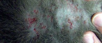
The main signs of demodicosis include:
- the appearance of small reddened areas on the surface of the skin and small ulcers;
- formation of characteristic scabs from scratching in the affected area;
- development of severe itching;
- hair loss, ending with complete baldness of the skin area;
- refusal to eat;
- the formation of cracks, bleeding wounds and swelling in the affected area;
- weakness and lethargy of the cat;
- enlarged lymph nodes.
A complication of demodicosis is the development of diseases of internal organs. In this case, signs of concomitant pathologies are added to the current clinical picture.
Diagnosis of cat scabies
To confirm demodicosis, differential diagnosis is necessary. The following diseases have similar symptoms :
- ringworm;
- folliculitis of bacterial origin;
- pyoderma;
- skin fungus;
- dermatosis;
- eczema;
- streptococcosis.
The veterinarian takes skin cells from the affected areas for analysis and sends them for laboratory testing. This helps not only to detect the presence of a tick, but also to determine the stage of its current development.
© shutterstock
If only adult parasites are detected in a cat, we can only talk about carriage.