Pets are susceptible to illnesses just like people. Eye disease in cats is common. Responsibility for the health of the pet lies with the person. The owner’s task is to recognize the signs of pathology in time and contact a veterinarian. The doctor will conduct an examination, make a diagnosis and prescribe treatment. You cannot ignore the symptoms and hope that the disease will go away on its own. Lack of timely treatment leads to deterioration of vision, extensive inflammation and blindness.
What eye diseases can a cat have?
Eye diseases in cats can be divided into two large groups:
- accompanied by inflammatory processes of the eye organs: conjunctivitis,
- keratitis,
- keratoconjunctevitis,
- iritis,
- inflammation of the nasolacrimal duct,
- blepharitis,
- panophthalmitis.
- cataract,
Diseases can occur against the background of another infectious disease or be the main cause of impairment of the animal’s health; acute and chronic forms of their course are possible.
Video: eye diseases in cats
Symptoms of cataracts
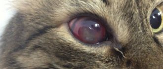
The second most common disease is cataracts. As a result of natural aging or various metabolic processes in the body, clouding of the lens occurs.
- A cloudy lens is unable to perform its main function - accommodation of the eye.
- A specific symptom that should alert a cat owner is the appearance of shine in the eyes.
At the same time, the pet also squints, trying to reflexively increase visual acuity.
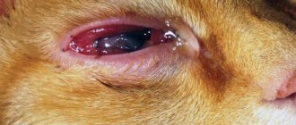
The photo of this eye disease shows a pronounced clouding of the eyeball, which every owner will notice.
Conjunctivitis - inflammation of the mucous membrane of the eye
The mucous membrane covers the eyeball and the inner surface of the eyelids, and the causes of its inflammation can be any health disorder: viral infection, allergies, mechanical damage. Depending on the degree of damage to the eye organs, there are several types of conjunctivitis:
- Catarrhal. Accompanied by redness and swelling of the eyelids, which can be turned out because of this. Constant lacrimation. Possible increase in temperature.
Conjunctivitis can be easily identified even in the early stages
- Purulent. Discharge of pus is added to the previous symptoms.
- Phlegmonous. In this case, the pus not only comes out, but also remains inside the eye.
- Follicular. Not only the conjunctiva becomes inflamed, but also the lymphatic follicles of the eye. In the case of follicular conjunctivitis, surgical intervention is required.
For a complete cure, it is necessary to eliminate the root cause of the disease so that the disease does not become chronic or lead to more severe consequences. The course of treatment begins with cleansing the eye mucosa from discharge and pathogenic microflora. To do this, you can use a solution of furatsilin (0.1 g per two glasses of water) or an infusion of calendula. But the main treatment is prescribed by a qualified veterinary clinic specialist.
Video: conjunctivitis in animals
Diagnostics
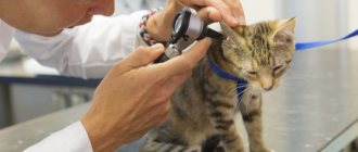
Before starting treatment, you should take your pet to see a veterinarian. The doctor must conduct a complete diagnosis to establish the correct diagnosis.
During the examination, the doctor must ask about the animal’s condition. The owner must talk about the pet’s behavior and accompanying symptoms. The specialist should also carefully examine the eyes of the cat.
To establish an accurate diagnosis, you will need to take stool and urine tests; it is better to bring the material with you right away. The veterinarian must take discharge from the eyes to determine the cause of the disease.
Keratitis - inflammation of the cornea
The causes of keratitis can be:
- mechanical influences;
- infectious diseases: calcivirus,
- toxoplasmosis.
There are two types of keratitis:
- superficial - only the outer epithelial layer is affected;
- deep - the process extends to the corneal parenchyma.
Both types can be accompanied by purulent discharge.
The symptoms of the onset of the disease are quite clear:
- lacrimation;
- the palpebral fissure is half-closed;
- the cornea begins to become cloudy, and with further development of the inflammatory process, the clouding intensifies;
- redness of the conjunctiva.
This is what the initial stage of keratitis may look like
If keratitis does not have complications, then the transparency of the cornea will be restored, otherwise a thorn will form on the eye.
When providing first aid, it is necessary to rinse the conjunctival sac with a disinfecting solution. To prepare it you can use:
- boric acid: half a teaspoon of powder is dissolved in one glass of cold boiled water;
- furatsilin: 0.2 g of powder per liter of warm water;
- herbal remedies: 1 tsp. chamomile or calendula is brewed with half a glass of boiling water, then cooled.
If there is pus, it must be completely removed from the eyelashes so as not to contribute to the spread of infection. The swab is moistened with a disinfectant solution and applied to the eye to soften the purulent crusts. Then the eyelids are cleaned.
Video: how to put drops in a cat’s eyes
Washing of the conjunctival sac is carried out according to the following scheme:
- the lower eyelid of the animal is slightly retracted;
- a solution is instilled into the opened cavity;
- the eyelid is released, the skin under it is lightly massaged;
- excess moisture is removed with a swab.
After this, you can treat the cornea with eye drops or hydrocortisone ointment. But the full course of treatment is prescribed only by a doctor after an accurate diagnosis has been established.
Video: what to do if a kitten’s eye is cloudy
How to properly treat a kitten's eyes
The duration and course of the disease depends on the careful implementation of the procedures prescribed by the veterinarian. There is no need to do it on your own, you just need to follow the doctor’s instructions.
If the kitten's eyes and muzzle are covered with crusts, they should be softened using Vaseline. The next stage of daily care is washing with antiseptic solutions or herbal decoctions.
- Place the animal on its back with its muzzle up, fix the position so that the baby does not kick. If the kitten struggles and scratches, you can wrap it in a towel.
- Prepare two bowls with the medicinal solution: one for the right eye, the second for the left. This precaution is necessary to prevent the infection (bacteria) from spreading. Gently drop the product into your left eye using a pipette, and then use a sponge soaked in the same solution to wipe your eyelids. Do not press hard on the eyeball, move from the outer corner of the eye to the inner.
- Perform the same manipulation for the right eye, using the contents of the second bowl.
- Do not place the dropper too close to the eye, as this may cause injury to the kitten. Each eye is wiped twice in one procedure. The course of treatment is usually 7-10 days.
After cleansing, the eyes are instilled, slightly pulling the eyelid. The kitten must blink so that the medicine is evenly distributed over the eyeball. If necessary (as prescribed by a doctor), apply the ointment according to the following scenario:
- place the animal on its side so that its muzzle “looks” up;
- lift the lower eyelid and apply a little ointment (usually tetracycline);
- massage the eyelid so that the drug is distributed;
- treat the other eye;
- course of treatment up to 10 days.
If you conscientiously perform all the procedures prescribed by the doctor, the kitten will recover quickly!
Third eyelid prolapse
The inner, or third, eyelid is a thin membrane that protects the cornea of the eye from mechanical influences and dirt. In a healthy pet it is almost invisible. If the third eyelid does not fold, this is a sign of pathology. However, the list of diseases that change the condition of this organ is quite large and includes not only eye diseases:
- eye injury or foreign body penetration;
Loss of the third eyelid in only one eye indicates that problems are related to the visual organs
- conjunctivitis of all types;
- inflammatory process in the membrane itself;
- manifestation of allergies, including to external parasites;
- helminth infection;
- problems with the gastrointestinal tract.
When the third eyelid appears, it is not recommended to carry out any treatment on your own. If the condition does not return to normal within the next few hours, you should seek veterinary help.
You can read more about the third eyelid in cats in the article “Third eyelid in cats: causes, symptoms and treatment.”
Video: third eyelid with herpes virus infection
How is demodicosis of the eye transmitted?
Demodectic mange of the eyes can be contracted at home - by sharing hygiene items, towels, bed linen, and cosmetics with the patient.
To prevent re-infection, you need to change your pillowcase and towel (or use paper towels) daily, and throw away contaminated cosmetics.
To treat each eyelid, you must use a separate (new each time) cotton swab.
1 Diagnosis and treatment of demodicosis
2 Diagnosis and treatment of demodicosis
3 Treatment of demodicosis
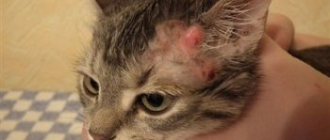
Blepharitis - inflammation of the eyelid
The first signs of blepharitis disease are redness and thickening of the conjunctiva. Inflammation may be accompanied by the release of pus, mucus, the appearance of bleeding ulcers, and severe lacrimation. If left untreated, the inflammatory process spreads to nearby organs.
The causes of blepharitis are different:
- mechanical eye injury or burn (thermal or chemical);
- manifestation of allergies to medications or food;
- fungal or bacterial infection;
- parasite infection.
To treat blepharitis, even at the initial stage, it is not enough to relieve the inflammatory process, since it is necessary to determine the initial cause of the disease, and this can only be done by a qualified specialist.
Blepharitis is often confused with conjunctivitis, but they are different diseases and require different treatments.
Panophthalmitis is a general infection of all tissues of the eye.
Panophthalmitis is a rare disease, but when diagnosing it, the question of preserving vision does not arise: the eye is removed so that the inflammatory process does not spread to the brain.
Panophthalmitis develops under the influence of pathogens of purulent infection (staphylococci, streptococci, sometimes E. coli), which enter the eye through a wound or perforation of the cornea in the absence of treatment for other eye diseases aggravated by purulent ulcers.
Signs of panophthalmitis:
- sharp pain in the eye;
- fear of light;
- lacrimation;
- desire to close eyes;
- swelling of the eyelids and conjunctiva;
- cloudy and purulent cornea,
- blurred vision.
At the first signs of panophthalmitis, you should contact a veterinary clinic.
With further development of the disease, the cornea melts and the eyeball moves forward. The eye loses its mobility. A large amount of pus and exudate is released.
Treatment of panophthalmitis can only be carried out in a veterinary clinic. First, the inflammatory process is removed using a novocaine blockade, then surgical removal of the eyeball is performed. If surgical intervention is performed on time, there should be no complications, and the animal lives for a long time, remaining active and healthy.
Video: purulent panophthalmitis in a cat
Main symptoms and treatment methods
For timely and adequate treatment, it is necessary to know the characteristic signs of eye diseases.
Features of conjunctivitis
One of the common diseases in pets is conjunctivitis. Inflammation of the mucous membrane occurs for the following reasons:
- injuries;
- infectious diseases;
- allergic reactions;
- infection with worms and ectoparasites;
- lack of vitamins in the body.
There are several forms of conjunctivitis: catarrhal, follicular, ulcerative and purulent. The main symptoms indicating the development of conjunctivitis are:
- redness of the mucous membrane;
- swelling of the eyelids;
- lacrimation;
- the appearance of pus;
- eyelid gluing;
- photophobia;
- cloudiness.
Purulent conjunctivitis is indicated by bilateral inflammation. In addition, the owner notes the following symptoms of the disease:
- general depression of the pet. He becomes lethargic, does not want to play and frolic;
- high body temperature. Normally, the body temperature of an adult animal is 38-39°C; in children, the figure may be half a degree higher (38.5-39.5);
- poor appetite or its complete absence also indicates a painful condition;
- the cat tries to avoid inspection and palpation, this process causes him pain in the area of the eyeball;
- in advanced cases, inflammation spreads to the cornea.
Treatment steps are as follows:
- A visit to the veterinary clinic to remove a foreign object from the eye.
- Washing can be done at home every 3-4 hours. For the procedure, use a warm solution of manganese, furatsilin, chamomile or calendula decoction. Herbs help reduce inflammation, eliminate swelling and itching. In addition, natural decoctions have anti-inflammatory properties. Manganese copes well with infection, but at the same time dries out the mucous membrane. Therefore, a pale pink solution is used. It is recommended not only to thoroughly dissolve the manganese crystals, but also to filter the resulting composition through several layers of gauze. This guarantees the absence of crystals in the water, the accidental entry of which can cause a burn to the mucous membrane. Furacilin is diluted in hot water (1 g per 5 l). Used for washing while warm.
- Applying ointment. At a pharmacy or pet store you need to buy any eye ointment, for example, Tetracycline, Syntomycin or Erythromycin. The composition is placed behind the eyelid. The procedure is carried out wearing gloves. The ointment should be applied carefully with a special glass rod. Applying with fingers or cotton swabs is unacceptable. After each application of the ointment, the stick is disinfected in boiling water. This is necessary in order not to spread the infection in the future. Some people use an ointment tube to apply the ointment. This must be done extremely carefully, otherwise bacteria will settle on the spout of the tube and, during the next treatment, will again get into the eyes.
- Instillation is carried out only in washed eyes. It is necessary to instill 2-3 drops every 4 hours.
- If the disease is caused by an infection, a course of antibiotic therapy is necessary.
- A solution of novocaine and hydrocortisone (ratio 1 ml/0.2 ml) helps relieve swelling. The composition is instilled into the conjunctival sac.
- Chronic pathology is recommended to be treated with silver-based drugs.
- In a veterinary hospital, a novocaine blockade is carried out. It reduces pain and improves tissue nutrition, which leads to a speedy recovery.
- For follicular conjunctivitis, surgical intervention is performed. After the operation, ointments and antibiotics are prescribed.
Keratitis and its types
Inflammation and clouding of the cornea in cats indicate the development of keratitis. The disease is acquired and requires proper treatment. The reasons for the development of keratitis are:
- mechanical injuries of the cornea. The ingress of solid dust particles, grains of sand, blades of grass, and fibers leads to mechanical damage;
- conjunctivitis. The combination of diseases leads to the formation of keratoconjunctivitis;
- thermal or chemical burns;
- infections and viruses. Typically, keratoconjunctivitis develops against the background of a general infection of the body (herpes, adenovirus). In this case, complex treatment is required, otherwise it will not be possible to get rid of keratitis;
- allergic reactions to food, dust, pollen, household chemicals;
- inflammatory processes in the lacrimal glands, leading to drying out of the cornea;
- lack of vitamins in food;
- genetic predisposition to the disease (in British, Sphynx, Persian and Siamese cats).
The appearance of the following symptoms should alert the cat owner:
- clouding of the cornea, its dullness and roughness;
- accumulation of blood vessels on the cornea;
- the discharge causes the fur around the eye to become wet;
- accumulation of pus;
- fear of bright light;
- the appearance of scars indicates the irreversibility of the process. The animal may go blind.
Treatment is prescribed after a thorough examination and confirmation of the diagnosis. The initial stage of the disease can be treated with drugs. In advanced cases, laser correction of the cornea is used.
The following groups of drugs are used as complex therapy:
- antibacterial agents (Tetracycline, Erythromycin, Gentamicin eye ointment, Tobrex);
- antiviral and immunomodulatory drugs (Cycloferon, Gamavit).
Keratitis requires long-term treatment and special care for your pet. If the veterinarian's instructions are not followed, the animal may go blind.
Third eyelid prolapse
The nictitating membrane in cats is a kind of protection provided by nature. The third eyelid is a whitish film located in the inner corner of the eye. The prolapse of this membrane can be noticed by the following signs:
- uncontrollable trembling, closing eyelids;
- increased lacrimation;
- redness around the eye;
- copious discharge of mucus or pus.
Prolapse of the nictitating membrane is associated with the following diseases:
- conjunctivitis;
- eye damage;
- gastrointestinal diseases;
- fungal pathologies;
- allergic reactions;
- helminthic infestations.
If you suspect third eyelid prolapse, you should wash your pet's eyes with a warm decoction of chamomile or calendula. If after a few hours the nictitating membrane has not returned to its place, a visit to the veterinarian is necessary. Surgery may be required.
Detection and treatment of blepharitis
There are several types of blepharitis in cats:
- simple;
- with the formation of ulcers;
- scaly;
- meibomian;
- demodectic;
- allergic.
If left untreated, simple blepharitis develops into ulcerative blepharitis. The plaque that appears along the growth of the eyelashes disappears, and ulcers appear in its place. The progression of the disease is accompanied by loss of eyelashes, further damage to the conjunctiva and eyes, and panophthalmitis develops.
The initial form of blepharitis is similar to the manifestations of conjunctivitis. The cat is worried about the following symptoms:
- lacrimation;
- itching;
- swelling and redness of the eyelids;
- reduction in eye size.
The causes of pathology include:
- viral diseases;
- tick-borne infection;
- lichen;
- injuries;
- allergic manifestations;
- the appearance of dandruff;
- endocrine and autoimmune disorders.
After the tests, long-term treatment will be required:
- the eyelids are treated with brilliant green, alcohol-ether;
- to soften the scales, use saline solution and petroleum jelly, then remove them with a cotton-gauze swab (necessarily sterile);
- Sofradex, Betnesol, Iris are prescribed for instillation;
- the conjunctival sac is filled with one of the suspensions - Gentamicin, Syntomycin, Methyluracil.
To prevent eye scratching, a protective collar is needed. You can wash your eyes with herbal decoctions (chamomile, calendula, hyssop, sage, St. John's wort).
Danger of cataracts
The development of cataracts is indicated by clouding of the lens. It rarely develops in young individuals; it occurs against the background of infectious diseases. Occurs more often in older pets. Some breeds (Persians, exotics) have a genetic predisposition to the disease. Cataracts can be suspected based on the following signs:
- the animal walks carefully and very slowly;
- spatial orientation is impaired;
- in an unfamiliar environment, the cat bumps into objects and furniture.
After confirming the diagnosis, the veterinarian prescribes the necessary course of treatment. The process of clouding of the lens is irreversible; doctors can stop the rapid progression with the help of medications.
To maintain vision, use Taufon and Quinax drops. They are instilled 2 times a day, 1 drop in each eye for a month. Then they take a month's break and repeat the course. Treatment is carried out constantly, otherwise the cat may go blind.
Consequences of glaucoma and its treatment
Glaucoma is characterized by an increase in eye pressure. According to the generally accepted classification, the disease can be congenital, as well as open- and closed-angle. There are primary and secondary visual impairment. Primary glaucoma has a poor prognosis. In secondary cases, it is important to identify the cause of the pathology and eliminate it. In this case, the chances of vision restoration increase.
An important symptom to look out for is an overly dilated pupil. The eyeball hardens and enlarges, and traces of hemorrhages are noted on the conjunctiva. The open-angle form is characterized by clouded areas of the cornea. The closed-angle form is characterized by a ring-shaped closed opacification.
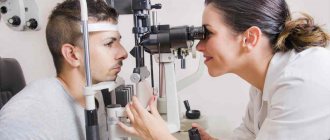
Pressure in the eyes reduces vision and leads to poor health. The pet needs consultation with an ophthalmologist and urgent treatment. To confirm the diagnosis, the doctor conducts an examination with a slit lamp and measures eye pressure with a Maklakov tonometer.
With open-angle glaucoma, the cat needs urgent prescription of drugs that reduce intraocular pressure. The following groups of drugs are used:
- diuretics (Mannitol);
- miotics (Pilocarpine, Physostigmine, Phospholine);
- adrenergic blockers (Timolol, Betaxolol);
- hyperosmotic drugs (Glycerin).
Corticosteroids may be an additional prescription. Conservative treatment methods have little effect on the development of the disease, so many pets undergo surgical intervention:
- cyclophotocoagulation;
- cyclocryotherapy;
- cyclodiathermy.
Veterinarians also use liquid nitrogen and retrobulbar injection of alcohol in the treatment of glaucoma. If vision cannot be restored, an intraocular prosthesis is installed. Removal of the eyeball may also be suggested.
Symptoms and causes of panophthalmitis
Purulent inflammation that affects all tissues of the eye is called panophthalmitis. Registered for complex wounds of the eyeball in cats. The main causes of the pathology are pneumococci, staphylococci, and E. coli that enter the eye from the outside during injury. The main signs of the disease:
- excessive tearing;
- uncontrolled closing of the eyelids;
- avoiding bright light;
- swelling of the eyelids and conjunctiva;
- clouding and swelling of the cornea;
- possible pinching of the mucous membrane of the eye.
The pet's vision sharply decreases and he is bothered by pain in the eye. As the disease progresses, other symptoms appear:
- anterior displacement of the eyeball;
- restriction of eye mobility;
- violation of the integrity of the sclera.
Treatment of panophthalmitis at home is impossible. The pet needs urgent hospitalization and surgery. The eyeball is removed and the eyelids are sutured. The sutures are removed after 24 hours, and the orbit must be lubricated with antiseptic agents for 7 days.
A dangerous complication of the disease is the transfer of purulent infection to the brain. In this case, the animal dies. Therefore, when the diagnosis of panophthalmitis is confirmed, the eye must be removed.
Combating inflammation of the nasolacrimal ducts
Excessive lacrimation and discoloration of the fur around the eyes are the main sign of an inflammatory process in the nasolacrimal ducts. The problem usually occurs in kittens, but can also affect adult animals. The causes of pathological lacrimation are:
- various eye diseases;
- blockage of tear ducts;
- foreign objects trapped in the lacrimal sac;
- inversion of the eyelids and curling of the eyelashes.
Treatment involves washing the canal or removing the lacrimal gland. If the amount of fluid released is minimal, conservative treatment is stopped. Obstruction is eliminated by rinsing through the nasal passages. Use disinfectant solutions of silver nitrate, furatsilin, boric acid, penicillin with novocaine (for pain relief).
In difficult situations, excision of the lacrimal sac or removal of the lacrimal gland is practiced. With excision, there is a possibility of the problem reoccurring due to the appearance of a scar.
Surgical intervention for entropion of the eyelid
Entropion of the eyelid occurs as a result of weakness of the ligaments that hold the skin folds. There are primary and secondary forms of pathology. The primary form refers to congenital ailments, while the secondary form occurs for a number of reasons already during the pet’s life. The main factors causing the development of the disease are:
- hereditary predisposition. Most often, the problem is identified in Persians, British, Scots, Maine Coons and Sphynxes;
- injuries;
- foreign objects in the eye;
- eye diseases accompanied by inflammation.
In rare cases, volvulus in kittens goes away on its own. But much more often, pet owners turn to specialists for help and treatment. The veterinarian performs surgical intervention, which allows you to get rid of the disease forever.
Dangerous consequences of entropion can include:
- chronic keratoconjunctivitis;
- corneal ulcer;
- ingrowth of eyelashes into the eyelid or cornea.
Characteristic signs of the disease:
- swollen, modified eyelid;
- profuse lacrimation;
- fear of light.
Causes of eyelid fusion
The anomaly can be congenital or acquired. The second form of the disease occurs against the background of head and eye injuries, eyelid burns, and also develops with chronic blepharitis. A characteristic sign of fusion is the inability to open the pet's eye, as well as the presence of scar tissue between the eyelids. Only a surgeon can help your pet.
Signs of lagophthalmos
Lagophthalmos is characterized by incomplete closure of the eyelids. The cat's palpebral fissure is visible even during sleep. Additional symptoms: lag of the lower eyelid from the eyeball, excessive production of tear fluid. The factors causing the pathology include paralysis of the facial nerve, eversion or abnormal structure of the eyelids. The only way to get rid of the problem is surgery.
Surgical treatment of ptosis
Involuntary drooping of the upper eyelid in animals indicates the development of ptosis. The cause may be muscle atrophy and facial nerve paralysis. Often, ptosis develops against the background of an inflammatory process. It cannot be done without surgical intervention.
Cataract - clouding of the lens of the eye
Cataracts begin with a slight decrease in the permeability of the lens of the eyeball. This leads to a decrease in the number of light rays hitting the retina. Vision deteriorates, the perceived image loses clarity. As the disease progresses, the clouding increases and vision deteriorates.
Senile cataracts are the most common, but the disease can also appear as a complication of other ailments:
- after an infectious disease;
- as a result of intoxication of the body;
- for diabetes mellitus;
- for diseases of the vascular system.
There are also cases of congenital cataracts. It is assumed that lens pathology in kittens occurs as a result of a pregnant female suffering from severe poisoning or helminth infestation.
The main symptom of the onset of the disease is deterioration of vision: the cat begins to navigate in space, relying on hearing and smell, rather than on vision. Taking a close look at your pet's eyes, you can see the beginning of clouding of the lens, lack of clarity in vision, and a possible change in the color of the pupil. All these signs indicate the presence of a disease and the need to contact a veterinary clinic.
Even at the initial stage, cataracts can be determined by carefully examining the cat's eyes.
After diagnosing the cause of the disease, the veterinarian prescribes a course of treatment that stops the process of pupil degradation. It is impossible to completely restore vision with cataracts, but complete blindness will not occur. If left untreated, the cat will not only become completely blind in that eye, but will also be at risk of developing an inflammation of the eye tissue.
In very advanced cases, the eyeball is removed to prevent death.
Video: cataracts in cats
Glaucoma - an increase in the size of the eyeball
In glaucoma, deformation in the tissues of the eye occurs due to increased intraocular pressure. This differs from dropsy, in which the eyeball enlarges as a result of an infectious process.
Symptoms of glaucoma:
- enlarged pupil (mydriasis);
- increased amount of discharge from the eyes;
- bending of the cornea and its clouding;
- redness and swelling of the eyelids;
- photophobia.
Mydriasis is one of the first signs of glaucoma
There are quite a few reasons that contribute to glaucoma:
- congenital defects in the structure of the drainage structure of the eye;
- eye diseases that have become chronic: conjunctivitis, uveitis (a general name for inflammatory processes of various eye tissues), tumors;
- mechanical damage to the organs of vision or head;
- diabetes;
- obesity;
- increased blood pressure;
- use of hormonal drugs;
- aging.
The disease has a genetic predisposition. The offspring of cats suffering from glaucoma develop this disease several times more often.
Some breeds of domestic cats get sick more often, these include:
- Siamese;
- Persian;
- Burmese;
- European shorthair.
Outbred pets are the most resistant to the disease.
If you suspect glaucoma, you should immediately contact a veterinary clinic. The disease is well diagnosed by modern methods of ophthalmological examination, including measuring intraocular pressure (IOP).
The use of medications is aimed at stabilizing IOP and relieving deformations that have occurred in the eye tissues. There is a practice of effective laser treatment, but this method is very expensive and not all clinics have the appropriate equipment. Possible surgical treatment methods include the creation of a tubular drainage to ensure the outflow of intraocular fluid.
In extreme cases, if there is no possibility of restoring vision, the eyeball is removed.
Entropion and eversion of eyelids
An inversion is the rotation of the outer edge of the eyelid and eyelashes towards the cornea; an inversion is the opposite effect: the edge of the eyelid turns outward.
An inversion creates discomfort for a pet, as the eyelashes constantly touch the cornea of the eye.
The reasons for such deformities are the same: tightening of the skin of the eyelids during the formation of scar tissue after healing of injuries, burns, and surgery. Inflammatory processes of the eye, conjunctivitis and simply aging of the body can also lead to this disease.
The disease is accompanied by severe lacrimation and redness of the conjunctiva.
Treatment often comes down to minor plastic surgery. If the disease is accompanied by inflammatory processes, then drug treatment is first carried out. In the absence of medical attention, the disease can lead to chronic keratoconjunctivitis.
Video: Maine Coon's eyelids are turned upside down
Obstruction of the lacrimal ducts
One of the causes of excessive lacrimation in a cat is stenosis - obstruction of the nasolacrimal duct.
The causes of this pathology may be different;
- a foreign body that has entered the canal and clogged it;
- conjunctivitis or other inflammatory processes in which the released pus can close the nasolacrimal duct;
- tear duct injuries;
- congenital insufficiency of the width of the mouth or neck of the lacrimal ducts.
Some cat breeds are predisposed to developing this disease, including:
- Scottish and British cats;
- exotics;
- Persians.
Obstruction of the nasolacrimal duct is determined using a special test. Before starting the procedure, the animal's eyes are thoroughly washed with an antibiotic solution to completely remove pus and other secretions. 1-2 drops of an organic dye solution (fluorescein) are instilled into the eye. During normal operation of the channels, after two minutes the internal cavity of the pet’s nose and mouth will acquire a greenish color. If this effect is obtained after ten minutes, then a partial blockage is diagnosed. Complete absence of staining indicates stenosis.
The course of treatment depends on the cause of the disease. First, the inflammatory process is relieved. If after this the functioning of the tear ducts is not restored, bougienage is performed - prompt removal of the plug blocking the duct.
Treatment
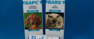
Most eye diseases are treated conservatively. To do this, use various drops and ointments containing antibacterial agents. Before placing or instilling them, wash the animal’s eyes with a weak solution of furatsilin or potassium permanganate.
The most popular are products based on Tetracycline, Gentamicin, Erythromycin and Levomycetin.
They try not to use Albucid to treat pets, as it burns strongly and the animal will not allow it to be instilled anymore.
After applying the ointment or drops, you should hold the cat in your arms for a while, otherwise it will wipe off all the medicine with its paw.
If there was an injury, Solcoseryl ointment (Actoveginovaya) helps well for healing; it is applied to the inside of the eyelid.
In advanced stages, antibiotics are administered intramuscularly or intravenously, depending on the condition of the animal and the tolerability of the procedure.
In order to exclude allergies to injected drugs, a course of antihistamines, for example, Suprastin or Tavegil, is prescribed.
They try to resort to surgical treatment in extreme cases or when it is not possible to preserve the organ of vision. For example, with panophthalmitis, this is often the only treatment option.