Osteochondrodysplasia (OCD) is a disease that sometimes affects Scottish Fold cats due to their genetic makeup. There is no cure for this disease, but cats are prescribed supportive therapy, which improves the quality of life. This article is for those who want to obtain primary information about osteochondrodysplasia or verify data received from a veterinarian.
The first mention of skeletal deformities in Scottish cats dates back to 1971, although the breed was registered in 1966. Those. Not all cats had problems, so we didn’t notice them right away.
Article continues after advertisement
IMPORTANT TERMS
HIP DYSPLASIA is a discrepancy between the articular surface and the head of the femur, leading to a violation of the congruence of the hip joint. More often appears at a young age. It is expressed in a disturbance in the cat's gait and the inability to jump.
OSTEOCHONDRODYSPLASIA is a malformation of bone and cartilage, leading to a lack of normal growth of bone tissue and to its deformation. Translated from Greek, “dysplasia” means “disorder of education”; this term refers to a disorder in the development of tissues and organs. The disease is characterized by skeletal deformities and progressive osteoarthritis.
ACHONDROPLASIA is one of the forms of osteochondrodysplasia. The bones do not grow to normal size, and as a result, the cat's paws are abnormally short - dwarfism develops.
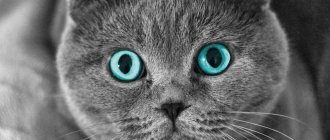
Scottish fold cats are at risk for this disease because... their breed-forming characteristic is the cartilage mutation gene, which deforms them. In other words, fold-eared cats (Scottish Folds and Highland Folds) have ears that “fall” because the cartilage has a fold. This defect is noticeable on the ears, but it also exists in other parts of the body. At the same time, the majority of Scottish fold cats live without knowing trouble, because... the defect is not developed enough to be visible. But it can also lead to the development of a disease called osteochondrodysplasia.
Opponents of the breed believe that all Scottish cats are sick, but this is definitely not the case. There is also an opposite opinion: a number of breeders claim that only those who have two Fd genes suffer from osteochondrodysplasia, i.e. children from incorrect crosses. Both points of view are not considered generally accepted. It would be more accurate to say that with proper crossing, when not two folds are brought together, but a fold and a straight (Scottish straight), the risk of the disease is minimized, because cats receive only one Fd gene. But life shows that the probability still remains (see the website of the Federation of Universities on Animal Welfare): it is difficult to predict who will be unlucky, even if the parents are clinically healthy.
The statement of a number of breeders that only heterozygous folds suffer from osteochondrodysplasia became the subject of lively discussions at one time, during which it was proposed to conduct a study of clinically healthy folds. But the research never began. As a result, the New Zealand Breed Standards Advisory Council (BSAC), in its report to the New Zealand Cat Fanciers (NZCF), noted:
Breeders underestimate the strength of the evidence that heterozygous individuals can develop OCD. Although research suggests that all lop-eared cats are susceptible to OCD, and anecdotal evidence also suggests, these studies do not show that parents with mild manifestations of the disease are likely to produce offspring with mild manifestations. The available information is not enough to definitively prohibit the breeding of Scottish Folds, but it is sufficient to understand the level of concern.
Due to the fact that heterozygosity of a fold does not guarantee its health, the Scottish Fold and Highland Fold breeds are considered extremely difficult to breed. All producers must be regularly screened for joint diseases and, if detected, the cat or female cat must be excluded from kitten breeding programs.
There is, in addition, the assumption of scientists that, because Most folds do not show any visible symptoms of OCD; there are one or more genes, in addition to the Fd gene, responsible for the development of this disease. Scientists are currently searching for these genes.
Symptoms, signs and development of the disease
Typically, owners notice the problem only when it is already severe. The following signs of osteochondrodysplasia are distinguished:
- lameness;
- limb deformity;
- enlarged head;
- nose too short;
- crooked teeth;
- slow growth;
- protruding jaw;
- ulceration of the skin under the growths;
- shortened hind legs;
- walking on bent legs;
- difficulties in jumping to heights and jumping off;
- the cat’s reluctance to walk and jump, pitiful meowing in such situations;
- stiffness and changes in gait;
- short, thick, sedentary tail;
- thin base of the tail.
The diagnosis of osteochondrodysplasia is made on the basis of behavioral symptoms, external examination, as well as x-ray examination, which shows abnormalities in the structure of the paws, spine and tail.
Article continues after advertisement
Diagnostics
Diagnosis of bone exostoses is not difficult. If these symptoms occur, you should consult an orthopedist. Based on the patient’s complaints, medical history and examination results, the doctor can already assume the presence of a neoplasm.
To clarify its size and other features, radiography is prescribed. It is carried out in 2 projections. Osteochondroma on x-rays is visualized as a shadow with smooth, clear contours and a pedicle or wide base connected to the bone. If there is no calcification in the cartilaginous layer, it will not be visible on x-ray.
To obtain additional data, the following may be assigned:
- CT scan, which makes it possible to detect the presence of bone marrow contents in exostosis and trace its connection with the medullary canal of the affected bone;
- MRI, which provides accurate data on the thickness of the cartilage cap and thereby clarifies the nature of the neoplasm (malignant tumors are characterized by a thickness of the cartilage tissue covering them of more than 2 cm).
If there is a suspicion of the development of oncology, patients are advised to undergo a biopsy - taking a fragment of exostosis for histological examination. In this way, benign osteochondroma is differentiated from malignant neoplasms.
If exostoses are detected in childhood and adolescence, it is imperative to conduct a full endocrine examination and determine the content of all significant hormones in the blood.
Treatment and supportive care
There is no treatment that can eliminate the disease once and for all. Cats with this problem are prescribed maintenance therapy, which can improve quality of life. However, it does not always improve so much that the pet can at least walk.
In cats suffering from pain, the pain can be controlled with non-steroidal anti-inflammatory drugs. However, this method is used only when there is severe pain. After all, anti-inflammatory drugs have their side effects, especially with long-term use.
For cats with little or no pain, omega-3 supplements are more suitable as an anti-inflammatory. They reduce inflammation, so they are recommended as a preventative measure for all folds, but fold-eared cats with problems definitely need them.
However, you need to choose a high-quality and expensive omega-3 drug, because There are a lot of fakes in pharmacies. Therefore, we recommend that you pay attention to similar drugs from the American website iHerb, where there are definitely no fakes, and all drugs have passed control. The fat should be either in capsules or in the form of a spray with a dispenser. In jars from which you need to pour, the fat quickly oxidizes and becomes harmful.
Omega-3 capsules from Now Foods
When you click the button, you will be taken to the iHerb store and using our affiliate link you will receive a 5% discount!
BUY
Alaskan Salmon Oil from Zesty Paws
When you click the button, you will be taken to the iHerb store and using our affiliate link you will receive a 5% discount!
BUY
Chondroprotectors are also required. Most often these are glucosamine and chondroitin sulfate. They are believed to help reduce cartilage degeneration and increase the amount of joint fluid. However, there are no studies that would confirm this 100%. However, doctors still prescribe these drugs because... The choice of treatment methods for OCD is, in principle, small. Also, these drugs can be administered to all folds as prophylaxis in courses of 1 month every 1 month.
Pet Naturals of Vermont
It contains an expensive ingredient that has proven itself in the fight against OCD - New Zealand green-lipped mussel. The glucosamine in this preparation comes from shrimp and crab. Chondroitin sulfate - pork. Fold, who lives with the author of the article, receives this particular drug and the effect is visible from it more than from many others.
When you click the button, you will be taken to the iHerb store and using our affiliate link you will receive a 5% discount!
BUY
Article continues after advertisement
Musculoskeletal support from Dr. Mercola
It contains collagen and hyaluronic acid (to support ligaments and joints), as well as natural painkillers and anti-inflammatory components - bromelain, methylsulfonylmethane, etc. Can be used simultaneously with other drugs from our list.
When you click the button, you will be taken to the iHerb store and using our affiliate link you will receive a 5% discount!
BUY
A number of veterinary experts advise using, among others, homeopathic medicines for chondrodysplasia: “Coenzyme compositum” (used for inflammatory and degenerative diseases) and “Discus compositum” (has metabolic, regenerative, analgesic, anti-inflammatory, antispasmodic and sedative effects).
Manual therapy with massage has also proven itself well, but it must be carried out by a professional.
In severe cases, surgery (osteotomy and arthrodesis) is indicated. The operation can give very good results if there are indications for it.
Radiation therapy in the treatment of OCD
There is also an experimental treatment originally from England, where such cats are irradiated. Similar experiments are being carried out in Japan. Radiation therapy has already been proven effective in certain individuals: it relieves pain and prevents further destruction of the joints. However, the problem is that such treatment is practiced in few places, and our clinics do not have the necessary equipment.
“In medicine, low-dose radiation therapy is usually used to treat inflammatory and degenerative diseases. Although the mechanisms of the analgesic effect have not yet been clarified, they are suggested to be related to the anti-inflammatory effect. Radiation is considered a promising treatment option for osteochondrodysplasia, but long-term evaluation of its effectiveness and late complications has not been adequately assessed. There is insufficient information on the clinical use of this treatment. Further studies with a larger sample size and longer follow-up period are required,” concluded the Japanese doctors who conducted the study.
It is worth noting that radiation therapy is still largely an attempt to relieve pain and slow down the progression of dysplasia for seriously ill animals. However, if it was possible to stop the pain syndrome in all cases, then it was not possible to stop the progression of the disease. There is still a lot of uncertainty in this technique.
What is bone exostosis
Osteochondral exostosis is a benign osteochondral formation on the outer surface of the bone. Most often, it is detected at a young age before the end of skeletal growth, including in children.
The formation of osteochondroma provokes a disruption of the natural processes of resorption (resorption) and the formation of new bone tissue (remodeling).
Exostosis is a growth formed by spongy bone and having a cortical layer (a strong, hard “shell”). It can grow on a stalk or be attached to a bone with a wide base. On the outside, the bone growth is covered with cartilage tissue, which has much in common with articular cartilage. Its thickness does not exceed 1 cm. The top layer of exostosis is also called a cartilaginous cap.
Osteochondroma is a consequence of impaired bone growth in the area of the epiphyseal plates, i.e., their displacement from the anatomically correct position with the “throwout” of cartilage tissue to the side. Subsequently, just like the metaphysis, it ossifies in the direction from the base to the apex, which leads to the formation of bone exostosis with a cartilaginous cap. At the same time, it continues to increase in size until the growth zones close, and can appear in absolutely any bone. The bony part of osteochondroma is formed from unevenly distributed bone beams. Between them there is adipose tissue with islets of hematopoiesis. Bone septa are formed due to enchondral ossification of cartilage. This raises the possibility of the presence of calcifications in the cancellous bone.
The metaphyseal areas of long tubular bones are most often affected:
- femoral (30%);
- shoulder (10-20%);
- tibial (15-20%).
Metaphysis (sometimes neck) is a small part of a tubular bone that is located between its epiphysis (head) and diaphysis (body). At 18-25 years of age, it stops growing and ossifies, which indicates the completion of bone formation.
Thus, articular exostosis is often observed - the formation of a growth on the upper (proximal) part of the tibia or lower (distal) part of the femur, i.e. the tumor is localized just below or just above the knee. Damage to flat bones, i.e. exostosis of the rib, scapula, pelvic bones, are less common. Osteochondromas of the bones of the fingers, toes, and clavicle are diagnosed even less frequently. The most rare occurrence is the formation of bone growths in the spine.
Also, bone exostosis can form on the lingual or buccal surface of the jaw. At the same time, it can take the form of a ridge, protrusion, mound, or take on bizarre shapes. In such cases, patients are advised to consult a dentist.
What to feed a cat with osteochondrodysplasia?
The basis of nutrition for sick cats is proper balanced feeding using additives that prevent destruction and inflammation of the joints. This is either natural food or factory feed with such components. Supplements and the diet itself should be rich in calcium, vitamins B and E, phosphorus, iodine and iron. Among professional food brands there are even special lines for cats with joints. Such feeds and supplements additionally contain chondroitin and glucosamine, which in combination mutually enhance each other.
If the cat eats a natural diet, then it should include raw cartilage, sinew and meat, rich in natural glucosamine and chondroitin. But the opinion about the need to cook jellied meats for such cats is wrong. All the substances that were originally in the cartilage and meat are no longer present in the digestion, and many pass into a form that is difficult to digest. Everything you need is already in the raw product.
Article continues after advertisement
It is also worth considering that cats with joint diseases are susceptible to obesity, because it is unpleasant or even painful for them to move. The owner must monitor the calorie content of the diet by regularly weighing the animal and identifying deviations in time. If the cat is already obese, it is necessary to select food for weight loss, because... excess fat threatens heart disease and greater stress on the musculoskeletal system. If your cat is of normal weight, still try not to overfeed her, because... a cat with reduced physical activity needs fewer calories.
Complications
Cartilaginous exostosis in children has a dangerous negative impact on the condition of the growth plates, which can result in shortening of the lower leg, thigh, forearm, shoulder, etc. This can also cause curvature of the arms and legs, increase the risk of fractures and cause disability.
Patients with severe forms of the disease feel inferior, which negatively affects their mental and emotional state.
The most dangerous complication of osteochondroma is its malignancy, i.e. transformation into a malignant tumor - chondrosarcoma. The highest risk of malignancy is with multiple exostoses. In this case, growths localized in the pelvic bones are more likely to undergo malignancy. Less commonly, exostoses of the ribs, scapula, and spine turn into malignant neoplasms. Single osteochondromas transform into a malignant tumor in no more than 1% of cases.
Atypical growth can be observed in any part of the exostosis: the cartilaginous cap, at the base or in the middle part.
How long do sick cats live?
It is unlikely that anyone will give you a specific prognosis for the life expectancy of cats with osteochondrodysplasia. Unless such responsibility will be assumed by the treating veterinarian who has fully examined the animal. And even then the forecasts are often wrong. Often, veterinarians mistakenly suggest euthanizing an animal or cutting off a limb, although, in fact, with the correct selection of medications and proper care, as well as the patience and love of the owner, the cat’s condition can be seriously improved. Therefore, do not be upset immediately as soon as you hear the diagnosis from the doctor. Be sure to consult with 1-2 more veterinarians. Only then make a decision!
The first signs of osteochondrodysplasia in Scottish fold cats can appear as early as a couple of months, so when choosing a kitten it is so important to pay attention to the behavior of the cat, the curvature of the paws, the presence of kinks in the tail, its mobility and length. It all starts with minor changes that may not be noticed if you don’t set such a goal. In most cats, the disease becomes noticeable later, when they have already found permanent homes.
The rate at which the disease progresses and its severity varies among individuals. The time of visible manifestation of the disease is the same. However, veterinarians recommend that all owners of fold-eared cats closely monitor the condition of the pet and, at the first manifestations, go to the veterinary clinic, because further quality of life will directly depend on how early treatment is started.
To increase the life expectancy of a sick cat, it is necessary, in addition to drug therapy, to balance the diet and prevent the appearance of excess weight, because. it creates additional stress on the musculoskeletal system. Pets with especially severe manifestations of the disease once again try not to move. Sometimes it is even difficult for such cats to reach the litter box, and therefore they may relieve themselves in the wrong place or on their own. If this has become a system, you can purchase special cat diapers. Then the cat will urinate and poop in them, thereby saving you from unnecessary problems.
If your cat is diagnosed with such a disease, we would like to advise you not to despair. For inspiration, watch a video about a rescued cat with osteochondrodysplasia, which, due to illness, was kicked out of the house by its owners in 40-degree frost, but a few days later it was rescued by volunteers, they came out - and now she lives happily in a new family.
However, in some cases it is still recommended to euthanize the cat. These are cases of severe progressive disease with pain. If the animal is suffering, cannot lead a normal life, is constantly on pills that do not help much, veterinarians advise stopping the cat’s suffering. And in this case they are most likely right.
Exostosis in a child
It is in children that osteochondroma is most often first diagnosed, which is due to its formation from cells of the epiphyseal plate that is present only until the end of the growth period, which is adjacent to the metaphysis. It is also called the bone growth zone, since it is a hyaline cartilage whose cells are in constant miotic division. As a result, new chondrocytes (cartilaginous tissue cells) are formed, forming the epiphyseal plate, and the old ones are shifted to the metaphysis and subsequently replaced by osteoblasts (bone tissue cells).
In infants, violation of the rules for the prevention of rickets, in particular excessive use of vitamin D preparations, increases the likelihood of the formation of exostoses.
After puberty, the growth plates gradually close and are replaced by bone tissue, transforming into a thin epiphyseal line. If hormonal imbalances occur during this period, it is possible that the growth zones may remain open, which creates the preconditions for the formation of osteochondromas.
Usually, until the age of 7-8 years, bone exostosis does not manifest itself in any way in a child and makes itself felt only during the period of intensive growth, i.e., at 8-16 years, since it also begins to grow actively. In young children, such growths are present in the metaphysis area immediately near the epiphyseal plate, but subsequently move away from it and approach the diaphysis. Therefore, by how far the bone exostosis in children is from the epiphysis, the time of its formation is determined.
The growth of the neoplasm continues until the end of the growth period.
Do straight-eared Scottish cats suffer from this?
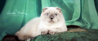
The straight-eared Scottish Straight and Highland Straight varieties of Scottish cats are no more prone to osteochondrodysplasia than any other breed. The disease does not only affect Scots, it is just more common among Folds. Straits do not have deformed cartilages and do not have the gene for lop ears, and it is this gene that makes cats vulnerable to osteochondrodysplasia. By the way, not only the Scots are considered vulnerable for this trait, but also the Ukrainian Levkoy, in the selection of which Scottish Fold cats were used. OCD also occurs in humans. Therefore, it cannot be said that this disease is exclusively of the Scottish breed.
Rate and share!
Surgery to remove exostosis
Surgical intervention is indicated for:
- large size of osteochondroma and the presence of persistent pain;
- development of complications (tendinitis, bursitis, vascular, neurological disorders, etc.);
- bone deformations as a result of the growth of exostosis;
- fracture of the base leg;
- malignancy of the tumor.
For children with bone exostosis, surgery is prescribed only in extreme cases. As a rule, it is carried out only if the testimony remains after reaching adulthood.
But if there are contraindications, surgery is not performed. This:
- purulent-inflammatory processes in the area of the upcoming intervention;
- exacerbation of chronic diseases;
- decompensated form of diabetes mellitus;
- acute infectious diseases.
Treatment of exostosis with surgery involves its complete excision with capture of the cartilaginous cap. There are several methods for removing osteochondral growth. A specific one is selected based on the location of the osteochondroma and its size. Usually preference is given to marginal resection of the growth, i.e., its removal within healthy tissue. Only in some cases is it necessary to resort to corrective osteotomy with concomitant resection of osteochondroma, but the cost of removing exostosis in this way is higher, since the operation also corrects bone deformation.
After surgery, complete recovery is observed in 98% of cases. Therefore, almost everyone who has once removed an exostosis forgets about it forever.
Marginal resection is a relatively simple type of surgery. Its essence is to expose the affected bone and remove the entire neoplasm in the front of healthy tissue along with the surrounding capsule using a sharp chisel, drill, oscillating saw or bur. After removal of the pathological formation, curettage of the maternal bone is performed using cutters and bur. In some cases, the extent of resection can be large and create the need for bone grafting using autografts or special implants. It is important to remove the entire cartilaginous cap and neoplasm, otherwise a relapse may develop.
Carrying out a corrective osteotomy involves cutting the mother's bone using an osteotome and removing the osteochondroma. After this, the bone fragments are fixed in the desired position using special plates, screws or knitting needles.
What ailments are there?
Diseases significantly shorten the life of fold-eared cats, so they may not live long depending on the disease. The most common:
- Osteochondrodysplasia. Characterized by skeletal deformation. Highland Fold and Scottish Fold breeds are at high risk of getting the disease. Straight-eared people are less susceptible. Proper treatment and care stops the disease and the cat can live to an old age.
- Hemophilia types A and B. Characterized by a blood clotting disorder and prolonged bleeding at the site of even a small cut. How many years Scottish cats with this diagnosis live depends on other anomalies of the body, since internal bleeding leads to rapid death.
- Eye diseases. A fold-eared or straight-eared cat often suffers from eversion or inversion of the eyelids, which provokes conjunctivitis, keratitis or blepharitis. Eye diseases do not affect a cat’s life expectancy if you consult a veterinarian in a timely manner and stop the inflammation.
- Ear ailments. The most common are otitis media, deafness and tumors due to the abnormal structure of the hearing organ. With proper care, your cat will live a long time. However, if the neoplasm is malignant, metastases may form and life expectancy will be significantly reduced.
- Deterioration of immunity. Pedigree cats have reduced body defenses, so they are susceptible to viral and bacterial diseases. Serious diseases (panleukopenia, rhinotracheitis) can “take away” your pet much earlier than expected.
- Metabolic disease. Spayed and neutered pets who suffer from hormonal imbalance suffer the most. This leads to obesity or diabetes.
How to increase the lifespan of a Scotsman
Thanks to many advances in veterinary medicine and the development of the pet business, the lives of pets have become much longer and more comfortable. To ensure that 15 years is not the limit for the life expectancy of a Scottish Fold, its owner must be responsible, starting with the choice of a kitten and throughout the pet’s life.
You should only purchase a fold-eared baby from a conscientious, reputable breeder or at a cat show.
This solution minimizes the risk of purchasing a sick animal. Particular attention should be paid to issues of maintenance and care. Even before the furry newcomer appears in the house, you need to decide on the type of food he will eat, whether it will be factory-made food or natural products. When choosing ready-made food, preference is usually given to the “super-premium” and “holistic” classes: these lines take into account the age, breed and individual characteristics of cats. A natural diet should be balanced, varied, meeting the needs of carnivores in general and Scottish Folds in particular: contain not only all nutrients, but also taurine, which Scottish cats, like all other cats, do not produce on their own.
- Routine and preventive examinations by a veterinarian are required, as well as timely vaccinations against rabies, panleukopenia, chlamydia, calcevirosis, and rhinotracheitis.
- Given the propensity of the Scottish population for cardiomyopathy and the difficulty in diagnosing this disease, ultrasound of the heart and chest should be included in the examination.
- Monitoring the condition of the gums and teeth must be constant: dental problems lead to diseases of the digestive system.
- If breeding a breed is not the purpose of purchasing an animal, it is advisable to sterilize it in due time.
- Regardless of whether the Scottish Fold is outdoors or in contact with other animals, periodic anti-parasitic measures are required.
- To maintain muscle tone and good shape, your pet needs to be provided with physical activity through games and training.
Such careful care and sincere love will allow the Scottish Fold to live to the respectable age of twenty.
Return to content
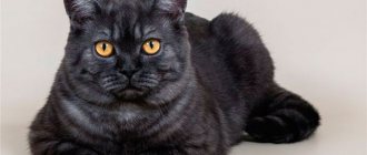
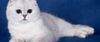
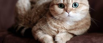
Appearance. What do Scottish Folds look like?
A striking feature of this breed is its erect ears. Among canine representatives there are many breeds whose ears do not stand up - Labradors, pugs, collies, spaniels, Airedale terriers, bulldogs, shelties, retrievers and others. The cat population cannot boast of such diversity; only the ears of the Scots, and with rare exceptions of the British, hang, and do not stand upright. This determines the popularity and unusualness of the breed.
Interesting fact: the second name of the breed, often found in veterinary reference books and books on animal care, is Scottish Fold.
The size of the cat is average: Scottish Folds have a medium-sized body with a rounded shape, with powerful and well-developed muscles, the shoulders and chest are well developed. The paws are thick, of medium length, massive and large, with delicate rounded pads. The color of the wide, bright eyes with mysterious sparkles varies depending on the color of the coat.
The jaw is well developed; a large oval or round head gently transitions into a wide neck; the ears are small, rounded towards the bottom; the nose is large and wide.
The coat is medium length, soft to the touch, like plush; tail of medium length, rounded at the end. The color can be any: breeders have bred seventy-five species of these wonderful animals.
Causes of the disease
OCD, the abbreviated name for feline osteochondrodysplasia, is a genetic disease. The gene that is responsible for the formation of the auricle can play a cruel joke on the animal’s body as a whole.
What we see is a beautiful crease in the ear, this is only part of the picture. Changes in cartilage tissue, disruption of its formation, can occur throughout the entire skeleton, and affect not only the ear.
And cartilage tissue plays an important role, primarily mobility. Any joint, intervertebral joint... will not be able to function normally without healthy cartilage.
When the formation of cartilage tissue is disrupted, then we get complications, and they will manifest themselves to varying degrees.
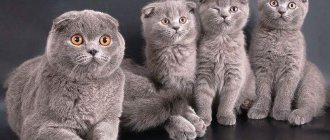
The likelihood of developing osteochondrodysplasia depends on the parents. If both parents are lop-eared (folds), then the likelihood of developing OCD in the offspring is very high. If one parent is fold and the other is straight (with straight ears), then the disease rarely appears, but it can also happen.
Violations also occur if one parent is a Scottish Fold and the other is of any other breed. Yes, such a combination can also happen when breeding is not carried out by professional breeders. The demand for fold-eared kittens is high, so many people breed them.
Case 1
An intact female Scottish Fold cat, less than 12 months of age, was admitted to the Veterinary University Hospital of Japan Veterinary and Life Sciences University (VMTH-NVLU) due to exercise intolerance due to chronic pain syndrome lasting several months. During palpation, the animal was practically motionless and demonstrated that this type of examination caused discomfort. The cat had short, massive and deformed limbs; Large plantar exostoses were noted, as well as changes in the shape of the claws. There was overstretching of the skin of the soles due to exostosis. There was a large thickness and low flexibility of the tail. There was no history of any treatment.
X-rays of all four limbs, tail (Fig. 1), abdominal organs, and chest were performed. X-rays of the thoracic limbs revealed fresh bone growths in the area of the carpal bones, metacarpus and phalanges. Shortening, thickening and disruption of the shape of the metacarpal bones in the distal section, as well as the phalanges, were noted. On the pelvic limbs, fresh bone growths were identified in the area of the tarsal and metatarsal bones, as well as the phalanges. These bones were also short, dense and misshapen. Radiographs of the tail revealed bone growths, as well as shortening and deformation of the caudal vertebrae. There was a narrowing of the intervertebral spaces, while the caudal vertebrae were in direct contact. X-ray examination of the chest and abdominal cavity revealed no other bone lesions.
Based on the animal's breed, symptoms and clinical manifestations, as well as radiographic findings, the cat was diagnosed with Scottish Fold osteochondrodysplasia. Due to the severity of the disease, the owner abandoned the animal. One of our employees agreed to become the new owner, from whom informed consent was obtained. As a measure of palliative treatment of chronic pain syndrome, radiation therapy was performed to the extremities on the right side. In order to evaluate the effectiveness, as well as complications, irradiation of the extremities was performed only on one side. The treatment plan was developed from X-ray CTa using treatment planning software based on 3D reconstructions.b During treatment sessions, the cat was anesthetized and placed in a supine position on a vacuum immobilization mattressc shaped to follow the contours of the patient's body. The limbs were wrapped in artificial skin (to adjust the radiation dose), but no blocks, wedges, or multileaf collimator were used. Palliative radiation was performed using a 4 MV linear accelerator.d The protocol included 6 fractionated doses of 1.5 Gy each on a Monday-Wednesday-Friday schedule for a total dose of 9 Gy.
Clinical manifestations resolved after 2 weeks from the start of radiation therapy. Radiography was performed (Fig. 1). No progression of the disease was detected. No acute complications, including alopecia, pigmentation, and wet desquamation, occurred. There were no radiographic differences between exposed and unexposed limbs. By 38 months, the animal was admitted due to severe pain in the extremities of the left half of the body that were not exposed to irradiation. Radiation therapy of the left thoracic and pelvic extremities was performed according to the protocol described above. The animal was observed for 60 months. At month 60, there were no late complications of radiation therapy, including aplopecia, bone or nerve necrosis, or tumor growth (Fig. 2). The cat willingly walked, ran and jumped. No changes in the morphology of bone lesions were detected according to radiography.
How to extend the life of a pet
Let's find out what procedures will help your pet live longer:
Timely vaccination
Please note: even if your pet lives exclusively at home and never goes outside, harmful microorganisms can enter the house via outdoor clothing and other items. Balanced diet. If you prefer to give your cat commercial food, it should be premium or super premium.
Natural nutrition should include a large amount of protein: lean meat, fermented milk products, deboned sea fish once or twice a week. Vegetables are good for you. They can be given raw or cooked, depending on the cat’s preferences. Your pet should always have access to clean water. After consulting with a veterinarian, you can purchase mineral supplements for your pet to strengthen bones and cartilage tissue. Hygiene procedures are important. About 2 times a month you need to clean the animal’s ears using cotton pads and a special gel (sold in pet stores). A short-haired pet should be brushed once a week using a brush with short, stiff, dense teeth. The long-haired variety of Scottish cats is combed daily using a comb with sparse long teeth. It is not advisable to use slicker coats for Scots. Sterilization increases the life of a pet by 2–3 years (regardless of gender).
Do not forget that how long your pet will live largely depends on you.
Diet
Since osteochondrodysplasia causes thinning of cartilage tissue, as well as deformation of the animal’s bone skeleton, a balanced diet and proper care are the key to making your pet’s life easier.
When choosing food for a sick animal, it is worth considering a side effect of low physical activity: obesity. In addition, Scottish cats, which are close relatives of British shorthairs, inherited from their common ancestors a breed tendency to quickly gain weight. Therefore, you need to control the calorie content of your food.
Cat food manufacturers have developed a special line of food products for animals with pathology of cartilage tissue. Such feeds contain all the necessary vitamins and microelements, such as calcium, selenium, phosphorus.
Scottish breeders recommend the following nutritional supplements: POLIDEX Multivitum, Hill's Prescription Diet and Holistic food, Primitive Feline Natural Cat Food.
If a Scottish cat suffering from OCD is accustomed to a natural diet, it is necessary to include in its diet foods rich in microelements and vitamins to strengthen cartilage tissue. However, it is not recommended to heat food, as it destroys important components of a healthy diet.
List of products needed for Scottish cats with OCD:
- Lean varieties of meat, which are given raw (to preserve trace elements in the composition), but are pre-frozen so as not to infect the animal with parasites.
- Fermented milk products (hard cheese, kefir, fermented baked milk) rich in calcium.
- Quail eggs.
- Special cat grass, rich in vitamins and minerals.
“Vulnerable” places of Scottish folds
Scottish Fold is classified as an undemanding strong breed. But every artificial species has “vulnerable” places.
What problems do owners of fold-eared cats encounter?
- Diseases of the musculoskeletal system. In adulthood, problems with the development of bone and cartilage tissue begin.
- Cats easily gain excess weight as a result of a sedentary lifestyle and an unbalanced diet. This gives impetus to liver and heart dysfunction, and the risk of serious problems increases.
- The ear is the weakest point of the Scottish Folds. Responsible owners of fold-eared cats regularly inspect their pets' ears and clean them of dirt and wax. Otherwise, an inflammatory process appears and enters the chronic stage.
- Long-haired cats suffer from the appearance of dense mats due to insufficient coat care. This reduces mobility and negatively affects muscle tissue throughout the body.
Be sure to read: Lilac British cat: features of the breed’s appearance, characteristics, behavior, care
There are diseases that are most often inherited by Folds.
What diseases do cat owners face:
- Osteochondrodysplasia. Health problems can be identified by lameness and hardening of the tail.
- Osteoarthritis is joint stiffness.
- Hypertrophic cardiomyopathy is a problem with the functioning of the heart.
- Polycystic kidney disease.
Information disclosure
None of the authors of this article received financial support from third parties or organizations that could unduly influence the content or lead to bias in the presentation.
Statement of Potential Conflict of Interest
: The authors declare no conflict of interest.
Statement on off-label use of antimicrobials
: The authors declare that there is no off-label use of antimicrobials.
Prevention measures
Hip dysplasia is an inherited disease. Cats with a history of dysplasia in their pedigree should not be allowed to be bred. If it is assumed that the kitten will participate in breeding, an in-depth analysis of its pedigree is necessary.
If your pet has a hereditary predisposition to articular dysplasia, it is important to carefully monitor his lifestyle. This will help reduce the risk of disease to a minimum.
- It is very important to adjust the animal’s diet by eliminating excess fats and carbohydrates. Food should be moderate in calories and contain sufficient minerals.
- It is necessary to monitor the cat's weight to prevent obesity.
- Regular veterinary examinations should not be neglected.
- It is necessary to provide the animal with the opportunity to move as much as possible and play active games.
It is in our hands to make sure that pets can always feel the joy of active movement. You just need to be a little more attentive to them so as not to waste time, and start treatment as early as possible.
Case 3
A one-and-a-half-year-old intact female Scottish Fold was admitted to the Veterinary University Hospital of the Japan Veterinary and Life Sciences University due to lameness on the right pelvic limb of one year's duration. The animal could not jump. The cat resisted palpation of the right pelvic limb and did not land on it when jumping. There was no history of any treatment.
X-rays of all four limbs, tail, abdominal organs and chest were performed. The presence of bone tissue growth in the area of the tarsal, metatarsal and calcaneal bones on both sides was determined. Osteophytes were detected in the area of the tarsal joints on both sides. Radiographs of the tail revealed shortening and deformation of the caudal vertebrae. No lesions were found in the thoracic extremities.
Based on symptoms and clinical manifestations, as well as radiographic findings, the cat was diagnosed with Scottish Fold osteochondrodysplasia. Radiation therapy was performed on both pelvic extremities according to the protocol described above. Clinical manifestations resolved after 1 month from the start of radiation therapy. No acute adverse effects were observed. When X-rays were performed regularly over 16 months, no progression of bone lesions was detected. At 48 months, new zones of deformation were noted in the area of the tarsal joints on both sides, but this did not affect the activity of the animal. Further follow-up was 72 months without additionally identified pathologies. No late complications were noted.
How is the diagnosis made?
An examination is sufficient for a preliminary diagnosis. If you have a fold-eared kitten (or an adult cat) and you observe the following symptoms, you may suspect OCD:
– began to limp for no particular reason.
– the gait has changed. Protects one or more limbs, hunches over.
– there is a delay in growth, a violation of proportions (short legs, tail...).
– you observe deformation of the joints, most often the hind limbs are affected.
– the tail loses mobility, becomes like a “stick”, and when trying to bend the animal experiences pain.
To clarify, an x-ray is taken. The image evaluates the density of bone tissue, whether there are anatomical changes in the joints, the presence of bone exostosis (growth) and other changes.
The doctor may also recommend additional tests: general blood test, blood biochemistry, ultrasound…. In order to exclude other diseases or identify disorders that often develop in parallel with osteochondrodysplasia.
For example, kidney disease, mineral metabolism disorders, etc.
Symptoms
The first thing you need to pay attention to is the general condition and mood of the cat. An unhealthy Scottish Fold will be lethargic, apathetic, lack of appetite, sometimes irritable reactions and unreasonable aggression
The nose becomes dry and very hot to the touch. Perspiration may appear on the fur. When measuring temperature, the readings will exceed 38.5 °C.
In specific diseases, the cat owner can focus on the symptoms.
Table 2. Symptoms of diseases of internal organs
| Type of disease | First symptoms |
| Viruses | Sneezing, discharge from mouth and nose, fever |
| Ear | The cat shakes its head, discharge from the ears (pus, fluid), putrid smell from the shells, redness of the skin in the shells, the cat scratches its ears |
| Ophthalmic | Redness, lacrimation, purulent discharge, swelling of the eyelids of varying degrees, the cat tries to scratch the eyes, poor orientation in space, cloudy pupils |
| Digestive | Refusal to eat, vomiting, nausea, constipation, diarrhea, undigested pieces in feces, increased thirst |
| Skin | Scabies, redness, bald patches, rashes, sores, blisters, peeling, nodules |
| Heartfelt | Shortness of breath, cough, swelling, increased frequency of sighs, low temperature, fainting |
| Genitourinary system | Sudden weight loss, pink urine, apathy, frequent urination, the cat meows when urinating, fussing |
| Bone | Disproportionately large head, crooked, protruding jaw, shortened limbs, curvature of the spine, twisted front legs, joints with a swollen effect, too massive joints, growths on them, slow or absent growth of the cat, shortened thick tail, inactivity, clumsiness of movements, attempts not to bend the paws when moving |
| Dental | Foam, pus, excessive drool from the mouth, bad odor from the mouth, yellow, brown, gray growths on the teeth, brittle teeth, refusal to eat (it hurts to eat) |
In the early stages, 2-3 mild symptoms may be observed. As the pathology develops, the number of manifestations increases. It is necessary to monitor your fold ear and consult a doctor at the first suspicion of abnormalities.
Forecasts
The lifespan of Scottish cats affected by osteochondrodysplasia is difficult to predict in advance. It largely depends on how willing the owners are to care for a disabled kitten.
For long-term prognosis, it is necessary to exclude other genetic diseases of Scottish cats, which can occur with irresponsible breeding as often as osteochondrodysplasia. These are polycystic kidney disease and hypertrophic cardiomyopathy, which can be detected using ultrasound.
With mild osteochondrodysplasia, cats can live for years, although they require constant supportive treatment and special housing conditions. For example, they may find it painful to go to the toilet, which leads to urination in the wrong places and chronic constipation.
If the sick kitten is homozygous for the lop-eared gene (both parents are Scottish Folds), the prognosis for life is extremely unfavorable. Bone deformation causes severe pain, which becomes increasingly difficult to relieve with age. The animal cannot move, and at some point the owners resort to euthanasia as a means of ending the suffering of their beloved pet.
Links
1. Niewald M, Seegenschmiedt MH, Micke O, et al. Randomized multicenter trial on the e?ect of radiotherapy for plantar
Fasciitis (painful heel spur) using very low doses–a study protocol. Radiat Oncol 2008;3:27.
2. Rodel F, Frey B, Manda K, et al. Immunomodulatory properties and molecular e?ects in in?amatory diseases of low-dose x-irradiation. Front Oncol 2012;2:120.
3. Hautmann MG, Neumaier U, Kolbl O. Re-irradiation for painful heel spur syndrome. Retrospective analysis of 101 heels. StrahlentherOnkol2014;190:298–303.
4. Koca T, Aydin A, Sezen D, et al. Painful plantar heel spur treatment with Co-60 teletherapy: factors in?uencing treatment outcome. Springerplus2014;3:21.
5. Arenas M, Sabater S, Hernandez V, et al. Anti-in?amatory e?ects of low-dose radiotherapy. Indications, dose, and radiobiological mechanisms involved. StrahlentherOnkol2012;188:975–981.
6. HublerM, Volkert M, Kaser-Hotz B, et al. Palliative irradiation of Scottish Fold osteochondrodysplasia. Vet Radiol Ultrasound 2004;45:582–585.
7. Wastlhuber J, Pederson NC. Diseases and Management in the Multiple-cat Environment. In: Pederson NC, ed. Feline Husbandry. California: Goleta; 1991:12–14.
8. Robinson R.Normal Genetics, Genetic Disorders, Developmental Anomalies and Breeding Programs. In: Pederson NC, ed.Feline Husbandry. California: Goleta; 1991:75–76.
9. Malik R. Genetic diseases of cats. J Feline Med Surg 2001;3:109–113.
10. Takanosu M, Takanosu T, Suzuki H, et al. Incomplete dominant osteochondrodysplasia in heterozygous Scottish Fold cats. J Small AnimPract2008;49:197–199.
11. Chang J, Jung J, Oh S, et al. Osteochondrodysplasia in three Scottish Fold cats. J Vet Sci 2007;8:307–309.
12. Malik R, Allan GS, Howlett CR, et al. Osteochondrodys- plasia in Scottish Fold cats. Aust Vet J 1999;77:85–92.
13. Mathews KG, Koblik PD, Knoeckel MJ, et al. Resolution of lameness associated with Scottish fold osteodystrophy following bilateral ostectomies and pantarsalarthrodeses: a case report. J Am Anim Hosp Assoc1995;31:280–288.
14. Kataoka T. Study of antioxidative e?ects and anti-in?amatory e?ects in mice due to low-dose X-irradiation or radon inhalation. J Radiat Res 2013;54:587–596.
15. Frey B, Hehlgans S, Rodel F, et al. Modulation of in?amation by low and high doses of ionizing radiation: implications for benign and malign diseases. Cancer Lett. Forthcoming 2015.
16. Emami B, Lyman J, Brown A, et al. Tolerance of normal tissue to therapeutic irradiation. Int J Radiat Oncol Biol Phys 1991;21:109–122.
17. Bomford CK, Kunkler IH, Hancock BW. Principles of Management and Dosage. Walter and Miller's Textbook of Radiotherapy, 6th edn. London: Churchill Livingstone; 2003: 307–323.
18. University of California. Davis: The Association; c2013–2015. Veterinary Genetics Laboratory; . Available at: https://www.vgl.ucdavis.edu/services/ScottishFold.php.
19. Gandol? B, Lyons LA, Adhikari B, et al. Inherited osteoarthropathy in Scottish Fold cats is associated with altered calcium channel function. In: Bailey E, Ciobanu D, Kim KS, eds. Plant{amp}amp; Animal Genome: Proceeding of the 13th international conference on the status of plant {amp}amp; animal genome research. SanDiego, CA; 2015: 151.
20. Tsang AK, Taverne A, Holcombe T. Marfan syndrome: a review of the literature and case report. Spec Care Dentist 2013;33:248–254.
21. Tuncbilek E, Alanay Y. Congenital contractural arachnodactyly (Beals syndrome). Orphanet J Rare Dis 2006;1:20.