5190Administration
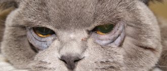
Inversion of the eyelid in a cat is a pathology in which a fold of skin turns inside the eye. Because of this, the cornea is constantly traumatically affected by eyelashes and hair covering the eyelid. When an inversion of the lower eyelid occurs in a cat, with prolonged absence of treatment, the formation of a corneal ulcer is observed. As a result, the animal loses vision and experiences constant pain. The pet's quality of life is seriously deteriorated, and the cat cannot remain cheerful and cheerful when an entropion has occurred and the eyelid is in an unnatural position.
Why does the eyelid curl up?
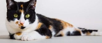
In cats, entropion of the eyelids occurs for various reasons. The disease is caused by various factors, the main one of which is a genetic predisposition that causes a violation in the structure, as a result of which the eyelid curls up. This eye disease can occur in pets of any breed, but it most often affects the exotic Sphynx, as well as British and Persian cats.
© shutterstock
A cat's eyelids may droop for the following reasons::
- Spasm of the muscles of the orbit of the eye - from such an effect the eyelid experiences inversion;
- Incorrect development of the eyelids, as a congenital defect, when their length is excessive;
- An eye injury in which entropion occurs mechanically. Often injury occurs during play or a fight with another cat or dog;
- Foreign body in the eye - volvulus occurs due to mechanical damage;
- Scar formation - usually this phenomenon occurs if the cat's eyelid has been thermally or chemically burned and its edge is injured. When the damage heals, a scar appears, which causes volvulus;
- Previous unsuccessful surgical manipulations in the eye area, in which the cat did not undergo proper restorative therapy - the structure of the eyelid is disrupted and entropion occurs;
- Paralysis of the facial nerve - against the background of this pathology, the cat loses the ability to control the muscles of the eyelids, and they droop or roll up;
- Age-related degenerative processes in muscles and skin cause, among other things, entropion of the eyelid;
- Neoplasms in the eyelid area of a malignant or benign nature - with such a disease, the eyelid changes (to varying degrees) its shape, which can provoke volvulus.
The upper eyelid inversion is experienced much less frequently than the lower one. The disease has primary and secondary forms. The primary one appears in kittens due to improper development of the eyelids, and the secondary one appears in adult animals against the background of provoking factors associated with muscle dysfunction or injuries.
What to do if you find a cat with entropion of the eyelids?
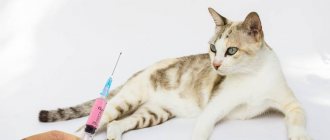
If you see signs of entropion in your pet, contact a veterinary ophthalmologist. Most likely, your pet will undergo a very delicate operation - eyelid surgery, and therefore make sure that it is a reference specialist, since in this case the result of the correction will be as good as possible. Next you need to reduce friction. To do this, it is recommended to apply ophthalmic lubricants (based on dexpanthenol or other carbomers) or ointments as often as possible (they will reduce the degree of damage to the cornea by slightly reducing friction). Unfortunately, it will not be possible to completely eliminate friction using therapeutic methods; gels will only soften it somewhat.
These drugs can be purchased at a human pharmacy or at a veterinary one, their essence is the same - to soften friction. It is contraindicated to use hormonal gels and ointments (SPVA, glucocorticosteroid drugs) for entropion of the eyelids in cats and corneal ulcers! We also do not recommend using anesthetic drops, because in this case the cat will no longer protect the eye (usually, when there is pain, they pull the eyeball into the orbit, trying to extend the third eyelid to protect the ocular surface). At such moments the friction will be the strongest.
Veterinarian ophthalmologist Yastrebov O.V.
Join us on social networks
Manifestations of the disease
If the owner knows the symptoms of the disease, then he will be able to identify bloat in the cat and provide timely assistance to the pet. First of all, when the eyelid rolls up, irritation of the cornea of the eye begins. Entropion of the eyelids is accompanied by other noticeable symptoms. Manifestations of the disease are presented in increasing order as it progresses :
- redness of the eyes;
- profuse lacrimation;
- swelling of the cornea of the eye;
- external reduction in eye size;
- change in the shape of the palpebral fissure due to the fact that the eyelid has rolled up;
- constant moisture in the fur around the eye;
- formation of a purulent mass in the corner of the eye;
- corneal ulcer;
- perforation of the eye.
At the last stage of the disease, a rolled eyelid causes such serious and irreversible changes in the cat's eye that it is impossible to save it. If you return the eyelid to the correct position in time, the problem will be completely eliminated.
© shutterstock
What's wrong with turning up the eyelids?
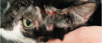
The fact is that, as it rolls up, the eyelid begins to rub its outer surface against the cornea of the eye. If the animal has fur, it will increase this friction and make it similar to friction with a hard brush. As a result, corneal damage develops: erosion, ulcer, descemetocele, or even a through defect. Animals that are completely devoid of hair on their eyelids (sphinxes, xoloitzcuintles) also suffer from the damaging factor, since the skin of the eyelid is much tougher than the conjunctiva. The cornea is especially severely damaged in short-haired animals - the same Sfinski or Devon Rex (short and stiff hairs cause the greatest harm). In serious stages of corneal damage, it can lead to perforation, so it is very important to provide timely treatment to your pet.
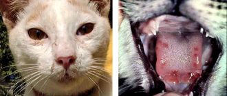
Friction is accompanied by discomfort and pain, so such animals cover the sore eye (or do not open it at all), there is copious discharge (white, yellow, watery eyes), itching, and redness of the eyelids. Vessels and defects visible to the naked eye - ulcers and erosions - may appear on the cornea.
The turning of the eyelids rubbed the corneal ulcer. Strong corneal vascularization is visible in the lateral third of the cornea
Diagnostics
The disease can be determined by a careful external examination. The animal's eyelid itself is directly noticeable, as well as swelling of the eye and purulent discharge.
Before surgical treatment, a complete examination is necessary. For this purpose, the animal is prescribed
- general blood analysis;
- biochemistry;
- Ultrasound of internal organs;
- Analysis of urine;
- ECG.
If all indicators are normal, then the cat will easily tolerate standard anesthesia, and the risk of complications will be minimal.
When a corneal ulcer occurs, after instillation of a fluorescent substance, the eye is examined using a slit lamp. If an infection of the eye is suspected, bacterial culture of the mucus and pus discharge on a nutrient medium is necessary to identify the pathogen and select antibiotics to which it is not resistant.
What medications can be used to relieve symptoms
In the preoperative period, during which preparation is carried out, to alleviate the symptoms of entropion, the ophthalmologist may additionally prescribe the following types of medications to the patient :
- Keratoprotectors , which protect the organs of vision from the development of dry eye syndrome and at the same time restore damage formed on the cornea from contact with eyelashes (blepharogel, emoxipin).
- Moisturizing drops that have a beneficial effect on the condition of the tear film of the eye and relieve irritation. As a rule, these are drops of Visine, Lacrisin, Systane.
- Gels with dexpanthenol , which promote both the healing of wounds formed during the course of the disease and tissue damage suffered during surgery.
In case of senile inversion of the eyelids, it is possible to prescribe retractable patches , which prevent the eyelid, which has stretched over the years, from curling up.
Treatment
The disease necessarily requires surgical intervention if it is not prohibited due to the general condition of the animal. If a sore eyelid is left untreated, the cat will not only go blind, but also lose an eye.
During therapy, one of two types of surgery can be performed. The first is the application of a temporary suture, and the second is eyelid surgery. The veterinarian will choose what kind of intervention the animal needs.
The first method is used in cats up to 7 months, when correction is possible due to the growth of the animal. To return the eyelid to a safe position, its lower edge is connected to the skin under the eye with a special mattress suture. The manipulation is simple and does not require anesthesia, which makes it much easier for the pet to tolerate.
© shutterstock
If the cat is older than 7 months, then the pathology can only be eliminated with eyelid surgery. This operation is performed under general anesthesia. During the intervention, a piece of excess skin is cut off, which is why the eyelid is curled inside the eye. The edges of the wound are then sutured, which returns the eyelids to their natural state.
Both types of surgery are performed only in the clinic.
Operation and consequences
It is important to properly prepare for surgery. For 12 hours you need to exclude food and give water in small quantities. Give the animal the last drink no later than 2 hours before the medical procedure. The cat will have to undergo a full examination to determine how he will tolerate the operation and anesthesia. Blepharoplasty for cats is performed under general anesthesia.
The animal will require special care for a week after the intervention. To prevent the pet from scratching the stitches, a special collar is put on it. The affected area is treated with antibacterial ointments (tetracycline or synthomycin). Iris or Tsiprolet eye drops are also used. An overly active animal can be given sedatives to reduce the risks. The sutures after blepharoplasty will be removed after 10 days.
Temporary sutures only require the use of eye drops. Such operations are most often easily tolerated by animals and rarely cause undesirable reactions.
Sometimes the stitches begin to fester. In rare cases, a veterinarian may damage the animal's visual organs during surgery. In addition, eyelid inversion occurs; this phenomenon goes away on its own.
Symptoms
The initial stages of the disease are manifested by frequent “winking”, mild photophobia, and increased lacrimation. As it progresses, the tears become cloudy and thick. An oblique gaze is typical, since direct gaze causes discomfort. The eyelids most often become inflamed, swollen, and hot to the touch.
In the absence of treatment, photophobia becomes stronger, the animal does not leave dark corners, since looking at light sources causes discomfort and increases lacrimation. The addition of a bacterial infection is indicated by the discharge of pus. Reflexively, the cat rubs its eyes with its paw, which increases the pain syndrome.
With further progression, keratitis and conjunctivitis develop. Most often, complications become chronic, which complicates further treatment.
Causes
The following causes of eversion and volvulus are distinguished:
- spastic eversion/volvulus (as a result of spasm of the orbicularis oculi muscle, blepharospasm)
- cicatricial ectropion/entropion (connective tissue formed after injury, burn, infection deforms the eyelid, leading to inversion or ectropion of the eyelid)
- paralytic eversion (facial nerve palsy causes the lower eyelid to sag)
- atonic ectropion (typical for elderly patients, when the skin becomes flabby and the eyelid sag under its own weight)
Symptoms
Knowing the signs of entropion increases the chances of early detection and prevention of complications by providing timely assistance to your pet. When the upper, lower or third fold is inverted, the cornea is first irritated and characteristic symptoms appear, which worsen as the pathology develops. The main ones include:
- redness, swelling of the eye with a decrease in size and a change in the shape of the eyelid itself;
- severe lacrimation, while the flowing liquid acquires a serous-mucosal component;
- moisture in the fur around the affected area;
- the appearance of pus in the corners;
- blepharospasm or spasmodic closure of the eyelid.
A possible complication of the pathology is the formation of ulcers on the cornea of the eye.
Signs of complications:
- corneal ulceration;
- perforation of the sclera;
- necrosis of corneal cells (typical for representatives of Persian breeds).
Basic information
Most cases of primary entropion of the eyelids occur in very young cats, who have just turned six months old. As a rule, a sick animal can only be cured with timely surgery. But in some cases, volvulus goes away spontaneously, without any medical intervention. But this only applies to young cats. It is important to understand that even in relatively severe cases you will still have to wait until at least four months of age to have surgery.
Stapling (procedure without incisions)
This blepharoplasty technique is used in animals in the following cases:
- when diagnosed with spastic entropion;
- when diagnosed with entropion of a growing pet;
- with contraindications to performing permanent blepharoplasty.
Stapling involves placing sutures or staples over your pet's skin, pulling the skin into a fold. This is how the position of the eyelid is adjusted to a physiologically correct one. This procedure requires constant corrections (once every 5-6 months) and monthly examinations of the patient. Eyelid surgery in pets is planned individually in each specific case. This procedure is modeled in advance and its results are predicted. Veterinary centers practice several surgical techniques to eliminate eyelid problems in animals. They can be used either separately or together.
- Eyelid surgery according to Kunt-Szymanowski. Used to eliminate macroblepharon, ectropion in various breeds of animals;
- Eyelid surgery according to Stades. Used to correct entropion of the upper eyelids in Shar Pei and Chow Chow at 5-6 months of age;
- Eyelid surgery according to Hot-Celsus. Used in dogs (English spaniel, bull terrier, collie, Rottweiler) and cats (Sphynx, Maine Coon, Persian and Scottish breeds);
- Eyelid surgery - Tacking technique. It is considered fast (lasts about 20 minutes), low-traumatic and effective. Used in puppies at an early (2-3 months) age;
- Eyelid surgery - Wedge technique. More used for excision of small eyelid tumors.
Thus, the comfortable condition and future life of the pet depends on the correctly chosen surgical technique and its timely implementation.
Diagnosis of the most common DISEASES OF THE EYELIDS
What can a general practitioner advise patients with chronic blepharitis? How to recognize malignant neoplasms of the eyelids? How to manage patients with entropion and ectropion?
In medical practice, eyelid diseases are often encountered, which a general practitioner can treat independently. Conventionally, they are divided into three large groups: inflammatory and infectious diseases, benign and malignant tumors and eyelid deformities.
- Inflammatory and infectious diseases
External stye.
Stye is an acute staphylococcal infection of the hair follicle and adjacent glands. Accompanied by painful swelling at the leading edge of the eyelid, which gradually resolves. Styes go away on their own, and antibiotics and surgery are rarely needed.
| Figure 1 (left). External styes go away on their own |
| Figure 2 (center). In case of recurrent meibomian cysts, meibomian gland cancer and BCC should be excluded |
| Figure 3 (right). Fatty cysts of the eyelids do not differ from similar types of cysts of any other location and must be removed in the same way |
Meibomian cyst.
The cause of the development of a meibomian cyst, or chalazion, is a blockage of the duct of the meibomian gland, the fatty gland of the cartilaginous plate of the eyelid. Each eyelid has about 30 glands that open at its posterior edge. The secretion of these glands serves to lubricate the edges of the eyelids and constitutes the outer lipid layer of the tear film.
The cyst contains sebum and becomes inflamed, forming walls of granulomatous tissue. It is a slowly growing painless tumor on the eyelid. A large cyst can put pressure on the cornea and cause astigmatic vision impairment. A chalazion is usually round in shape, hard to the touch and has the appearance of a granuloma when the eyelid is everted. If relapses occur, a biopsy should be performed to rule out meibomian gland carcinoma and basal cell carcinoma. Most cysts go away without treatment, but if they persist, they are excised and scraped out under local anesthesia, then antibiotic ointment is applied and the eye is tightly sealed with a bandage for several hours.
Internal stye.
It is an acute staphylococcal infection of the meibomian gland. The contents of such a lesion are released on the posterior edge of the eyelid, so this type of disease more often requires surgical intervention than external barley.
Other cysts.
Moll cysts are clear swellings found on the anterior margin of the eyelids. Zeiss cysts are not transparent, but are otherwise similar to Moll cysts. Their contents are aspirated using a needle or catheter.
Fatty cysts of the eyelids do not differ from similar cysts of any other location and also need to be removed.
Blepharitis.
Blepharitis is a chronic disease of the eyelid margin, often affecting the conjunctiva. The cause of this disease is not fully understood, but staphylococcal infection and seborrhea play an important role in the pathogenesis.
Seborrheic blepharitis indicates dysfunction of the Zeiss glands and often accompanies seborrheic dermatitis.
Dysfunction of the meibomian glands leads to the development of posterior blepharitis, in which lipid secretion increases, causing burning pain and blurred vision. This condition is often accompanied by anterior blepharitis.
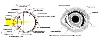
| Figure 4. Eyelid: vertical section |
Blepharitis is a common disease, although often missed by doctors, since the clinical signs are too subtle, despite the complaints of patients whose condition, as a rule, worsens in the morning.
Staph infection affects the base of the eyelashes, causing chronic irritation, itching and soreness, moderate photophobia and weeping. The blood vessels at the edges of the eyelids dilate. Clusters of dry flakes at the base of the eyelashes are called lace collars. After removal of such collars, small ulcers often remain on the edge of the eyelid, which tend to deepen and hypertrophy.
Sometimes the eyelashes curl inward, causing a foreign body sensation and damage to the cornea. Hypersensitivity to staphylococcal exotoxin can lead to inflammation of the inferior tarsal conjunctiva and punctate corneal erosions, usually affecting the lower third of the cornea.
Seborrhea can be oily or dry. It is believed that the excess neutral lipids produced in this condition are broken down by Corynebacterium acnes into irritating fatty acids.
Symptoms and signs of seborrheic blepharitis are similar to those of staphylococcal blepharitis, but less pronounced. The eyelids seem greasy and the eyelashes stick together. Soft scales are easily removed.
In posterior blepharitis, oil globules can sometimes be seen on the posterior edge of the eyelid, and the conjunctiva is moderately injected.
| Figure 5 (top left). Blepharitis |
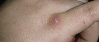
| Figure 6 (top center). Molluscum contagiosum |
| Figure 7 (top right). Dermoid cyst |
| Figure 8 (top). Basal cell carcinoma |
Styes, meibomian cysts and marginal keratitis often develop against the background of blepharitis.
Treatment of blepharitis consists of constant monitoring of the patient’s condition and minimizing the inconvenience that arises from it, since a cure is not possible. The mainstay of treatment is eyelid care, which includes removing crusts and toxic substances from the edges of the eyelids.
To do this, take a flannel or cotton ball soaked in a weak solution of baby shampoo or soda. This procedure is carried out twice a day, and less often when the condition stabilizes (as necessary).
If there are signs of a staphylococcal infection, antibacterial ointment is applied to the edges of the eyelids.
P
For severe exacerbations, when the diagnosis is beyond doubt, 0.5% eye drops with prednisolone and neomycin or 0.5% hydrocortisone eye ointment can be prescribed. They quickly relieve discomfort and inflammation, but should not be used long-term due to the risk of developing iatrogenic glaucoma and cataracts.
Dry eyes are characteristic of staphylococcal blepharitis, and in this case artificial tears help. It is also important to treat other areas of seborrheic dermatitis, especially the scalp and eyebrows. Difficulties may occur in the treatment of posterior blepharitis, so it is better to refer such patients to the ophthalmology department. It may be necessary to re-examine the glands, which is best done using a slit lamp.
- Tumors
Benign tumors.
Molluscum contagiosum, a viral infection most often found in children, can also affect the eyelids. Desquamation of virus-laden cells causes follicular conjunctivitis and, rarely, superficial keratitis. The lesions are painless and look like pearls with a depression in the center. Treatment consists of squeezing or cauterization, but uncomplicated lesions should not be touched; they go away on their own.
Common benign skin tumors, including warts, squamous papillomas, dermoid and fatty cysts, actinic keratoses and seborrheic keratoses, also affect the eyelids. Less common are keratoacanthomas and cutaneous horn.
The principles of treatment are the same as for tumors of other locations, but any lesion requiring excision that crosses the edge of the eyelid should be referred to an ophthalmologist.
Hemangiomas.
Hemangiomas often occur in children, most often in the first six months of life. These flat and red lesions disappear spontaneously by age five, requiring no treatment unless they obstruct the visual axis. If in doubt, send your child to the eye department.
Xantellasma
- this is the accumulation of lipids in the medial part of both eyelids, they are slightly raised above the skin level and have a yellowish color; as a rule, they indicate an increase in blood lipid levels. They can be excised, but usually they appear again.
- Malignant tumors
Basal cell carcinomas (BCCs) are the most common malignant tumors of the eyelids. As a rule, they affect the lower eyelids and medial corner of the eye. The typical age of patients is 60 years and older.
| Figure 9. BCC: The appearance of BCC varies from a pearl-like nodule to a typical “gnawed out” ulcer. The sclerosing type is a flat formation with poorly defined boundaries |
The appearance varies from a pearl-like nodule to a typical “gnawed” ulcer. The sclerosing type is a flat formation with poorly defined boundaries
These tumors do not metastasize, but are prone to local invasion (especially those located in the medial corner of the eye). The method of choice in the treatment of such tumors is their removal, involving 3 mm of the eyelid margin, followed by histological examination.
Squamous cell carcinomas (SCC) account for only 5% of the total number of malignant eyelid tumors. They appear as an ulcerated area, nodule, or papilloma. They are characterized by faster growth than BCC and the ability to metastasize to the preauricular and submandibular lymph nodes.
Treatment is the same as for BCC, but a larger area is excised.
Sebaceous gland carcinoma is a rare tumor of the meibomian glands. It is a separate nodule of a slightly yellowish color; it can easily be mistaken for a relapse of chalazion. Multifocal tumors resemble chronic blepharitis with areas of inflammation along the eyelid margin.
This tumor metastasizes, with a five-year mortality rate of 30%, perhaps in part due to diagnostic difficulties. Treatment is surgical, the same as for PCI.
- Deformations of eyelids and eyelashes
| Figure 10 (top). Squamous cell carcinoma accounts for only 5% of malignant eyelid tumors |
Ectropion.
It is an inversion of the lower eyelid, leading to weeping and chronic inflammation of the conjunctiva. Gradually the conjunctiva hypertrophies and thickens.
A common form of involutional ectropion, in which the muscles and tendons weaken and the horizontal dimension of the lower eyelid lengthens. Due to the fact that the eyelid is not adjacent to the eyeball, there is an excessive amount of tears and damage to the cornea with the development of keratitis and a foreign body sensation.
Treatment depends on the degree of eyelid weakness. Mild weakness in the medial part of the eyelid is corrected by applying a thermal cautery to the conjunctiva. In more severe cases, eyelid shortening is required. Other causes of ectropion include scarring, both congenital and caused by facial paralysis.
| Figure 11 (bottom). Entropion |
Entropion.
Entropion is an inward turning of the eyelid, causing discomfort, photophobia, and weeping.
The most common form is involutional, or senile, entropion. The eyelid turns inward as its cartilaginous plate thins and atrophies, as a result of which the upper edge bends more than the lower one. The fatty layer of the palpebral fissure also becomes thinner, and enophthalmos is often observed, aggravating the patient’s condition.
Temporary relief comes from attaching the eyelid to the cheek with bandages or removing the eyelashes to prevent damage to the cornea. However, the radical treatment method is surgery.
Trichiasis.
Eyelashes that curl inward and scratch the cornea cause discomfort, photophobia and weeping, as in entropion. Treatment consists of epilation by electrolysis or cryotherapy at least every three months. The latter can lead to depigmentation of the eyelid margin, especially noticeable in dark-skinned patients.
Districhiasis.
In this condition, the patient has a second row of eyelashes on the inner edge of the eyelid. They can be congenital or appear as a result of scarring of the eyelids. Treatment is the same as for trichiasis.
Note!
- For recurrent meibomian gland cysts, it is necessary to rule out meibomian gland carcinoma using a biopsy.
- Treatment of blepharitis consists of strict monitoring of the patient’s condition and minimizing the inconvenience that arises from it. Therapeutic measures are based on eyelid care; if there are signs of staphylococcal infection, antibacterial ointment is applied to the edges of the eyelids. For severe exacerbations, prednisolone and neomycin drops quickly relieve discomfort and inflammation, but they should not be used for a long time
- The most common eyelid tumors are basal cell carcinomas. As a rule, they affect the lower eyelids and medial corner of the eye. The typical age of patients is 60 years and older. These tumors do not metastasize, but are prone to local invasion, especially those located in the medial corner of the eye
- Treatment of ectropion depends on the weakness of the eyelid. Slight weakness of the middle part of the eyelid is corrected by applying a thermal cautery to the conjunctiva. Medial conjunctival repair involves removing a diamond-shaped area of tissue just below the reshaped area. In more severe cases, eyelid shortening is required
- With entropion, temporary relief comes from attaching the eyelid to the cheek with bandages or removing the eyelashes. The radical method of treatment is surgery