Inflammation of the mammary glands
- Fact 1:
It is quite difficult to detect mastitis at the initial stage. - Fact 2:
If you delay a visit to the veterinarian, then, most likely, taking medications and massage will no longer be enough - Fact 3:
The hemorrhagic form is the most severe. Extensive bleeding occurs in the skin and mammary glands - Fact 4:
Diagnosing the disease yourself is difficult
Mastitis is an inflammation of the mammary glands, either one or several at once. Animals during lactation, pregnancy or false pregnancy are at risk. The development of the disease goes through several stages. The initial one heals quickly. Treatment at home is possible. The disease, which has taken a severe form, often requires immediate surgical intervention.
The first signs of the disease
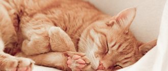
It is quite difficult to detect mastitis at the initial stage. Firstly, many pregnant or lactating cats do not like touching their bellies and try to avoid them. Secondly, in pets with thick hair it is not so easy to see the nipples, which become swollen and red as the disease develops.
The first signs indicating any deviation are often changes in the cat’s behavior:
- she begins to lick her breasts frequently;
- sometimes refuses to eat;
- After feeding the offspring, she looks lethargic and apathetic.
In this case, you need to examine the mammary glands. If the nipples and the tissue around them are red and swollen, and the body temperature has increased, we can talk about the development of pathology. It is better to immediately contact a veterinary clinic.
Treatment of the disease in the initial stages, as a rule, is limited to conservative methods, does not require surgical intervention and ends successfully. If you delay a visit to the veterinarian, then, most likely, taking medications and massage will no longer be enough.
Factors contributing to successful mating
Domestic cats become sexually mature at 6–8 months, depending on the breed and individual characteristics. Neither males nor females are ready for full mating in the first “hunt”. Stray animals mate at this age, which leads to death or infertility in an average of 30% of females, a reduction in sexual productivity in 50% of males - a natural way of controlling the birth rate and population of wild animals.
It is believed that the female is ready to bear kittens at the age of 1.3–1.5 years. For successful “cooperation” with a cat, the owners of the female need to take into account several points:
- The search for a “spouse” may take a long time; you need to start 4–6 months before the planned mating. Remember that animals may not like each other, so have a backup plan.
- Three months before mating, cats must be examined for infectious diseases, and tested for viruses a month before.
- The cat is not vaccinated for 14–20 days.
- Preventive deworming is carried out 14 days in advance.
- Future partners do not bathe and trim their nails immediately before mating.
- The meeting takes place on the territory of the cat; appropriate conditions must be prepared for the female.
Important! If your pet is not of breeding value and you do not plan to have offspring, the animal must be sterilized at the age of 7–12 months. The period is considered optimal - the animal’s hormonal levels have established, but it is not yet ready to mate. According to international rules for keeping pets, all animals that do not represent breed value or have genetic abnormalities must be sterilized.
Causes
There are many reasons why mastitis begins to develop. At risk are animals with weak immunity, low body weight, or those who have just acquired any disease.
Other factors that provoke the development of the disease:
- bruises;
- hypothermia;
- mammary gland injuries;
- infection or fungus - inflammation occurs when bacteria penetrate through micro cracks in the skin or due to the influx of infected blood from internal organs during a systemic disease;
- complications during or after childbirth;
- stagnation of milk due to early weaning of kittens, small number of cubs in the litter, death of offspring;
- false pregnancy.
As a rule, mastitis develops under the influence of several factors, and is not a consequence of one.
When is self-medication unacceptable?
Cat injections for mastitis are prescribed for purulent, fibrinous or hemorrhagic forms. If the pathology is caused by a bacterial infection, then antibiotics are necessary. The most effective medications in this category are considered to be antibiotics of the penicillin (Ampicillin, Amikacin) or cephalosporin (Cefaloridin, Cefadroxil) group.
The drugs are well tolerated by animals and do not cause complications. If the disease was caused by a fungal infection, then for medicinal purposes it is necessary to use antifungal drugs (Amphotericin, Griseofulvin).
Kinds
There are several forms of the disease. The initial stage is usually accompanied only by redness and slight swelling of the nipple, and a slight increase in body temperature. It often goes unnoticed by humans. If left untreated, it progresses to more severe forms of mastitis:
- Serous . The mammary glands enlarge significantly. They become hot and hard, and the cat reacts painfully to touch. When pressed, liquid with white flakes is released from the nipple.
- Catarrhal . When pressed, a liquid is released, the consistency of which is similar to sour milk.
- Fibrinous . Characterized by increased body temperature and enlarged lymph nodes. On palpation, “crunching” sounds are heard.
- Purulent - a complication of previous forms. An unpleasant-smelling fluid mixed with blood and pus is released from the chest. In most cases, at this stage the disease becomes chronic. The mammary glands fill with connective tissue, which can lead to a lack of milk in the future.
The hemorrhagic form is the most severe. Extensive hemorrhage occurs in the skin and mammary glands, staining the tissues bright red. The animal's temperature rises and it becomes feverish. Surgery required.
Atypical fibroepithelial hyperplasia of the nipples in a Sphynx cat
E. Ozenc, MF Bozkurt, Faculty of Veterinary Medicine, Afyon Kocatepe University, Afyonkarahisar, Turkey
Fibroepithelial hyperplasia (FEH) of feline mammary glands (also known as fibroadenomatous hyperplasia or feline mammary hyperplasia) was first described in 1973 [1, 6]. This condition is characterized by non-tumor proliferation of interlobular canals and surrounding mammary gland stromal cells [5]. FEG develops in the tissues of the mammary glands under the influence of progesterone from the corpus luteum and is most often observed in young, mature or non-adult, unsterilized cats [1, 7, 9, 6]. Clinical cases of FEG have also been reported in cats of any age receiving synthetic progestins [7, 8, 9, 10]. To our knowledge, to date, no cases of FEG in the nipple area have been described in the veterinary literature. The purpose of this study is to evaluate a clinical case of cat nipple FEG in terms of clinical and pathological parameters.
Despite the fact that the Sphynx is known as a breed of hairless cats, they can have fur on the nose, tail and toes [14, 4]. In Europe, the number of Sphynx cats tended to increase, and at the moment this cat breed is one of the most popular in the world [3, 2]. Despite the fact that studies have been published on the topic of skin diseases, muscular dystrophy and heart disease in Sphynx cats [12, 17, 3, 15], there are no reports in the veterinary literature on hyperplasia and tumors of the mammary glands in this breed.
Description of a clinical case
A three-year-old Sphynx cat was taken to the clinic of the Department of Obstetrics and Gynecology of the Faculty of Veterinary Medicine at Afyon University due to tumors found in the nipples. The anamnesis revealed that the cat had kitted twice. The neoplasms first looked like redness, and then over the course of 6–12 months they took on the appearance of masses hanging from the ends of the nipples. Clinical examination revealed the presence of these neoplasms on all nipples except the right inguinal (Figure 1A). When examining the lobes of the mammary glands, no inflammation or neoplasms were detected.
| Photo 1. A) Macroscopic appearance of atypical fibroepithelial hyperplasia of the nipples. B) A ligature is placed between the nipple and the tumor. B) The tumor was cut above the ligature and removed. D) Nipples and mammary glands two months after surgery |
Nipple tumors were removed under general anesthesia. The cat's premedication consisted of 0.045 mg/kg atropine sulfate (Belladone, 2 mg/ml, Alke, Istanbul, Turkey). General anesthesia was performed with 2 mg/kg xylazine hydrochloride (Alfazyne® 2%, Ege Vet, Izmir, Turkey) and 10 mg/kg ketamine hydrochloride (Alfamine® 10%, Ege Vet, Izmir, Turkey). The surgical site was prepared and disinfected while the animal was in the supine position. To remove the tumor, a ligature is placed on the nipple between the nipple and the beginning of the tumor (photo 1B). Resection of the mass was performed over the ligature, after which the mass was removed so that the ligature remained on the nipple (Figure 1B). During a control study carried out in the second month after surgery, no pathologies of the nipples and mammary glands were detected (photo 1D). However, a year after the operation, a similar neoplasm was discovered above the right inguinal lobe of the mammary gland, where it had not previously been observed. It was removed under general anesthesia using a similar technique, and all lesions were subsequently sent to the pathology laboratory.
On gross examination, the right thoracic, right anterior abdominal, and right posterior abdominal nipples were 0.7 cm, 1.3 cm, and 1.3 cm in diameter, respectively; the left thoracic, left anterior abdominal, left posterior abdominal and left inguinal nipples had diameters of 1.4 cm, 0.6 cm, 1.5 cm and 0.9 cm, respectively, and were distinguished by a hard consistency upon palpation. In addition, the surfaces of the nipples were ulcerated, the skin was 2–5 mm thick and brownish-black in color. The surfaces of the sections had a homogeneous gray color.
Tissues were fixed in 10% neutral buffered formaldehyde, processed and embedded in paraffin, cut into 5-micrometer sections, and stained with hematoxylin-eosin (HE) and trichrome.
| Photo 2. A, B) Histopathological appearance of atypical fibroepithelial hyperplasia of the nipples. GE and trichrome B) Histopathological appearance of purulent inflammation and ulceration of the skin. GE D) Vimentin-positive connective tissue cells around the adenoid region. AEC chromogen, Gill hematoxylin D) GMA-positive myoepithelial cells at the base of areas of hyperplasia E) JAPC-positive gland cell nuclei, AEC chromogen, Gill hematoxylin |
On histopathological examination, cells initially derived from the mammary tubules were observed, which, however, penetrated beyond the tubules to give rise to areas of developing epithelial hyperplasia. These cells were completely surrounded by a large area of connective tissue consisting of fibrocytes and collagen-rich fibroblasts (Figure 2A, 2B). All of these changes were accompanied by foci of ulceration consisting predominantly of neutrophilic leukocytes, red blood cells, and bacteria (Figure 2B).
Immunohistochemical studies were performed using the following antibodies to distinguish between connective and muscle tissues: vimentin (1/200 dilution, Abcam, ab28028), desmin (1/50 dilution, Genetex, GTX28592) and smooth muscle actin (SMA, 1/100 dilution, SantaCruz, SC-53142). Proliferating cell nuclear antigen antibody (NPC, 1/500 dilution, Abcam, ab18197) was used to detect changes in cell proliferation. For this purpose, the antigen was isolated by exposure to microwaves on a swab soaked in a citrate solution. After incubation with primary antibodies, biotinylated anti-rabbit and anti-mouse IgG (BA-1100, BA2000, Vectorlab) were used as secondary antibodies. A conjugated avidin-biotin complex (ABA) kit (PK-6100, Vectorlab) was used along with peroxidase enzyme. Tissue samples were stained with 3-amino-9-ethylcarbazole (AEC, Invitrogen, 00200) substrate. Gill's hematoxylin (I) was used to stain the background. Slides were covered with watery mounting medium. All slides were examined and photographed under a light microscope.
Immunohistochemical analysis showed that the cells surrounding the hyperplastic epithelium were positive for vimentin (Figure 2D) and negative for desmin and GMA. Myoepithelial cells at the base of the hyperplastic areas were positive for GMA (Figure 2E). Proliferative activity, measured by JAPC staining, reached 50% in hyperplastic cells (Figure 2G).
Based on the research results, these nipple neoplasms were diagnosed as atypical fibroepithelial hyperplasia.
Discussions and conclusions
This is the first description of an atypical presentation of feline fibroepithelial nipple hyperplasia in cats. The literature mentions fibroadenomatous changes in the mammary glands in cats as a result of exogenous or endogenous intake of progesterone into the body of young and pregnant animals [13]. These lesions could involve one or more mammary glands and were capable of rapid growth, thereby causing clinical problems. In this case, no pathological neoplasms were found in the tissues of the mammary glands. FEG was detected only at the ends of the nipples. In addition, the animal was not pregnant and there was no history of progesterone use. This clinical case is characterized by eight nipple neoplasms observed over a two-year period. Since the cat was three years old, we believe that similar neoplasms are also possible in older animals.
| Photo 3. Cat litter |
Acrylic, polyester and cotton fabrics are often used as bedding materials for cats. In this case, plush polyester fur was used as bedding (photo 3). The chemical mercaptobenzothiazole, which has irritant properties, is used in the production of synthetic polyester fibers. In humans, direct contact with this material or the substance used in its production can cause skin pathologies, such as, for example, contact irritant dermatitis [11]. Sphynx cats are hairless, and we assume that lying on plush bedding led to the development of local irritation and itching in the nipple area, which subsequently developed into inflammatory changes and FEG. The widespread use of this type of bedding for the comfort of cats may pose a health risk to the animal and a potential hazard that should be the subject of further research.
Clinically, fibroadenomatous changes are usually presented as painless, dome-shaped, dense, limited areas of breast tissue [16]. Microscopically, fibroadenomatous changes often have an edematous or mucinous fibrous stroma [5]. However, in this case, we observed that the proliferative tubular epithelial cells were completely surrounded by dense fibrous connective tissue containing large numbers of collagen fibers and fibrocytes. The presence of this dense fibrous connective tissue is most consistent with the atypical presentation of feline fibroepithelial hyperplasia. We believe that atypical FEG developed due to the proximity of the nipples to the ground, as well as their chronic irritation in response to friction with the litter.
This report describes the first case of an atypical presentation of fibroepithelial hyperplasia in the nipple region of a cat. We observed bilateral FEG, which can also be observed in older animals.
Literature
1. Allen HL (1973): Feline mammary hypertrophy. Veterinary Pathology 10, 501–508.
2. CFA (2014): Sphynx. Avialable online at: https://www.cfa.org/Breeds/BreedsSthruT/Sphynx.aspx Accessed January 2, 2014.
3. Chetboul V, Petit A, Gouni V, Trehiou-Sechi E, Misbach C, Balouka D, Carlos Sampedrano C, Pouchelon JL, Tissier R, Abitbol M (2012): Prospective echocardiography and tissue Doppler screening of a large Sphynx cat population : reference ranges, heart disease prevalence and genetic aspects. Journal of Veterinary Cardiology 14, 497–509.
4. Gandolfi B, Outerbridge CA, Beresford LG, Myers JA, Pimentel M, Alhaddad H, Grahn JC, Grahn RA, Lyons LA (2010): The naked truth: Sphynx and Devon Rex cat breed mutations in KRT71. Mammalian Genome 21, 509–515.
5. Goldschmidt M, Pena L, Rasotto R, Zappulli V (2011): Classification and grading of canine mammary tumors. Veterinary Pathology 48, 117–131.
6. Gorlinger S, Kooistra HS, van den Broek A, Okkens AC (2002): Treatment of fibroadenomatous hyperplasia in cats with aglepristone. Journal of Veterinary Internal Medicine 16, 710–713.
7. Hayden DW, Johnston SD, Kiang DT, Johnson KH, Barnes DM (1981): Feline mammary hypertrophy/fibroadenoma complex: clinical and hormonal aspects. American Journal of Veterinary Research 42, 1699–1703.
8. Hayden DW, Barnes DM, Johnson KH (1989): Morphological changes in the mammary gland of megestrol acetate-treated and untreated cats: a retrospective study. Veterinary Pathology 26, 104–113.
9. Johnston SD, Kustritz MVR, Olson PNS (2001): Chapter 34 Disorders of the mammary glands of the queen. In: Johnston SD, Kustritz MVR, Olson PNS (eds.): Canine and Feline Theriogenology. 1st ed. W. B. Saunders Company, Philadelphia. 474–485.
10. Loretti AP, Ilha MR, Ordas J, Martin de las Mulas J (2005): Clinical, pathological and immunohistochemical study of feline mammary fibroepithelial hyperplasia following a single injection of depot medroxyprogesterone acetate. Journal of Feline Medicine Surgery 7, 43–52.
11. Maiphetlho, L (2007): Contact dermatitis in the textile industry. Current Allergy and Clinical Immunology 20, 28–35.
12. Martin PT, Shelton GD, Dickinson PJ, Sturges BK, Xu R, LeCouteur RA, Guo LT, Grahn RA, Lo HP, North KN, Malik R, Engvall E, Lyons LA (2008): Muscular dystrophy associated with alpha- dystroglycan deficiency in Sphynx and Devon Rex cats. Neuromuscular Disorders 18, 942–952.
13. Misdorp W (2002): Tumors of the mammary gland. In: Meuten DJ (ed.): Tumors in Domestic Animals. 4th ed. Iowa State Press, Ames, Iowa, USA. 575–606.
14. Robinson R (1973): Canadian hairless or Sphinx cat. Journal of Heredity 64, 47–49.
15. Silverman SJ, Stern JA, Meurs KM (2012): Hypertrophic cardiomyopathy in the Sphynx cat: a retrospective evaluation of clinical presentation and heritable etiology. Journal of Feline Medicine and Surgery 14, 246–249.
16. Sontas BH, Turna O, Ucmak M, Ekici H (2008): What is your diagnosis? Feline mammary fibroepithelial hyperplasia. Journal of Small Animal Practice 49, 545–547.
Volk AV, Belyavin CE, Varjonen K, Cadiergues MC, Stevens KB, Bond R (2010): Malassezia pachydermatis and M nana predominate among the cutaneous mycobiota of Sphynx cats. Journal of Feline Medicine and Surgery 12, 917–922.
Source: Veterinary Medicina 59, 2014 (5). This is an Open Access article distributed under the terms of the Creative Commons Attribution License (https://creativecommons.org/licenses/by/2.0), which permits unrestricted use, distribution, and reproduction in any medium, provided the original work is properly cited
SVM No. 5/2014
Rate material
Like Like Congratulations Sympathy Outrageous Funny Thoughtful No words
How to tell if your pet is sick
You need to monitor your pet's behavior and well-being very carefully. Especially during such an important period of life as pregnancy and feeding offspring. This is always stressful for the body, which means the cat is more susceptible to various infections and diseases.
Symptoms of mastitis in the first stage:
- redness of the nipples;
- slight swelling and compaction;
- increase in body temperature;
- the occurrence of microcracks;
- discharge of a curdled mass when pressing on the chest.
The pet reacts painfully to any touch and often licks its tummy.
As mastitis moves to the next stage, redness and swelling increase, the temperature consistently exceeds the norm by at least 1C, and the tissue around the mammary glands becomes very hard. When pressed, a yellowish or grayish liquid with drops of blood or pus is released.
In these cases, immediate contact with a veterinarian is required. If this is not done, the disease becomes more severe. The animal shows the first signs of intoxication of the body - apathy, fever, weakness.
What changes occur before childbirth?
It is very important for the owner to know the due date, because it is unknown how this process will go, and it may be necessary to provide assistance to the pet. You can find out when this period is approaching using the following signs:
- About 3 days before the onset of birth, the cat begins to look for a secluded corner, she looks for a suitable place, and begins to build a nest. To prevent this process from happening on your bed, it is best to prepare for it in advance. You can offer her a box covered with a warm cloth and a sheet on top.
- The cat's nipples sharply increase in size, from which colostrum is secreted. Mucus discharge is observed from the genitals.
- The temperature drops sharply by about 1 degree and can reach 37 degrees.
- 10 hours before the birth of the offspring, the pet hides in its nest. There is no need to disturb her; you can help at this stage by providing fresh water.
Diagnostics
It is difficult to diagnose the disease on your own. Therefore, at the slightest change in the behavior of a nursing or pregnant cat, it is better to consult a veterinarian. In order to understand that something is wrong with your pet, you need to carefully examine its mammary glands and abdomen. If the nipples have increased in size, become dense and hot to the touch, and look inflamed, we can talk about the development of mastitis.
You can make sure the diagnosis is correct by gently pressing on the nipple. You need to do palpation yourself carefully, since the inflamed glands cause severe pain to the animal. If uncharacteristic discharge appears, you should definitely consult a doctor.
Another sign indicating negative changes in the body is increased body temperature. In the first stages, it is 1–2 °C higher than normal.
What to do
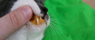
The first thing to do is to carefully examine the nipple yourself. To understand the nature of the disease, whether there is a tumor under the nipple and to see the cat’s reaction.
The next step is to take your pet to a doctor for examination . Only a specialist can accurately determine the cause of the tumor and decide what needs to be done. It may be necessary to remove breast tumors surgically if the cancer is in its early stages. Or, if it is mastitis, and the tumor is the remains of milk. You will need to take a course of antibiotics. In any case, both diagnosis and treatment are prescribed only by a veterinarian
Treatment
If mastitis is detected in a timely manner and a visit to a veterinary clinic is performed, surgical intervention can be avoided.
Often only conservative methods are sufficient. In order to choose the right treatment regimen, the owner is usually asked to tell how the birth and pregnancy went, and they are asked general questions about the cat’s health. This allows you to identify the cause of mastitis and prescribe the correct treatment. This means that it will facilitate and speed up the recovery process for your pet after an illness. Before you start taking medications, your cat will have to undergo tests. To do this, a drop of fluid from the mammary glands is taken for laboratory testing. These actions reveal the nature of inflammation.
After the preparatory stage and receipt of test results, the doctor prescribes treatment. If mastitis becomes severe, antibiotics are prescribed. Such measures make it possible to stop the inflammatory process and prevent its spread to surrounding tissues. As an auxiliary therapy the following is prescribed:
- massage;
- expressing milk;
- washing inflamed areas with infusions of medicinal plants (chamomile, sage, oak bark);
- treating nipples with products for external use that relieve inflammation and external symptoms.
It is recommended that cats reduce the amount of water they consume. Temporarily switch to feeding dry ready-made formulas.
Only the most severe forms require surgical intervention, when mastitis develops in several mammary glands at once, they become clogged, and impurities of blood and pus appear in the milk. In this case, kittens are taken away from their mother without fail.
Caring for your cat after surgery
The result of treatment depends not only on timely surgery. Surgical removal of the tumor is only the initial stage of the fight against cancer. Postoperative care plays an important role. A pet who has undergone surgery must be provided with complete rest. In addition, you must follow your veterinarian's instructions and follow all recommendations regarding your cat's diet.
The seam must be regularly treated with a special product, which is also prescribed by the veterinarian. In addition, the animal must be given a number of medications, strictly observing the time of administration and dosage. This will help avoid complications.
For some time after removal of the tumor, the cat must wear a special blanket. It is designed to protect the seam and prevent inflammation. The owner must carefully ensure that the blanket is held securely and does not come undone.
Scheduled inspections are carried out weekly. To assess the animal’s condition and understand how its recovery process is going, the veterinarian will take the necessary tests. However, even the most experienced doctor cannot say exactly how long a cat will live after surgery.
How to avoid illness
In order to avoid developing mastitis, you need to take some precautions that will help avoid problems in the future. Very often the cause of the disease is hypothermia, so a pregnant or lactating cat should live in a warm and dry room without drafts. It is better to move a pet living outside for a while indoors.
The second common cause of mastitis is infection. Kittens may accidentally scratch or damage the skin around their nipples while feeding. This, in turn, will lead to infection in the wounds and inflammation. You can avoid this outcome by regularly removing animal bedding and washing the kittens’ paws. A person should wash their hands thoroughly before touching a cat's belly.
A preventive measure that is of great importance is a proper balanced diet. You can choose special dry food for your pet. It would be good if it belongs to the premium class. Before purchasing a mixture, it is better to consult a veterinarian or breeder so as not to harm the health of the animal.
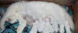
It is very important to wean kittens correctly. When babies begin to eat on their own, the cat's mammary glands need to be bandaged. This way you can reduce the amount of milk produced and protect your pet from infection.
Diagnostic definition of pregnancy
Some owners are interested in the question of whether it is possible to determine whether conception has occurred in an animal using a human test. Veterinarians give a clear answer that the test is not suitable for mustaches. It is possible to determine whether the female managed to become pregnant using ultrasound only after the first 2 weeks. After 5 weeks, the veterinarian will perform a palpation test and tell you how many kittens she will give you.
You can find out whether your pet managed to get pregnant using a clinical test for relaxin. This test can provide reliable information only 3 weeks after the expected day of the onset of an interesting situation; it requires blood plasma. The result will be ready in 10 minutes. You can determine whether your attempt to get pregnant was successful using an x-ray 5 weeks after mating.
Diagnostic measures for suspected cancer
It is impossible to independently determine the nature of a lump on a cat’s chest. Only a professional doctor can do this. In a veterinary clinic, the doctor carefully examines the animal’s mammary glands and other parts of the body; by palpation will determine the characteristics of the lump, its volume, consistency, skin temperature, etc. Then he will prescribe laboratory and hardware diagnostic methods, including :
- urine and blood tests;
- X-ray;
- Ultrasound.
© shutterstock
If cancer is suspected, the cat will undergo a biopsy by pinching off a piece of the lump under anesthesia and sending the pathogenic tissue for examination. Specialists will determine whether malignant cells are present in it.
Therapy for cancer
Treatment for mammary cancer in cats is usually surgical. Not only the tumor on the mammary gland is removed, but also the entire number of glands. And sometimes it is necessary to amputate both rows with deep involvement of the subcutaneous tissue, which is called a total mastectomy. If there are affected lymph nodes, they are also removed.
Such radical measures are necessary in order to reduce the risk of the oncological process spreading from the lump to neighboring tissues. After all, one remaining cancer cell is enough for the process to resume.
In addition to surgery for mammary cancer in cats, chemotherapy is sometimes indicated. It is carried out under the supervision of a specialist. This treatment should be taken very seriously, doing everything strictly according to the protocol. Chemotherapy exhausts a cat's body.
A large number of medications are required that will have a supporting effect and will not allow other diseases to take hold. Chemistry almost completely kills the immune system. During this period, the cat can die from any draft. The animal needs good care.
Chemotherapy alone is rarely used to treat breast cancer . More often it is combined with surgical removal of the lump and nearby tissues. Sometimes radiation is used instead of chemotherapy. In some cases, all three methods are combined.
Specialists undertake to treat malignant lumps in cats in the early stages, when the prognosis is still quite favorable. In advanced situations, any therapy is useless. She will only torment the cat. If stage 4 has already occurred, veterinarians usually recommend euthanizing the unfortunate animal.
© shutterstock
Symptomatic picture
With cancer, the appearance of lumps on a cat's chest may not be immediately detected. At the first stage, the nodules are very tiny. Their maximum size is 1-1.5 cm, and since they are hidden under the fur, the owner does not pay attention to them. The behavior of a cat in the initial stages of mammary cancer also remains normal. She eats normally and is as active as before.
© shutterstock
Alarming symptoms often appear only at the 3rd stage, when the lump reaches a size of 3-5 cm. It is characterized by the following features :
- clear boundaries;
- immobility;
- dense consistency;
- the temperature of the skin over the tumor is not increased;
- slight soreness.
With advanced mammary cancer, the lump becomes covered with ulcers that open and bleed, causing severe pain to the cat. The animal looks depressed, loses appetite and sleep, and loses a lot of weight. Lumps may appear on other parts of the body - limbs, neck, head. These are metastases.
Malignant bumps differ from benign ones, first of all, by their rapid growth. If the tumor was small a week ago, but has now reached several centimeters, the cat should be urgently shown to a doctor. The sooner treatment is started, the better.