Pleuritis is an inflammation of the pleura that occurs in animals of any age, sex and breed. The pleura is a serous membrane that lines the inside of the chest cavity and also covers the lungs. The pleura forms symmetrical right and left sacs, the space between which is called the mediastinum. The mediastinum contains the trachea, esophagus, heart, large blood vessels, lymph nodes, and nerves.
A special feature of the pleura is that between its layers there are slit-like cavities that contain a small amount of serous fluid. It is needed to reduce the friction force of the pleura during breathing.
In addition, each pleural layer has microscopic slit-like openings in the mediastinum. Under unfavorable conditions, this contributes to the rapid transition of pathological processes from the pleura to the lungs and vice versa.
Classification of pleurisy
By origin, pleurisy is primary and secondary.
Primary pleurisy is a lesion of the pleura itself, when the process of inflammation initially develops in its tissues.
And secondary pleurisy is a complication of the disease of neighboring organs. In this case, first, for example, lung diseases, tumor processes, etc. arise, and after this the inflammation spreads to the pleura. Secondary pleurisy is much more common than primary pleurisy.
According to the course, pleurisy can be acute, subacute, or chronic.
A cat usually suffers from acute pleurisy for up to 14 days, subacute can last up to 1.5 months, and chronic pleurisy lasts for several months or even years.
Based on the localization of the process, a distinction is made between limited, that is, local pleurisy, and diffuse, which spreads in different places of the pleura.
Unilateral pleurisy can affect the right or left side of the chest cavity, and bilateral pleurisy can affect both.
Depending on the nature of the process and the presence of effusion, pleurisy can be wet or dry.
With wet pleurisy, inflammatory fluid is released into the pleural cavity. The composition may be dominated by blood, pus, protein, microorganisms and fragments of decayed tissue.
With dry pleurisy, the exudate is rich in protein - fibrinogen. It easily curls up and settles on the surface of the pleura, while liquid does not accumulate.
Epidemiology
The literature regarding this pathology contains a small number of retrospective studies. However, this allows us to get an idea of the extent of its manifestation.
If we talk about breed predisposition, then, probably, chylothorax of an idiopathic nature is found in the Afghan hound, although the authors themselves (TW Fossum et coll., 1986) do not attach much importance to this. Most cases of chylothorax are found in medium and large breed dogs. There is only one case of this pathology described in a poodle (NC Myers et al., 1996). As for cats, the greatest predisposition was noted in European and Himalayan breeds (TW Fossum et al., 1968–1989).
Chylothorax can occur regardless of age, but its idiopathic nature is more often noted at 5–6 years of age (TW Fossum et coll., 1969-1989).
Causes of pleurisy in cats
Factors that cause inflammation can enter the pleura from the lungs, through the bloodstream or lymphatic system.
These include:
1. Pathogens: viruses (for example, FIP), bacteria (cocci), fungi, protozoa, helminths.
2. Inflammatory processes that spread to a number of underlying organs. For example, from the lungs (pneumonia, abscesses), pericardium, etc.
3. Tumors of the lungs, esophagus, mediastinal lymph nodes or other organs of the chest cavity.
4. Injuries and operations on the pleura and chest.
5. Pathologies of the abdominal organs:
- pancreatitis;
- renal failure (uremic pleurisy);
- When the diaphragm ruptures, organs are displaced from the abdominal cavity into the chest cavity and compress the lungs, causing inflammation of the pleura.
6. Taking medications, for example, furadonin.
7. Predisposing factors: spring - autumn period, hypothermia, overwork, unbalanced nutrition and transportation, weakened immunity, stress.
Prevention of hydrothorax
Prevention of thoracic hydrops in cats is aimed at preventing factors that can lead to this disease (mechanical injuries, exhaustion of the body, infectious and parasitic diseases). Pay attention to several useful recommendations that, in some cases, will help avoid the development of hydrothorax in cats:
- Vaccinate your pets against infectious diseases in a timely manner at the veterinary clinic. It is important to consider which infections often affect animals in the region where you live.
- Carry out antiparasitic treatment (deworming) 1-2 times a year. This applies not only to those cats that have the opportunity to go outside, but also to pets.
- Always consult a veterinarian for diseases of the cardiovascular system, kidneys and liver. This will avoid the possibility of developing thoracic hydrops in cats as a concomitant condition with the disease.
- Pay attention to your pet's diet . It must be healthy and balanced. To provide additional prevention of hydrothorax, try to fortify it with vitamins C and K.
Remember that the main thing with chest dropsy is to promptly seek help from a veterinarian!
Symptoms of pleurisy in cats
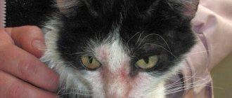
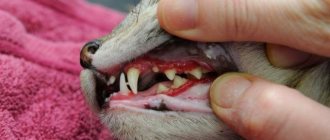
- Depression, elevated body temperature; the mucous membranes of the oral and nasal cavities, the conjunctiva are bluish in color. In severe cases, exhaustion occurs.
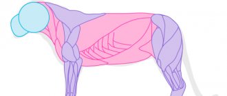
- Respiratory system disorders: - shallow rapid breathing, increased heart rate, dry painful cough; - shortness of breath with exudative pleurisy with difficulty breathing; - if the layers of the pleura grow together as a result of inflammation, then abdominal breathing is observed, which is carried out due to contraction of the diaphragm and the abdominal wall; - with unilateral exudative pleurisy with a large amount of fluid, the chest becomes asymmetrical during breathing, and the cat takes a forced position that makes breathing easier;
- Pain in the chest when feeling and listening;
- Brown, foul-smelling urine, dry feces.
Symptoms
What are the symptoms of this disease? Let us list the most typical ones, allowing us to more or less accurately make at least a preliminary diagnosis:
- Difficulty, or sharply rapid, shallow breathing.
- The cat takes a sitting position and stretches its head up. A very specific symptom, indicating the accumulation of a large amount of exudate. It is important to remember that in a different position the animal simply cannot breathe.
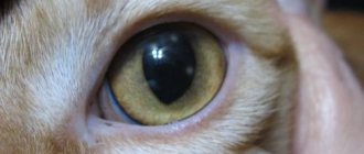
- Cyanosis (blue discoloration of all visible mucous membranes).
- The cat begins to breathe through its mouth.
- Loss of appetite.
- Lethargy, apathetic state.
For many cats, the most characteristic symptom is a wide open mouth, rapid and very shallow breathing. As a rule, it is easier for them to inhale (when exhaling, a strong pain reaction is noted). The development of “abdominal” breathing is also very characteristic.
Diagnosis of pleurisy in cats
First of all, the doctor collects information about the animal’s life history and disease. This is followed by a clinical examination, thermometry, and auscultation (listening to breathing). With dry pleurisy, auscultation reveals a pleural friction rub. Putrefactive pleurisy corresponds to the sound of splashing.
A necessary study is x-ray diagnostics, which shows the presence of pathological fluid in the chest cavity. In addition, the image usually shows the general condition of the heart and lungs.
Pleurisy in cats - radiograph in the right lateral projection
To assess the severity of the condition and the severity of the body's response to the disease, a general clinical blood test will be needed. It pays attention to the increase in neutrophilic leukocytes.
To study the nature of the exudate, thoracentesis may be required - a puncture of the chest wall.
Also, as necessary, diagnostic measures are carried out for concomitant diseases.
Diagnostics: examination, x-ray and thoracentesis
Diagnosis of pulmonary pleurisy begins with a visual examination of the cat, measuring body temperature, listening to the sternum area with a phonendoscope for the presence of wheezing and noise. The veterinarian will definitely take a general blood test to determine the level of neutrophil leukocytes (during inflammatory processes in the body, their level is usually higher than normal).
One of the effective methods for diagnosing pleurisy in a cat is radiography, which is a method of dynamic research using x-rays. In real time, using this device, the veterinarian is able to monitor the condition of the animal’s internal organs and select the most suitable projection for the image in order to determine the presence or absence of inflammatory processes in the lungs.
The most important condition for making the most accurate diagnosis is a procedure called “thoracentesis”, which consists of puncture of the pleura through the intercostal space for the purpose of diversion, aspiration and further study of the pathological contents. In addition, this type of diagnosis allows you to normalize respiratory function in real time absolutely painlessly, so the procedure is performed without sedation or local anesthesia and is well tolerated by pets.
Treatment of pleurisy in cats
First of all, to treat pleurisy, you will need antibiotics, sulfonamides, analgesics, multivitamins, diuretics, as well as other drugs, based on the symptoms of concomitant diseases.
For purulent and putrefactive pleurisy, thoracentesis is used. It is necessary to remove exudate, followed by washing the pleural cavity with antiseptic solutions.
Prerequisites for a speedy improvement in well-being and recovery are rest and a balanced diet.
Pyothorax in animals
Pyothorax in animals or thoracic empyema is an inflammation of the pleura, characterized by the accumulation of purulent effusion in the chest cavity.
Pyothorax is the result of a bacterial or fungal infection of the pleural cavity. In most cases, pyothorax is characterized by a moderate to significant amount of pleural exudate.
In animals with pyothorax, many pathogens can be cultured, but there is a high frequency of diseases in which a single anaerobic pathogen is identified. The most commonly cultivated species are Bacteroides and Fusobacterium, as well as Pasteurella multocida. Streptococci, staphylococci, various species of Corynebacterium, Clostridium, Enterobacteriacae, Mycoplasma and even some types of fungi are also often found.
The causes of purulent inflammation of the pleural cavity may be:
- penetrating wounds of the chest cavity,
- bacterial pneumonia,
- penetration of foreign bodies,
- perforation of the esophagus,
- spread of infections from the cervical or lumbar spine and mediastinum,
- hematogenous and lymphogenous spread of bacteria,
- perforation of the chest wall,
- osteomyelitis,
- inhalation of cereal awns and their subsequent migration into the bronchi and pleural space.
Type of pleural cavity in a cat with pleurisy
No reliable breed or gender predisposition to the development of pyothorax in small domestic animals has been identified. It is believed that young intact cats involved in fights and receiving chest wounds have an increased risk of developing pyothorax, however, recent research has shown that the most common cause of feline pyothorax is invasion through the lung microflora of the oropharynx. Adult dogs of large breeds (especially hunting dogs) may be predisposed to the development of pyothorax due to the increased frequency of inhalation of foreign plant material (plant spines) and the receipt of penetrating wounds to the chest. Cats with multiple housing may also be predisposed to pyothorax.
The course of the disease depends on the form and severity of the process. Secondary pleurisy can last for months and years (tuberculosis). Purulent and putrefactive pleurisy often ends in the death of the animal during the first decade of the disease.
Symptoms
Pyothorax often has an insidious course, and the appearance of clinical signs may not be evident for a long time. Clinical signs appear due to restrictive processes and include:
- inspiratory dyspnea,
- rapid shallow breathing,
- dyspnea (impaired frequency and depth of breathing, accompanied by a feeling of lack of air.),
- orthopnea (difficulty breathing when lying down).
Additional clinical signs are exercise intolerance, lethargy, anorexia and fever. Chronic or severe infection results in septic shock, dehydration, exhaustion, and hypothermia.
Features of clinical manifestations in dogs:
- depression, anorexia, fever;
- shortness of breath, shallow breathing, abdominal type;
- with dry pleurisy, pain in the intercostal spaces, friction noises coincide with excursions of the chest.
- with effusion pleurisy, splashing noises during auscultation, during percussion - horizontal dullness, regardless of changes in posture;
- body temperature rises by 1-1.5 ° C;
Features of clinical manifestations in cats:
- depression, decreased appetite;
- cyanosis of mucous membranes;
- temperature rises by 1-2°C;
- urine is brown with a foul odor, stool is dry;
- shortness of breath, frequent abdominal breathing;
- upon palpation, the animal becomes restless and groans;
- when a cat lies down, the chest is compressed, which interferes with breathing, so the cat is afraid to lie down;
- The slightest stress leads to a sharp deterioration of the condition.
Diagnostics
The diagnosis is made on the basis of a blood test, chest x-ray and thoracentesis results, followed by cytological and microbiological examination of the resulting fluid.
Laboratory examination reveals pronounced neutrophilic leukocytosis, degenerative shift to the left, anemia of chronic inflammation. Also, when examining blood and urine, signs of secondary infection of organs (hepatitis, pyelonephritis) may be detected.
With thoracentesis, the effusion is not transparent, the color ranges from white to amber and red, the protein content is usually more than 3.5 g/dl. Cytological examination reveals a large number of degenerative neutrophils. Macrophages and reactive mesothelial cells are present in the effusion in varying quantities, depending on the causative agent and the chronicity of the pyothorax. Culture of effusion is indicated in all animals with pyothorax, but positive results are not always achievable, especially when infected with anaerobic organisms.
Left-sided pyothorax in a cat
During X-ray examination, due to the fact that the liquid has a high ability to absorb rays, a typical picture is observed. It is characterized by a sharp division of the projection of the entire pulmonary field into two parts, lower and upper. In the upper part, the shadows of the vertebrae and ribs stand out in contrast, and somewhat condensed root and pulmonary patterns are visible. The lower part of the chest is represented by a continuous, extensive, deeply intense and homogeneous darkening, the upper border of which has a horizontal and sharply contoured edge. Against the background of this uniform dense shading, formed due to pleural effusion, in contrast to pneumonic shading, not even the shadows of the ribs protrude. With extensive effusions, the cardiac silhouette is also not visible.
Treatment
The basis of treatment for pyothorax is drainage.
After the diagnosis is made, a thoracostomy tube is installed through which periodic lavage ( 2-3 times a day) with warm saline is carried out with aspiration of the contents an hour after insertion. The introduction of antibiotics into the lavage solution does not provide any advantages over their systemic administration. The duration of lavage for pyothorax can take up to 5-7 days.
Supportive care is often necessary, including intravenous fluids and nutrition (via a nasal feeding tube or gastrostomy tube) to replace lost nutrients.
The final choice of antibiotic is made based on the results of the culture test, and a combination of antibiotics is prescribed while waiting for the results. It should be remembered that anaerobic microflora is not always determined by culture. The duration of antibiotic therapy for pyothorax is 4-6 weeks.
If the condition does not improve, further investigation should be performed for underlying diseases (eg, feline leukemia virus, viral immunodeficiency, foreign body) or encapsulated abscesses in the lungs or pleura; they can develop as a result of insufficiently timely or insufficiently effective treatment. If an abscess is present, it must be opened after thoracotomy.
In animals with pyothorax, if conservative treatment is not effective, an attempt is made to identify and surgically correct the source of infection (foreign body, lung abscess, volvulus of the lung lobe). Surgical correction may also be indicated to resect the involved tissue and remove debris.
The prognosis for pyothorax is favorable. In animals treated only with systemic antibiotics without lavage, there is a high probability of recurrence of pyothorax. With the development of fibrinous pleurisy, the prognosis may not be favorable.
What types of cancer most often lead to pleurisy?
The prognosis of pleurisy in oncology depends on the organ affected by cancer. The highest survival rate has been established for malignant tumors of the breast and ovaries. The prognosis for pulmonary pleurisy in oncology is much worse. To determine the outcome, the LENT scale is used, which takes into account not only the condition, but also the patient’s functional capabilities.
Most often, pleurisy develops with cancer:
- lung (24-50% of patients);
- ovaries (up to 10%);
- breast (48%).
Less commonly, a complication occurs in the presence of a malignant process in the colon, stomach and pancreas - from 1 to 6% of cases. The prognosis and therapeutic options are determined by the prevalence and nature of the tumor process. The degree of sensitivity to drug therapy matters.
If a malignant tumor is sensitive to cytostatics, as in ovarian, breast and lung cancer, then with timely adoption of therapeutic measures it is possible to prevent relapses of exudation. If systemic therapy is ineffective, pleurodesis is performed to improve the prognosis.
Prognosis for recovery
If you start timely treatment, severe consequences can be avoided. In advanced cases, death is possible. Only a veterinarian can select individual therapy for your pet after passing all the necessary examinations.
Self-medication is strictly prohibited. The accumulated liquid is pumped out only in a medical facility. Otherwise, your blood pressure may rise sharply, causing your heart or breathing to stop (heart or respiratory failure).
A favorable prognosis is possible with timely detection of the disease and effective therapy, which can last for a fairly long period (from one to two months). There is chronic pleurisy, it is treated throughout life. In this case, the owner must carefully monitor the cat’s diet and give medications daily to support the cardiac and respiratory systems.
Preventive actions
It is possible to prevent the development of pathology, the main thing is to avoid hypothermia of the pet. You cannot take your animal outside during the cold season and it is not recommended to bathe it. The owner must carefully monitor the diet: products must contain a large amount of protein; vitamin and mineral supplements can be used as complementary foods.
For preventive purposes, the owner is obliged to bring the cat annually for a routine examination. Monitor your pet to ensure that it does not suffer a chest injury. The healthiest foods to give to an animal are: legumes, fish, lean beef. It is necessary to exclude fatty pork, salty and fried foods from the diet.