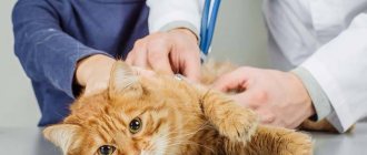
Bladder catheterization in a cat is the only way to save the life of a pet in case of acute urinary retention. Most often, such an emergency condition of an animal occurs due to urolithiasis. With this pathology, stones disrupt the normal flow of urine and cause bladder overflow. If catheterization is not carried out in a timely manner to remove the accumulation of urine from the organ, it will stretch to the limit, and at some point the walls of the bladder will burst due to excessive stretching. It is almost impossible to save a cat in such an emergency situation.
Sometimes catheterization is needed to flush the bladder for therapeutic purposes. Placement of a catheter for any purpose is carried out only by a doctor.
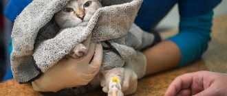
© shutterstock
Installing an intravenous catheter in a cat
Catheter selection
Stiletto catheters are most commonly used in small animal veterinary medicine. In such catheters, a stainless steel stylet needle is located inside the catheter and protrudes from it by 1-2 mm. The stiletto is used to puncture the skin and veins. After the catheter is inserted into the vein cavity, the stylet is removed from the catheter. These types of catheters are available in a variety of sizes and usually have an outer diameter ranging from 24 to 14 G.
and length from ¾ to 50mm.
The cephalic catheter is the most common IV catheter in dogs and cats:
- 24 G for puppies and kittens
- 22 G for cats and small dogs
Always use personal protective equipment and hygiene.
Hands should be thoroughly washed or disinfected before and after venipuncture.
The danger of the procedure: what is it?
If you do not follow all medical recommendations during catheterization and during further care of the cat, then unpleasant consequences are possible. Often after the procedure, mechanical damage to the urethra occurs, against the background of which an inflammatory reaction is recorded. Catheterization may also be complicated by lack of urination. To eliminate the problem, you will need to take antispasmodics. If the procedure is performed by an experienced veterinarian, then no complications will arise and the procedure will bring a positive result and the cat will no longer have difficulty eliminating urine.
A set of necessary tools
- IV catheter model with feeding kit and red fluid bag;
- intravenous catheter;
- plug or T-port;
- medical tape - two strips, cut to size before starting work;
Additional equipment you will need with a live animal:
- clipper and blades;
- 70% isopropyl alcohol;
- tampon/cotton wool;
- needle;
- syringe;
- sterile saline solution;
- gloves;
Call a veterinarian Moscow
+7(495)162-70-70
Catheter placement
- Your assistant should close the vein by applying pressure to it as close to the catheterization as possible.
- Insert the catheter with the needle at a slight angle to facilitate penetration through the skin into the vein with an upward bevel.
- Once blood appears in the needle hub, hold the needle in place while advancing the catheter from the needle into the vein.
Indications for catheterization
Inflammation of the kidneys and intestines are the main indications for catheterization. A blocked bladder increases potassium levels (hyperkalemia), which affects the heartbeat and can lead to the death of your pet. Once a urinary obstruction is identified, emergency treatment and stabilization are required.
Catheterization is performed for:
- Diagnosis of urolithiasis.
- One-time urine collection for analysis if excretion through cystocentesis is not possible.
- Inclusions of contrast agent during examinations.
- Obstruction therapy.
If your cat has chronic kidney disease, the urethra may need to be dilated. This procedure is designed to provide a permanent opening that allows crystals, mucus plugs, or small stones to pass out of the urethra.
In most cases, a dose of general or local anesthesia is administered before the procedure. Infection of the renal pelvis, kidney stones, and chronic renal failure are possible consequences of recurrent urethral obstruction.
Secure the catheter in place
- Using 15-25mm wide medical tape, place a 10cm long strip, adhesive side up, on the underside of the catheter hub with a mark approximately 1.5cm to the left of the catheter.
- Carefully wrap the tape around the catheter hub and wrap the remaining tape around the animal's limb.
- Perform 2 additional wraps of 15-25 inch wide tape starting midway down the catheter under the hub and wrapping around the leg and continuing proximally over the catheter hub.
- Wrap another 25mm wide piece around the middle under the catheter hub. Make sure it is wide enough so that the injection cap does not come into contact with your skin or hair.
- A successfully secured catheter should prevent any movement of the catheter into or out of the vein.
Installing a drip (part 1)
There are various ways to administer fluids, but the most common method is to use a "drip bag" (a bag containing the fluid to be administered) and a length of "drip tube" attached to a needle that is placed under the skin (or into a vein through a catheter) and most cats are very tolerate fluid therapy well.
The IV bag must be suspended at least 1 m above the cat's level so that the liquid can flow under the influence of gravity. It usually takes a few minutes for the fluid to be administered and it is often helpful to cuddle and pet the cat during this period or offer the cat some food to distract it.
1.Preparation of accessories.
- liquid bag
In most cases, a liquid called Ringer's solution is used.
- sterile IV set
Remove protective packaging.
3.Attach the liquid tube to the bag.
- There will be a large spike at the top end of the fluid line that will fit into the bottom hole of the fluid bag. Remove the rubber covering of the spike and insert it into the fluid bag. Do not let the tenon touch anything before inserting it.
- Rotate the spike so that it fits snugly against the bag of liquid.
4.Fill the dropper about halfway.
There is a drip chamber at the top of the fluid line that allows you to see how fast the fluid is flowing. Squeeze the liquid bag until the chamber is about half full.
Run liquid through the line.
- To pass liquid through the tube, remove the protective cap from the bottom of the tube. Loosen the roller clamp and watch the liquid flow through the tube and out of the bottom hole.
- Do not allow anything to touch the lower fluid line opening. This is the place for the needle.
- Close the roller clamp after a few seconds to avoid wasting too much liquid.
- Place a protective cap at the bottom of the tubing to connect the IV to the catheter or needle.
Hang the bag of liquid.
The bag of liquid should hang about a meter above your cat. If you don't have special equipment, you can place the top opening of the liquid bag on a clothes hanger.
a) In order to use the intravenous method of administering fluids, the catheter is connected to the drip system.
b) To use the subcutaneous fluid injection method, a needle is used to deliver fluid under the cat's skin near the spine.
Attach a new needle.
The bottom of the needle (where it attaches to the fluid line) may have a plastic coating. Remove this cover, if present, and attach the connector on the needle to the lower fluid line hole. The needle will have a different covering to protect the top end (the actual needle that is inserted under the skin). Do not remove this covering until you are ready to begin injecting fluid.
Call a veterinarian Moscow
+7(495)162-70-70
Technique
If pathological processes are detected in an animal, it may be necessary to rinse the bladder. The doctor will perform the procedure as follows:
- To relieve pain, the male needs sedation or general anesthesia. Clinicians use local aerosol analgesics (Lidocaine Asept (spray), Carmolis).
- Sterile lubricant is placed on the tip of the tube. An atraumatic tip lubricated with a synthetic lubricant is inserted into the lumen of the urethra. If the device cannot pass through the excretory duct, the veterinarian will drain the animal's bladder by placing a needle on the outside and releasing the urine using a syringe.
- Once the first 2 cm of the catheter is in the penile urethra, the non-dominant hand is used to pull the preputial opening caudally to continue insertion of the tube.
- When regulating water and electrolyte metabolism in an animal, parenteral fluid therapy is performed. The infusion volume is 45-68 ml of crystalloid per kilogram of body weight over 30 minutes.
- Removal of obstructive materials is carried out using solutions that destroy plugs and mucus clots.
- Surgery may be required to remove stones from the organ to prevent inflammation from recurring.
- When the device completely enters the excretory channel, liquid begins to be released from the tube. The clinician determines the condition of the organ by color, consistency and smell.
- Blood clots are eliminated using a solution of novocaine.
- Cats with life-threatening arrhythmias secondary to hyperkalemia are treated immediately with sodium bicarbonate (stimulation of intracellular potassium metabolism) or calcium gluconate (stabilization of the heart and changes in threshold potential).
If necessary, leave the instrument for several days. The veterinarian prescribes medication and diet therapy at home.
Fluid administration (part 3)
Open the fluid supply tube.
- Monitor the fluid bag to ensure the correct amount is injected.
Be careful not to form a lump.
- With the subcutaneous method of fluid administration, the formation of a lump is normal. It will subside as your cat's body absorbs the liquid.
- If the lump is large and dense, and not the entire amount has entered the skin, you need to make a puncture with another needle in a different place.
Keep an eye on the drip chamber.
- If you notice that fluid is not flowing into the chamber, the needle may be pressed against the skin and blocking the flow. Gently move the needle under the skin without removing it from the tent.
- You may need to do this several times to ensure your cat receives the required amount of fluid.
Remove the needle.
- Once your cat has received the required amount of fluid, close the roller clamp to stop the flow of fluids.
- Slowly remove the needle by backing it out of the tent and cap it.
- Pinch the skin for a few seconds to make sure no liquid leaks out of the tent (a few drops of liquid and blood are normal).
- Remove the used needle from the IV set and install a new sterile needle into the IV set.
Caring for the animal after catheterization
After completing the manipulations, the veterinarian will examine the condition of the tube. If kinks or dents are detected in the system, there is a possibility of injury. The dye used in the procedure is excreted from the body by the kidneys. Antispasmodics are prescribed to relieve unpleasant symptoms.
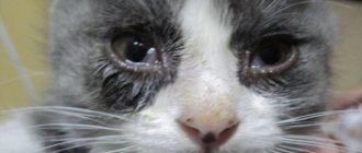
At home, care involves observation. Before increasing the daily amount of fluid consumed, you should consult a veterinarian, because... the animal may have problems urinating. A burning sensation in the urethra occurs when urine is released the first few times. If your pet's discomfort lasts longer, it is a sign of infection. Post-obstructive diuresis is a common complication requiring immediate hospitalization.
To diagnose the causes of discomfort and examine your pet, you need to call a doctor at home.
All information posted on the site is provided in accordance with the User Agreement and is not a direct instruction to action. We strongly recommend that before using any product, you must obtain a face-to-face consultation at an accredited veterinary clinic.